

 PDF
PDF Citation
 Print
Print


INTRODUCTION
Unruptured intracranial aneurysms are relatively common vascular lesions, represents 3–5% of the adult population worldwide [28]. Unruptured aneurysms often remain asymptomatic. Once rupture occurs, they usually result in devastating subarachnoid hemorrhage with a high mortality and morbidity. On the other hand, potentially serious complications related to treatments should not be neglected. Thus, identifying reliable indicators of risk for aneurysm rupture is crucial for clinical decision making. The PHASES (Population, Hypertension, Age, Size of aneurysm, Earlier SAH history from another aneurysm, Site of aneurysm) score, a clinical prediction model based on patient characteristics and aneurysm morphology, was proposed for assessing the rupture risk [3,9]. However, this score does not take into account physiological changes of the aneurysmal wall.
Recently, magnetic resonance vessel wall imaging (MR-VWI) has emerged as a valuable noninvasive tool for assessing aneurysmal wall pathology. Aneurysm wall enhancement (AWE) has been suggested as a specific marker of unstable or ruptured aneurysms [7,11,24]. However, the association between the AWE and risk predictors of aneurysms rupture, like the PHASES score, is not well described. Therefore, this study aims to investigate the AWE extent detected on quantitative MR-VWI and clinical risk factors to predict high rupture risk of unruptured aneurysms based on the PHASES scores.
Go to : 
MATERIALS AND METHODS
Population
This study was approved by the Institutional Review Board of Huashan Hospital Affiliated to Fudan University (IRB No. KY2019-009). Patients with unruptured intracranial aneurysms diagnosed by 3D MR-VWI were prospectively enrolled in our center from February 2016 to October 2017. The exclusion criteria were the following : 1) lack of MR-VWI examination, 2) dissecting or fusiform aneurysms confirmed by MRVWI, 3) a previously treated aneurysm, and 4) insufficient magnetic resonance imaging (MRI) imaging quality to observe aneurysm wall characteristics. Patients’ age, gender, risk factors (such as hypertension, earlier subarachnoid hemorrhage (SAH) from another aneurysm), examination time and clinical manifestations were obtained from medical history.
Imaging protocol
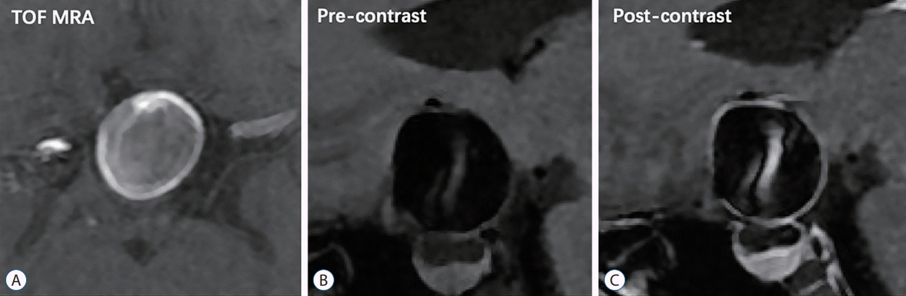
All patients were consecutively scanned utilizing a 3T MR scanner (Discovery MR750; GE Healthcare, Milwaukee, WI, USA) with 32-channel head coil. The images from three different MR scanning sequences were investigated as shown in Fig. 1 with 400% enlargement. As shown in the enlarged image of the aneurysm in Fig. 1C, the sac wall from the 3D CUBE T1 was significantly highlighted by the contrast agent. Location and maximum diameter were obtained through 3D-time-of-flight (TOF)-MR angiography (MRA) reconstruction images. The imaging protocols included 3D TOF MRA sequence, T1-weighted 3D CUBE fast-spin-echo sequence (field of view, 20×20×16 cm3; repetition time/echo time, 600/13.7 ms; matrix, 288×288×160 interpolated to 512×512×320; spatial resolution, 0.4×0.4×0.4 mm; section thickness, 1.0 mm; pixel bandwidth, 62.5 kHz; echo chain length, 24; acquisition time, 256 seconds) before and after administration of Gd-BT-DO3A (Gadovist, Bayer Schering Pharma, Berlin, Germany) at 0.1 mmol/kg. All raw data were transferred to GE workstation for analysis using the same location at all 3D sequences to ensure data comparability.
PHASES score
The risk of aneurysm rupture in Chinese people is assumed to be similar to Japanese. Thus, populations from China had three points for geographical region [5,14]. PHASES risk scores in this dataset were divided into two levels based on the rupture risk (high risk ≥10 points, intermediate-low risk <10 points).
Morphology
Aneurysm morphological characteristics recorded included : 1) neck width (the largest cross sectional diameter of the aneurysm neck); 2) presence of daughter sac (defined as being <50% of the parent aneurysm size); and 3) presence of multiple lobes (defined as being >50% of the parent aneurysm size) [20].
Wall enhancement extent and type
The WEI was measured quantitatively through 3D CUBE T1 sequencing pre- and post-contrast enhancement [22]. Aneurysm wall signal acquisition was obtained using the workstation software ring signal tool, selecting the most obvious AWE and the maximum signal (Fig. 2). Reference signal was acquired from a specific location in the right frontal lobe white matter with T1 weighted image (T1WI) sequence pre- and post-enhancement. Signal intensity of the aneurysm wall was calculated as wall signal divided by the same sequence reference signal.
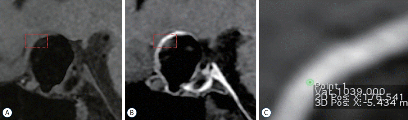
WEI = (enhanced T1WI wall signal intensity – unenhanced T1WI wall signal intensity) / unenhanced T1WI wall signal intensity
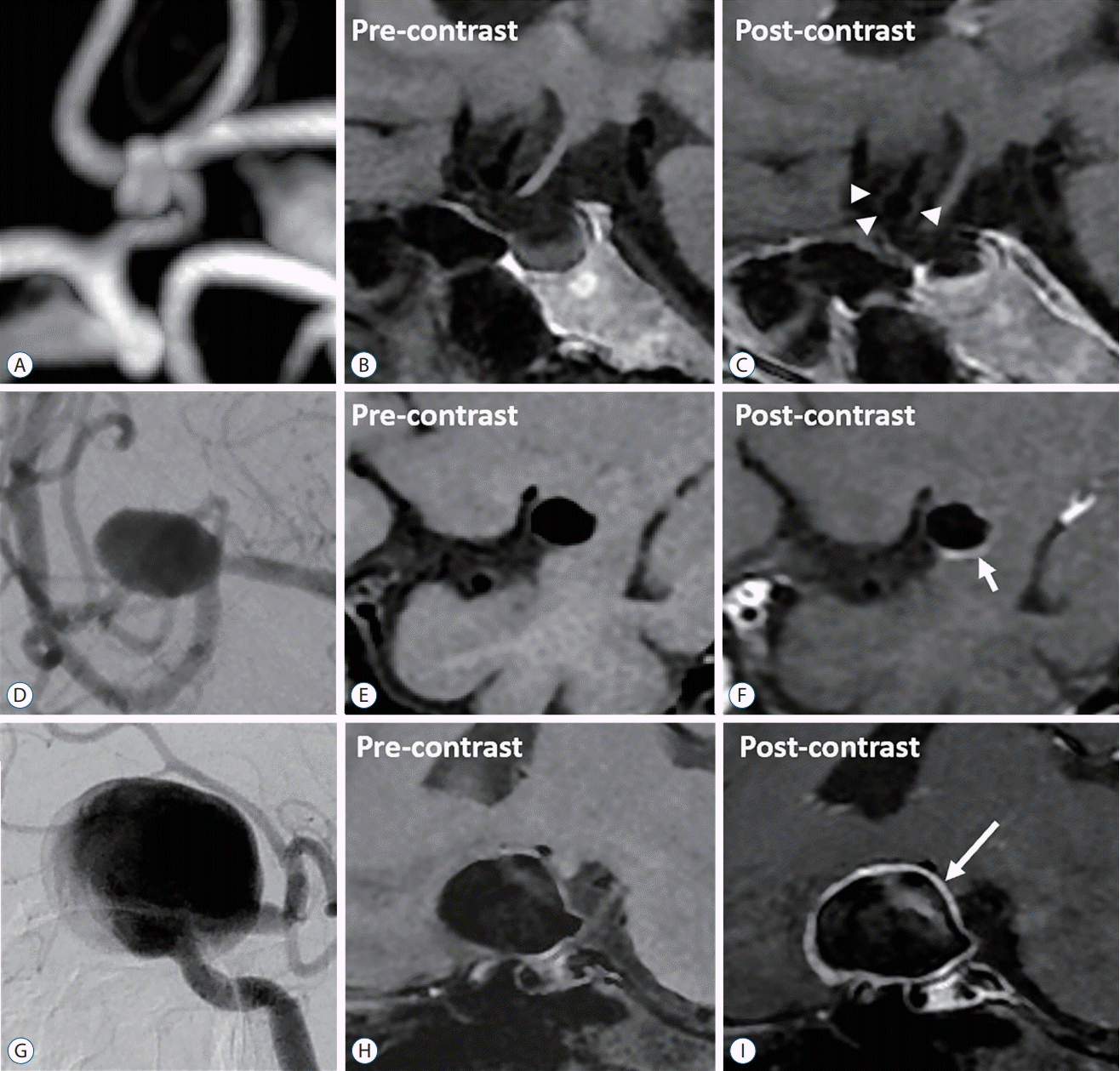
Wall enhancement types were classified into three groups : circumferential, partial, and no enhancement (Fig. 3).
Statistical analysis
SPSS ver. 22 software (SPSS Inc., Chicago, IL, USA) was used for statistical analysis, and p<0.05 was considered statistically significant. The continuous variables were described as mean±standard deviation, while the categorical variables were rendered as numbers and percentages. Independent T or ×2 testing were used to determine the factors related to high ruptured risk aneurysms. p<0.05 potential independent variables in the univariate analysis were included into multivariate regression analysis using a forward process to predict aneurysms at higher risk of rupture. The variables cutoff values with the best sensitivity and specificity for differentiating high risk aneurysms from intermediate-low aneurysms were identified by assessing the receiver operating characteristic (ROC) curve.
Go to :
RESULTS
From February 2016 to October 2017, a total of 133 patients with 157 intracranial aneurysms underwent MR-VWI examination in our center. We excluded 25 patients harboring 30 aneurysms, 24 of which were proved to be dissecting aneurysms, while six were excluded due to poor MRI image quality. Finally, 108 patients with 127 unruptured saccular aneurysms were enrolled for analysis. Of these patients, 74 were women and 34 were men. Mean age of the patients were 58.5± 10.6 (range, 17–80). Sixty-four patients (59.3%) had hypertension, while 20 patients (18.5%) had a previous SAH from another aneurysm (Table 1).
Table 1.
PHASES predictors distribution between high and intermediate-low risk group
Values are presented as mean±standard deviation or number (%). PHASES : Population, Hypertension, Age, Size of aneurysm, Earlier SAH history from another aneurysm, Site of aneurysm, SAH : subarachnoid hemorrhage, ICA : internal carotid artery, MCA : middle cerebral artery, ACA : anterior cerebral artery
![]()
Thirty aneurysms had PHASES score ≥10 (higher risk for aneurysm rupture), and 97 aneurysms had PHASES score <10 (intermediate-low risk for aneurysm rupture). Univariate analysis showed that neck (4.5±3.3 mm vs. 3.4±1.7 mm, p=0.002), wall enhancement (100.0% vs. 62.9%, p<0.001), enhancement type (p<0.001), and WEI (1.6±0.6 vs. 0.8±0.8, p<0.001) were significantly associated with for high rupture risk (Table 2). Compared with the partial, the circumferential type presents higher proportion of rupture risk, however, there was no statistical difference (25.9% vs. 45.5%, p=0.056). The WEI of circumferential type presents higher than the partial (1.6±0.6 vs. 1.2±0.7, p=0.011). However, there was no association between PHASES score and hyperlipidemia, smoking status, drinking, and aneurysms with irregular shape. Multivariate regression analysis revealed that WEI was the most important factor in predicting high rupture risk (odds ratio, 2.6; 95% confidence interval, 1.4–4.9; p=0.002). The ROC analysis can efficiently differentiate high risk aneurysms (area under the curve, 0.780; p<0.001) which have a reliable WEI cutoff value (1.04; sensitivity, 0.833; specificity, 0.67) predictive of high rupture risk (Fig. 4).
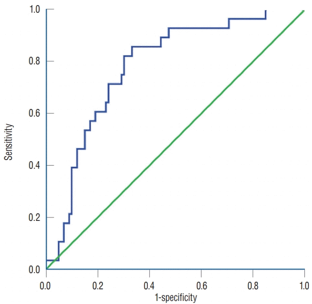 | Fig. 4.High rupture risk (PHASES score ≥10) was efficiently differentiated on ROC curve (AUC, 0.780; p<0.001). Higher WEI than cutoff value (1.04; sensitivity, 0.833; specificity, 0.67) predict high rupture risk. PHASES : Population, Hypertension, Age, Size of aneurysm, Earlier SAH history from another aneurysm, Site of aneurysm, ROC : receiver operating characteristics curve, AUC : area under the curve, WEI : wall enhancement index.
|
Table 2.
Compare clinical rupture risk factors between high and intermediate-low risk group
![]()
Go to :
DISCUSSION
In this study, we prospectively compared MR-VWI detected wall enhancement extent, type and clinical risk factors between intracranial aneurysms at high and intermediate-low risk for future rupture. The neck width, wall enhancement extent including quantitative WEI and type were found to occur in aneurysms at higher risk of rupture based on PHASES score. The WEI was the independent risk factors to predict the risk of aneurysm rupture. The promising WEI cutoff value (1.04; sensitivity, 0.833; specificity, 0.67) was proposed to predict high rupture risk aneurysms.
Intracranial AWE has been proposed as an imaging marker for rupture risk assessment. Edjlali et al. [6] demonstrated that circumferential aneurysmal wall enhancement was more frequently observed in unstable (ruptured, symptomatic, or undergoing morphologic modification) than stable (incidental and nonevolving) aneurysms (87% vs. 28.5%, respectively). Circumferential enhancement in ruptured aneurysms was higher than in unruptured aneurysms. Wang et al. [29] investigated 106 intracranial aneurysms with 19 ruptured aneurysms, including 10 patients with multiple aneurysms. Enhanced ratio (>61.5%) and aneurysm with partial wall enhancement are better predictors of rupture. Nagahata et al. [21] studied 61 ruptured and 83 unruptured aneurysms, of which wall enhancement was observed in 73.8% of ruptured aneurysms and in 4.8% of unruptured aneurysms. Atherosclerosis, inflammation, neovascularization, and the presence of vasa vasorum were proposed to be possible mechanisms of wall enhancement [13,25]. Miyata et al. [19] studies that vasa vasorum formation in an adventitia is associated with rupture of intracranial aneurysm. Endothelial disruption damage or inflammatory healing response could also explain this type enhancement in ruptured aneurysms [12,18]. Such enhancement pattern in high rupture risk aneurysms could be associated with a larger range and greater extent of enhancement compared with intermediate-low risk aneurysms in our results. The larger wall enhancement range have an increased trend of high risk of rupture consistent with previous reported studies. However, statistical difference was not found in this present study which might be related with uneven data distribution and small sample size. The circumferential type have greater intensity than the partial. High wall enhancement extent with quantitative evaluation can predict aneurysm rupture risk. Wall enhancement might be an indicator of ruptured aneurysms for managing patients with subarachnoid hemorrhage, especially in multiple aneurysms [8,18]. This enhancement would not reveal in wall itself but to the interface enhancement between wall and the surrounding brain tissue in ruptured aneurysms. Fresh thrombus in or around the ruptured point might be enhanced in these acute ruptured cases. In our study, wide-necked aneurysms were found to have a high risk of rupture. The wider neck with increased size could be explained this phenomenon. As previously demonstrated, the wall enhancement was independently associated with aneurysm size in unruptured intracranial aneurysms [2,15,16]. Further prospective and pathological investigation is needed to prove the exact mechanisms of this enhancement effect.
In this present study, ROC analysis can efficiently differentiate high risk aneurysms from intermediate-low risk based on PHASES score which provides 5-year absolute aneurysm rupture risk. We concluded a reliable WEI cutoff value (1.04; sensitivity, 0.833; specificity, 0.67) which was proposed to quantitatively differentiate high risk aneurysm based on 3D MR-VWI under reference of frontal parenchymal signal intensity to predict a ruptured state. Omodaka et al. [22] reported a WEI cutoff value (0.53) to differentiate ruptured from unruptured status in circumferential enhanced aneurysms. Due to the signal intensity variability on high resolution-MR imaging with different parameters setting, this cutoff value cannot be generalized. However, our study definitely indicated that higher WEI are correlated to increased rupture risk. Aneurysm daughter sac assumed to be the ruptured site was locally enhanced in some aneurysms. Intracranial AWE was proposed as a characteristic of ruptured aneurysms. Previous studies used qualitative assessment to reveal that wall enhancement was more frequently occurred in ruptured aneurysm [6,18,21]. Vergouwen et al. [27] reported gadolinium enhancement of aneurysm wall on MRI predict the increased risk of aneurysm instability in the longitudinal design study. Pathophysiologically, vascular wall enhancement is related to the inf lammatory reaction which plays a crucial role in the growth and rupture of intracranial aneurysms in the vascular wall [10,23,25]. In hemodynamic aspects, local changes in hemodynamic stress can activate the inflammatory reaction of endothelial cells, mononuclear cell aggregation and activation, and a variety of inflammatory molecules release from the vessel wall [1,23,26]. These eventually cause the release of matrix metalloproteinases and vessel wall cells apoptosis, wall degenerative reconstruction, leading to wall thinning and aneurysm rupture [4,17,23,26]. Therefore, the higher WEI predict higher PHASES score suggesting that higher WEI (>1.04) may provide additional information of aneurysm instability to improve current size-based rupture risk assessment metrics.
Study limitations
Limitations for this study are as follows. First, patient selection bias and limited number of cases from a single institution might be affecting this study consequence. Second, some confounding factors such as enhanced intra-aneurysm flow stagnation, surrounding veins, dura, venous sinus and hematoma may obscure wall enhancement. Third, the heterogeneous distribution aneurysms may be confounding factor in this study. Fourth, the causal relationship was not being concluded between the aneurysm enhancement and aneurysm rupture due to the retrospective design study. A further larger prospective study is needed to evaluate between AWE and rupture risk.
Go to :
 XML Download