Abstract
Glioblastoma (GBM) is a disease without any definite cure. Numerous approaches have been tested in efforts to conquer this brain disease, but patients invariably experience recurrence or develop resistance to treatment. New surgical tools, carefully chosen samples, and experimental methods are enabling discoveries at single-cell resolution. The present article reviews the cell-of-origin of isocitrate dehydrogenase (IDH)-wildtype GBM, beginning with the historical background for focusing on cellular origin and introducing the cancer genesis patterned on firework. The authors also review mutations associated with the senescence process in cells of the subventricular zone (SVZ), and biological validation of somatic mutations in a mouse SVZ model. Understanding GBM would facilitate research on the origin of other cancers and may catalyze the development of new management approaches or treatments against IDH-wildtype GBM.
Isocitrate dehydrogenase (IDH)-wildtype glioblastoma (GBM) is a disease with a dismal prognosis, a distinction that has led to GBM being dubbed the “emperor of all cancers”. The disease progression is rapid, and even with standard therapy [53] and supra-total resection [52], the median survival of patients from diagnosis is approximately 14–20 months [53,63].
There are continuing efforts by researchers in diverse disciplines, including epidemiology and molecular biology, to identify the cause of GBM. Epidemiological research on GBM suggests numerous risk factors that should be taken into account, such as irradiation of the head [8,23,46,50,55] and the allergic history of patient [4]. Biological approaches for determining the cause of GBM include attempts to identify molecular signatures in patient samples (e.g., DNA mutations) [61], the use of in vivo mouse models [58], and investigation of progenitor cells of GBM [3,13,43].
This article reviews the proof-of-concept that GBM originates in the subventricular zone (SVZ) from the microscale perspective such as a molecular biology with a cellular level resolution. The first sections present a brief overview of the disease and recapitulate the importance of mutations in the development of GBM. The remaining sections describe the cell-of-origin of GBM and the cancer genesis patterned on firework from SVZ [43] and discuss a new hypothesis driving GBM research.
There are plenty of discussions and arguments about the existence of the cell-of-origin from the early era of neuro-oncology whether the disease originates from embryonic cells or dedifferentiated cells [51]. The brain cancer model induced from the mutation of specific cells of brain is blurring the gap between those two sides [2] by leading an edge of this field with a subsequent drug discovery targeting GBM [58].
The cell-of-origin of GBM has been suspected to have characteristics of a stem cell, and this cell type has also been regarded as the source of tumor recurrence [22]. There have been many trials conducted to isolate such treatment-resistant insidious cells from the tumor tissues [3,34,41]. Those cells were labeled as tumorspheres [27,35,36] and their behaviors and characteristics have been validated from multiple sources [14,25,30,39,42,49,64]. As the patient-derived tumor cells are heterogeneous in the sample dimension (patient age, the size and location of the sample in the original tumor mass, sample control, purity of samples, or previous treatments), the mutation-induced tumor model has been accepted as another robust and predictable platform in the repeated experiments and clearly demonstrates the consequences of mutation [1,20,43] better than the orthotopic xenograft by primary tumorsphere [31,35,38,42].
An epiphany came from the experience of 5-aminolevulinic acid (5-ALA) fluorescence without any tumor cells in the tumor specimen by pathologic examinations [45]. During surgery to remove GBM, high fluorescence of 5-ALA was found in the walls of ventricles [45], which are non-coincidentally exposed after supra-total resection (or a so-called planned lobectomy) [52,53]. Moon et al. [45] described interesting patterns of 5-ALA fluorescence in the ventricular wall of brain tumor patients. What they found the most interesting was the ventricular walls with positive 5-ALA fluorescence without magnetic resonance imaging enhancement and no tumor cells in the pathology, a finding that perplexed surgeons [45].
Concurrence of 5-ALA positive tissues with no tumor cells in the ventricle sample [45] helped and guided us to find the origin of GBM in the in vivo model [43]. This 5-ALA glittering tumor negative samples from the ventricular wall or the SVZ has a pattern of mutations that has not been previously discovered. By comparing those mutation patterns, a novel way of understanding the disease could be revealed. The tumor-private mutations in the tumor tissue might facilitate the development of a new targeted therapy. In addition, the anatomical location of those cells would mature the Big Bang theory [7,60] into the Firework theory of subventricular abnormal cells [43].
Epidemiologic studies have linked a number of pollutants to the incidence of GBM [48]. Ionizing radiation to the head at a younger age increases the risk of glioma and meningioma [8,23,46,50,55]. An allergic or atopic disease has been associated with a lower risk of GBM, and consistently with lower-grade glioma [4]. DNA mutations and its consequent syndromes increase the risk of acquiring GBM [32,48], such as Turcot syndrome (from mismatch repair-deficiency) [15], Li-Fraumeni syndrome (from the germline mutation of TP53) [37] and neurofibromatosis type 1 (from NF1 gene) [26,59].
However, there are additional unproven and controversial risk factors. One example is non-ionizing radiation from cellular phones. Studies have not shown a consistent increase in the risk of glioma [24]. Another example is Radon exposure which reported to be associated with the risk of glioma in a Danish cohort [9], but neither radon exposure nor background radiation was associated with glioma incidence in a cohort of British children [28]. Inconsistent results have been obtained with respect to the association of GBM development with exposure to air pollutants or particulate matter, owing in part to difficulties in measuring exposure history [5]. There is hardly any or no evidence linking insecticides, rubber processing, pesticides, farming, solvents, metal fumes, or jet engine manufacturing to the incidence of glioma [48,54].
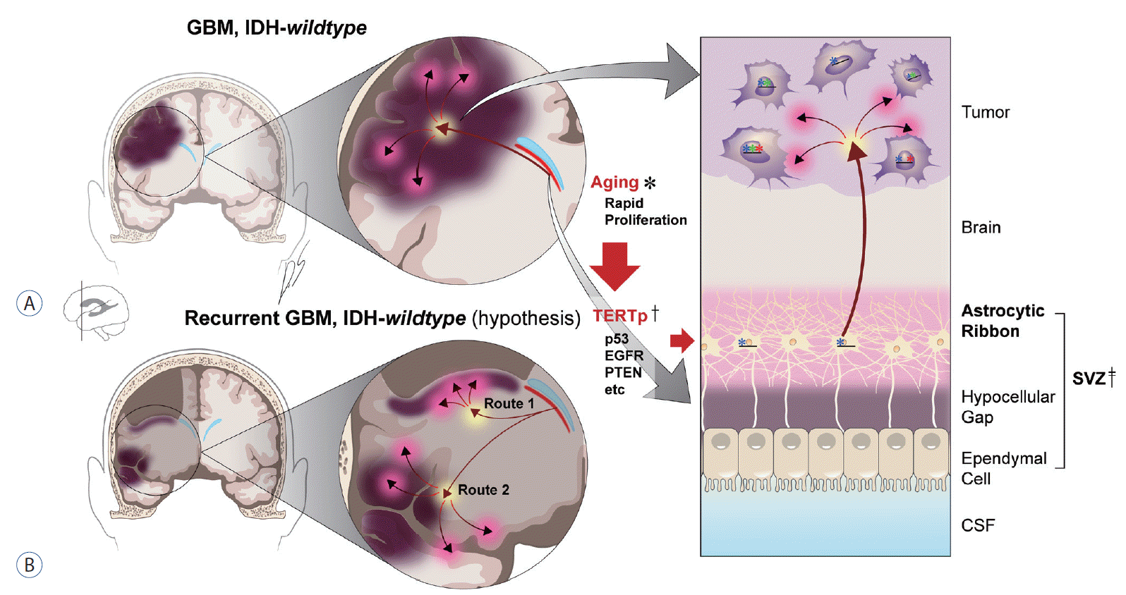
Two senescence-associated mutational patterns were found in GBM : mutational signatures 1 and 5 [43]. These two types dominated the mutation profile of SVZ samples of the astrocytic ribbon (SVZ‡ in Fig. 1A, 33% [7.8 genes out of 23 total genes] and 45% [10.4 genes out of 23 total genes] for signature 1 and 5 respectively in the SVZ samples) [43].
The average number of genes for mutation signature 1 jumped up from 7.8 genes (SVZ) to 81.9 genes in tumor samples of the Severance GBM cohort (10.5 fold increase) [43]. While the fold change of average number of genes that contribute to mutation signature 5 (fold 0.71, calculated from 10.4 genes of SVZ to 7.4 genes of the tumor) remains relatively stable as that of other signatures (fold 1.18, calculated from 4.8 genes of SVZ to 5.7 genes in the tumor) [43]. These findings suggest that GBM arises from the senescence process, especially if we focus on the number of genes of mutational signature 1 and 5 from SVZ to tumor, or alternatively from other causes that are associated with age.
Accumulation of mutations in both the stem cells and the tumor region raises questions regarding the origin of the mutations. Are mutations in both tumors and SVZ related? Are they coincidental? Do mutations in the SVZ reflect contamination from the tumor? To deconvolute this complex pattern of mutations, the authors compared shared mutations (i.e., those in common to both the SVZ and tumor) and tumor-exclusive mutations (i.e., those not found in the SVZ) [43]. There were several shared mutations in the tumor-free SVZ and GBM bulk tumor, including those in the TERT promoter [43]. An analysis of variant allele frequencies (VAFs) and single-cell sequencing results revealed a unidirectional pattern from SVZ to bulk tumor. A two-dimensional VAF plot in a previous report [43] showed unique patterns of shared mutations between the tumor and the SVZ, with low-level driver mutations being located near the y-axis or tumor-axis. However, the tumor-contaminated SVZ-tumor pair showed more genes that are spread along the identity line as opposed to the uncontaminated SVZ-tumor pair [43] (Fig. 1A).
Single-cell sequencing of the DNA of bulk tumors also validated the result showing directionality from the SVZ to the tumor [43]. Twenty-four clones were derived from one IDH-wildtype GBM patient (GBM245) [43]. The genes on the table of Lee et al. [43] are subset of those genes of directionality. This surprising pattern of mutation was later confirmed from the intraventricular injection of plasmids to induce mutation in the SVZ [16,43].
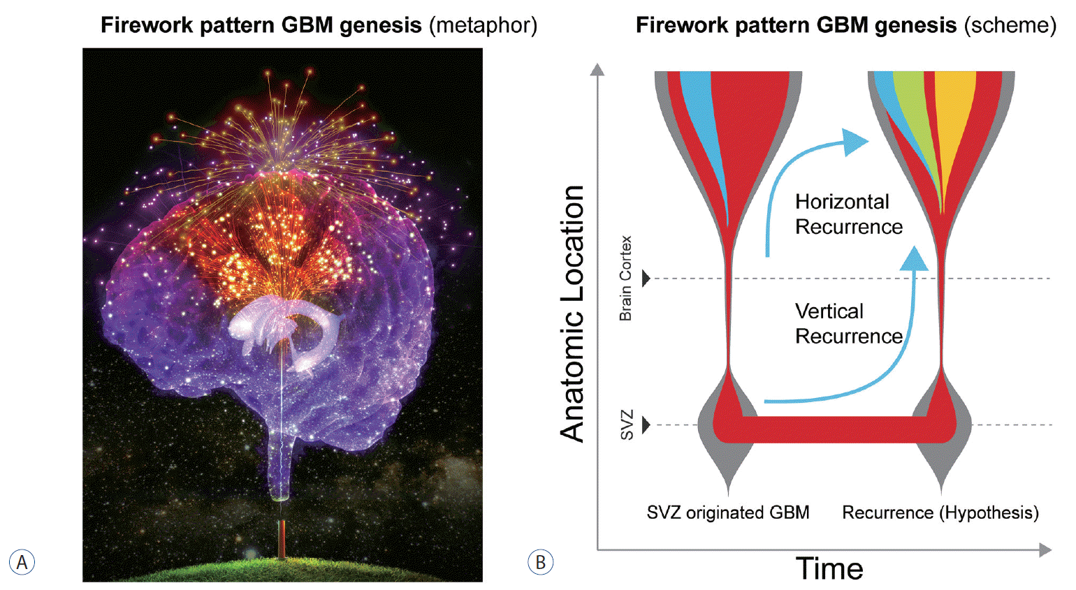
Mutations oftentimes start with TERT promoter mutations in cells of the astrocytic ribbon in SVZ under the ventricle (TERTp† in Fig. 1A). With additional mutations in oncogenes, the mutated cells from SVZ migrate to the cortex and manifest as the GBM (Fig. 1A, right) [43]. The authors searched for mutations in triple-matched samples of tumor-free SVZ, GBM tumor, and normal brain cortex (or blood) [43]. The authors have hypothesized that the SVZ, which includes neural stem cells, is suspected of being the origin of tumors [43]; mutations then progress from the SVZ to the tumor mass. In addition to this directionality, the number of mutations increases dramatically after the cell-of-origin reaches the cortex. As described in the previous study of Lee et al. [43], about 20 different somatic mutations of SVZ that contribute to the mutational patterns increased to around 100 mutations in the tumor. Furthermore, mutational signature 1 dominated most mutations in tumors, whereas mutational signature 5 was preserved at about the same level between the SVZ and the tumor [43]. This pattern is slightly different version of the Big Bang concept and, considering the anatomic location of the progression, the authors refer to it as a “Firework” pattern (Fig. 2).
Deep under the ependymal layer of the SVZ is a ribbon-like feature called the astrocytic ribbon [57] (SVZ‡ in Fig. 1A). This area is populated by neural stem cells in both mouse and human brains [65]. Using molecular techniques described below, we introduced three types of mutations into the population of cells in the SVZ. These mutation-bearing cells migrated to the cortex, ultimately forming high-grade gliomas [43] (Fig. 1A), presenting with firework-like GBM genesis (Fig. 2A).
A mass of GBM is created in the cortex by the mutation in the SVZ cells [43]. Crossbred mice (postnatal day 2–3 pups) carrying LSL-tdTomato and LSL-EGFRviii [71] were used for these experiments [43]. A Cre-containing CRISPR/Cas9 plasmid was electroporated into one side of the ventricle in the brain to knock out Pten and Trp53 (mouse analog of human TP53) genes using single guide RNA (sgRNA) for these genes. After injection into the ventricular space with a sharp needle, the electroporation device was applied to deliver the plasmid into the SVZ [43]. In the presence of the mutations, brain tumors developed in 90% of mice (9/10), with 67% of tumors developing in a region distant from the mutation-arising SVZ, and these mice showed a median survival of around 20 weeks [43].
One of the next steps in the research of IDH-wildtype GBM should be related with the recurrence pattern of GBM (The hypothesis on the recurrence of GBM). Careful selection of samples without tumor in the SVZ is the beginning point of this research. Recent article by Watts group [61] is one example of selecting the tumor-invaded SVZ (median tumor content 22.1%, rather than tumor-free SVZ) by the IDH-wildtype GBM and its results and implications should be carefully contemplated in the research of vertical recurrence from the cell of SVZ (Fig. 2B).
GBM recurs after irradiation of tumor margins at varying depth [69]. Although irradiating the SVZ appears to have a prognostic benefit for GBM patients [29,47], it does not cure these patients or prevent exacerbation of symptoms. There are at least two scenarios for recurrence of IDH-wildtype GBM. In the first case, GBM-initiating cells already present in the parenchyma of the brain restart the tumor after a dormant period (Fig. 2B, horizontal recurrence). In the second, dormant cells in the SVZ re-migrate to the tumor bed area, where they create the recurrent tumor (Fig. 1B, the only hypothesis in this article; Fig. 2B, vertical recurrence). We hope to resolve the second phenomenon using tumor-free SVZ samples from GBM patients.
Many previous attempts to treat GBM have failed and the list harbors 5-f luorouracil and methotrexate [10]. A breakthrough in treating GBM came with the application of alkylating agents [18,62,67]. Nitrosoureas proved the most effective at the time, and it became the most commonly used drugs for the management of brain tumors, including GBM [56,70]. Their antitumor effects were widely accepted and even led to the development of an implantable chemotherapeutic system impregnated with the nitrosourea derivative carmustine [11].
However, these nitrosoureas had toxicity issues, and temozolomide subsequently gained popularity because of its limited adverse effects compared with earlier alkylating agents [6]. But even temozolomide proved to be ineffective in treatment of recurrent GBM similarly to other alkylating agents [33,44,66]. And the therapeutic efficacy of other novel options is limited in Bevacizumab [19,21,68], IDH-targeting drugs [12] and immunotherapy [17,40].
After the repeated efforts to treat the tumor itself, the origin of GBM emerged as the next generation target. Ultimately, in vitro and in vivo models will accelerate the screening for drugs that can preemptively target the initial process of cancer genesis with firework pattern from SVZ.
Mutated neural stem cells, that are present in the astrocytic ribbon in the SVZ, may migrate to the brain cortex and generate IDH-wildtype GBM with/or without any molecular or environmental cue [43]. While this concept has been demonstrated scientifically, elucidating the detailed mechanism underlying the GBM genesis process will require additional investigation. Our ongoing research efforts would focus on identifying the cell-of-origin to develop a potential treatment option for IDH–wildtype GBM, the “Emperor of cancer”.
Notes
AUTHOR CONTRIBUTIONS
Conceptualization : SJY, JHM, EHK, JHC, JHL, SGK
Data curation : SJY, SGK
Funding acquisition : SGK
Methodology : SJY, HJK, JHL, RJC, JKS, JHL, SGK
Project administration : SGK
Visualization : SJY, DSJ, SGK
Writing - original draft : SJY, SGK
Writing - review & editing : SJY, JP, HJK, JHL, EJ, RJC, JKS, JHM, EHK, JHC, JHL, SGK
ACKNOWLEDGMENTS
This work was supported by grants from the Korean Health Technology R&D Project, Ministry of Health & Welfare, Republic of Korea (HI17C2586), the National Research Foundation of Korea (NRF) grant funded by the Korea government (MEST) (NRF-2019R1A2C3004155), and the NRF grant funded by the MSIP : Ministry of Science, ICT and Future Planning, Republic of Korea (NRF-2017M2A2A7A01071).
References
1. Alcantara Llaguno S, Chen J, Kwon CH, Jackson EL, Li Y, Burns DK, et al. Malignant astrocytomas originate from neural stem/progenitor cells in a somatic tumor suppressor mouse model. Cancer Cell. 15:45–56. 2009.
2. Alcantara Llaguno S, Sun D, Pedraza AM, Vera E, Wang Z, Burns DK, et al. Cell-of-origin susceptibility to glioblastoma formation declines with neural lineage restriction. Nat Neurosci. 22:545–555. 2019.
3. Alcantara Llaguno SR, Wang Z, Sun D, Chen J, Xu J, Kim E, et al. Adult lineage-restricted cns progenitors specify distinct glioblastoma subtypes. Cancer Cell. 28:429–440. 2015.
4. Amirian ES, Zhou R, Wrensch MR, Olson SH, Scheurer ME, Il’yasova D, et al. Approaching a scientific consensus on the association between allergies and glioma risk: a report from the glioma international case-control study. Cancer Epidemiol Biomarkers Prev. 25:282–290. 2016.
5. Andersen ZJ, Pedersen M, Weinmayr G, Stafoggia M, Galassi C, Jørgensen JT, et al. Long-term exposure to ambient air pollution and incidence of brain tumor: the european study of cohorts for air pollution effects (escape). Neuro Oncol. 20:420–432. 2018.
6. Bae SH, Park MJ, Lee MM, Kim TM, Lee SH, Cho SY, et al. Toxicity profile of temozolomide in the treatment of 300 malignant glioma patients in korea. J Korean Med Sci. 29:980–984. 2014.
7. Barthel FP, Wesseling P, Verhaak RGW. Reconstructing the molecular life history of gliomas. Acta Neuropathol. 135:649–670. 2018.
8. Braganza MZ, Kitahara CM, Berrington de González A, Inskip PD, Johnson KJ, Rajaraman P. Ionizing radiation and the risk of brain and central nervous system tumors: a systematic review. Neuro Oncol. 14:1316–1324. 2012.
9. Bräuner EV, Andersen ZJ, Andersen CE, Pedersen C, Gravesen P, Ulbak K, et al. Residential radon and brain tumour incidence in a danish cohort. PLoS One. 8:e74435. 2013.
10. Bredel M. Anticancer drug resistance in primary human brain tumors. Brain Res Brain Res Rev. 35:161–204. 2001.
11. Brem H, Piantadosi S, Burger PC, Walker M, Selker R, Vick NA, et al. Placebo-controlled trial of safety and efficacy of intraoperative controlled delivery by biodegradable polymers of chemotherapy for recurrent gliomas. The polymer-brain tumor treatment group. Lancet. 345:1008–1012. 1995.
12. Calvert AE, Chalastanis A, Wu Y, Hurley LA, Kouri FM, Bi Y, et al. Cancer-associated idh1 promotes growth and resistance to targeted therapies in the absence of mutation. Cell Rep. 19:1858–1873. 2017.
13. Chen J, McKay RM, Parada LF. Malignant glioma: lessons from genomics, mouse models, and stem cells. Cell. 149:36–47. 2012.
14. Choi J, Lee JH, Koh I, Shim JK, Park J, Jeon JY, et al. Inhibiting stemness and invasive properties of glioblastoma tumorsphere by combined treatment with temozolomide and a newly designed biguanide (hl156a). Oncotarget. 7:65643–65659. 2016.
15. Dipro S, Al-Otaibi F, Alzahrani A, Ulhaq A, Al Shail E. Turcot syndrome: a synchronous clinical presentation of glioblastoma multiforme and adenocarcinoma of the colon. Case Rep Oncol Med. 2012:720273. 2012.
16. Fernández ME, Croce S, Boutin C, Cremer H, Raineteau O. Targeted electroporation of defined lateral ventricular walls: a novel and rapid method to study fate specification during postnatal forebrain neurogenesis. Neural Dev. 6:13. 2011.
17. Filley AC, Henriquez M, Dey M. Recurrent glioma clinical trial, checkmate-143: the game is not over yet. Oncotarget. 8:91779–91794. 2017.
18. Fine HA. The basis for current treatment recommendations for malignant gliomas. J Neurooncol. 20:111–120. 1994.
19. Franceschi E, Lamberti G, Paccapelo A, Di Battista M, Genestreti G, Minichillo S, et al. Third-line therapy in recurrent glioblastoma: is it another chance for bevacizumab? J Neurooncol. 139:383–388. 2018.
20. Friedmann-Morvinski D, Bushong EA, Ke E, Soda Y, Marumoto T, Singer O, et al. Dedifferentiation of neurons and astrocytes by oncogenes can induce gliomas in mice. Science. 338:1080–1084. 2012.
21. Gilbert MR, Dignam JJ, Armstrong TS, Wefel JS, Blumenthal DT, Vogelbaum MA, et al. A randomized trial of bevacizumab for newly diagnosed glioblastoma. N Engl J Med. 370:699–708. 2014.
22. Goffart N, Kroonen J, Rogister B. Glioblastoma-initiating cells: relationship with neural stem cells and the micro-environment. Cancers (Basel). 5:1049–1071. 2013.
23. Inskip PD, Sigurdson AJ, Veiga L, Bhatti P, Ronckers C, Rajaraman P, et al. Radiation-related new primary solid cancers in the childhood cancer survivor study: comparative radiation dose response and modification of treatment effects. Int J Radiat Oncol Biol Phys. 94:800–807. 2016.
24. INTERPHONE Study Group. Brain tumour risk in relation to mobile telephone use: results of the interphone international case-control study. Int J Epidemiol. 39:675–694. 2010.
25. Jeong H, Park J, Shim JK, Lee JE, Kim NH, Kim HS, et al. Combined treatment with 2’-hydroxycinnamaldehyde and temozolomide suppresses glioblastoma tumorspheres by decreasing stemness and invasiveness. J Neurooncol. 143:69–77. 2019.
26. Jeong TS, Yee GT. Glioblastoma in a patient with neurofibromatosis type 1: a case report and review of the literature. Brain Tumor Res Treat. 2:36–38. 2014.
27. Kang SG, Cheong JH, Huh YM, Kim EH, Kim SH, Chang JH. Potential use of glioblastoma tumorsphere: clinical credentialing. Arch Pharm Res. 38:402–407. 2015.
28. Kendall GM, Little MP, Wakeford R, Bunch KJ, Miles JC, Vincent TJ, et al. A record-based case-control study of natural background radiation and the incidence of childhood leukaemia and other cancers in great britain during 1980-2006. Leukemia. 27:3–9. 2012.
29. Khalifa J, Tensaouti F, Lusque A, Plas B, Lotterie JA, Benouaich-Amiel A, et al. Subventricular zones: new key targets for glioblastoma treatment. Radiat Oncol. 12:67. 2017.
30. Kim EH, Lee JH, Oh Y, Koh I, Shim JK, Park J, et al. Inhibition of glioblastoma tumorspheres by combined treatment with 2-deoxyglucose and metformin. Neuro Oncol. 19:197–207. 2017.
31. Kim S, Alexander CM. Tumorsphere assay provides more accurate prediction of in vivo responses to chemotherapeutics. Biotechnol Lett. 36:481–488. 2014.
32. Kinnersley B, Mitchell JS, Gousias K, Schramm J, Idbaih A, Labussière M, et al. Quantifying the heritability of glioma using genome-wide complex trait analysis. Sci Rep. 5:17267. 2015.
33. Kitange GJ, Carlson BL, Schroeder MA, Grogan PT, Lamont JD, Decker PA, et al. Induction of mgmt expression is associated with temozolomide resistance in glioblastoma xenografts. Neuro Oncol. 11:281–291. 2009.
34. Kong BH, Moon JH, Huh YM, Shim JK, Lee JH, Kim EH, et al. Prognostic value of glioma cancer stem cell isolation in survival of primary glioblastoma patients. Stem Cells Int. 2014:838950. 2014.
35. Kong BH, Park NR, Shim JK, Kim BK, Shin HJ, Lee JH, et al. Isolation of glioma cancer stem cells in relation to histological grades in glioma specimens. Childs Nerv Syst. 29:217–229. 2013.
36. Kong BH, Shin HD, Kim SH, Mok HS, Shim JK, Lee JH, et al. Increased in vivo angiogenic effect of glioma stromal mesenchymal stem-like cells on glioma cancer stem cells from patients with glioblastoma. Int J Oncol. 42:1754–1762. 2013.
37. Kratz CP, Achatz MI, Brugières L, Frebourg T, Garber JE, Greer MC, et al. Cancer screening recommendations for individuals with li-fraumeni syndrome. Clin Cancer Res. 23:e38–e45. 2017.
38. Kwak J, Shim JK, Kim DS, Lee JH, Choi J, Park J, et al. Isolation and characterization of tumorspheres from a recurrent pineoblastoma patient: feasibility of a patient-derived xenograft. Int J Oncol. 49:569–578. 2016.
39. Laks DR. The assessment of glioblastoma tumorspheres reveals molecular determinants of proliferation and therapeutic response. Ann Arbor: University of California;2015. p. 245.
40. Lamberti G, Franceschi E, Brandes AA. The burden of oncology promises not kept in glioblastoma. Future Neurol. 13:1–4. 2018.
41. Lathia JD, Mack SC, Mulkearns-Hubert EE, Valentim CL, Rich JN. Cancer stem cells in glioblastoma. Genes Dev. 29:1203–1217. 2015.
42. Lee CH, Yu CC, Wang BY, Chang WW. Tumorsphere as an effective in vitro platform for screening anti-cancer stem cell drugs. Oncotarget. 7:1215–1226. 2015.
43. Lee JH, Lee JE, Kahng JY, Kim SH, Park JS, Yoon SJ, et al. Human glioblastoma arises from subventricular zone cells with low-level driver mutations. Nature. 560:243–247. 2018.
45. Moon JH, Kim SH, Shim JK, Roh TH, Sung KS, Lee JH, et al. Histopathological implications of ventricle wall 5-aminolevulinic acid-induced fluorescence in the absence of tumor involvement on magnetic resonance images. Oncol Rep. 36:837–844. 2016.
46. Neglia JP, Robison LL, Stovall M, Liu Y, Packer RJ, Hammond S, et al. New primary neoplasms of the central nervous system in survivors of childhood cancer: a report from the childhood cancer survivor study. J Natl Cancer Inst. 98:1528–1537. 2006.
47. Nourallah B, Digpal R, Jena R, Watts C. Irradiating the subventricular zone in glioblastoma patients: is there a case for a clinical trial? Clin Oncol (R Coll Radiol). 29:26–33. 2017.
48. Ostrom QT, Bauchet L, Davis FG, Deltour I, Fisher JL, Langer CE, et al. The epidemiology of glioma in adults: a “state of the science” review. Neuro Oncol. 16:896–913. 2014.
49. Park J, Shim JK, Kang JH, Choi J, Chang JH, Kim SY, et al. Regulation of bioenergetics through dual inhibition of aldehyde dehydrogenase and mitochondrial complex I suppresses glioblastoma tumorspheres. Neuro Oncol. 20:954–965. 2018.
50. Pearce MS, Salotti JA, Little MP, McHugh K, Lee C, Kim KP, et al. Radiation exposure from CT scans in childhood and subsequent risk of leukaemia and brain tumours: a retrospective cohort study. Lancet. 380:499–505. 2012.
51. Ramon Y Cajal Agüeras S. Pío del río-hortega: a visionary in the pathology of central nervous system tumors. Front Neuroanat. 10:13. 2016.
52. Roh TH, Kang SG, Moon JH, Sung KS, Park HH, Kim SH, et al. Survival benefit of lobectomy over gross-total resection without lobectomy in cases of glioblastoma in the noneloquent area: a retrospective study. J Neurosurg. 2019; [Epub ahead of print].
53. Roh TH, Park HH, Kang SG, Moon JH, Kim EH, Hong CK, et al. Long-term outcomes of concomitant chemoradiotherapy with temozolomide for newly diagnosed glioblastoma patients: a single-center analysis. Medicine (Baltimore). 96:e7422. 2017.
54. Ruder AM, Carreón T, Butler MA, Calvert GM, Davis-King KE, Waters MA, et al. Exposure to farm crops, livestock, and farm tasks and risk of glioma: the upper midwest health study. Am J Epidemiol. 169:1479–1491. 2009.
55. Sadetzki S, Chetrit A, Freedman L, Stovall M, Modan B, Novikov I. Long-term follow-up for brain tumor development after childhood exposure to ionizing radiation for tinea capitis. Radiat Res. 163:424–432. 2005.
56. Salcman M. Survival in glioblastoma: historical perspective. Neurosurgery. 7:435–439. 1980.
57. Sanai N, Tramontin AD, Quiñones-Hinojosa A, Barbaro NM, Gupta N, Kunwar S, et al. Unique astrocyte ribbon in adult human brain contains neural stem cells but lacks chain migration. Nature. 427:740–744. 2004.
58. Shi Y, Lim SK, Liang Q, Iyer SV, Wang HY, Wang Z, et al. Gboxin is an oxidative phosphorylation inhibitor that targets glioblastoma. Nature. 567:341–346. 2019.
59. Shibahara I, Sonoda Y, Suzuki H, Mayama A, Kanamori M, Saito R, et al. Glioblastoma in neurofibromatosis 1 patients without idh1, braf v600e, and tert promoter mutations. Brain Tumor Pathol. 35:10–18. 2018.
60. Sottoriva A, Kang H, Ma Z, Graham TA, Salomon MP, Zhao J, et al. A big bang model of human colorectal tumor growth. Nat Genet. 47:209–216. 2015.
61. Spiteri I, Caravagna G, Cresswell GD, Vatsiou A, Nichol D, Acar A, et al. Evolutionary dynamics of residual disease in human glioblastoma. Ann Oncol. 30:456–463. 2019.
62. Stenning SP, Freedman LS, Bleehen NM. An overview of published results from randomized studies of nitrosoureas in primary high grade malignant glioma. Br J Cancer. 56:89–90. 1987.
63. Stupp R, Mason WP, van den Bent MJ, Weller M, Fisher B, Taphoorn MJ, et al. Radiotherapy plus concomitant and adjuvant temozolomide for glioblastoma. N Engl J Med. 352:987–996. 2005.
64. Sung KS, Shim JK, Lee JH, Kim SH, Park S, Roh TH, et al. Success of tumorsphere isolation from who grade IV gliomas does not correlate with the weight of fresh tumor specimens: an immunohistochemical characterization of tumorsphere differentiation. Cancer Cell Int. 16:75. 2016.
65. Tabata H, Yoshinaga S, Nakajima K. Cytoarchitecture of mouse and human subventricular zone in developing cerebral neocortex. Exp Brain Res. 216:161–168. 2012.
66. Tseng WL, Hsu HH, Chen Y, Tseng SH. Tumor recurrence in a glioblastoma patient after discontinuation of prolonged temozolomide treatment. Asian J Neurosurg. 12:727–730. 2017.
67. Walker MD, Green SB, Byar DP, Alexander E Jr, Batzdorf U, Brooks WH, et al. Randomized comparisons of radiotherapy and nitrosoureas for the treatment of malignant glioma after surgery. N Engl J Med. 303:1323–1329. 1980.
68. Wenger KJ, Wagner M, You SJ, Franz K, Harter PN, Burger MC, et al. Bevacizumab as a last-line treatment for glioblastoma following failure of radiotherapy, temozolomide and lomustine. Oncol Lett. 14:1141–1146. 2017.
69. Wernicke AG, Smith AW, Taube S, Mehta MP. Glioblastoma: radiation treatment margins, how small is large enough? Pract Radiat Oncol. 6:298–305. 2016.
70. Wilson CB, Gutin P, Boldrey EB, Drafts D, Levin VA, Enot KJ. Single-agent chemotherapy of brain tumors. A five-year review. Arch Neurol. 33:739–744. 1976.