

 PDF
PDF Citation
 Print
Print


INTRODUCTION
THE SHORT HISTORY OF FCD
Table 1.
| Focal dysplasia type | Subtype | Main neuropathological features |
|---|---|---|
| Mild MCD | Type I | Heterotopic/excess neurons in layer I |
| Type II | Heterotopic/excess neurons outside in layer I | |
| FCD type I | Type Ia | Cortical dyslamination only (±MCD features) |
| Type Ib | Cortical dyslamination+giant* or immature neurons† | |
| FCD type II (Taylor-type) | Type IIa | Cortical dyslamination+dysmorphic neurons‡ |
| Type IIb | Cortical dyslamination+dysmorphic neurons and balloon cells |
In Palmini’s classification, ‘giant neurons’ were described focusing on cell size. In the other hand, ‘dysmorphic neurons’ were described focusing abnormal morphology and aggregated coarse Nissl substance. The abnormal neurons in Fig. 5B are similar to the ‘giant neurons’ and cells in Fig. 6B are more similar to the ‘dysmorphic neurons’ in Palmini et al.’s classification. In the current classification, the definition of dysmorphic neuron has two morphological features, both large size and the characteristic Nissl substance. According to Palmini et al.’s classification [11],
* giant neurons are neurons of increased size (compared with layer V pyramidal neurons) with central nuclei.
† The immature neurons are round cells with immature nucleus, not dysmorphic or giant. They could be seen in heterotopic nodules.
![]()
THE CURRENT ILAE CLASSIFICATION SYSTEM OF FCD
Overview
FCD type I
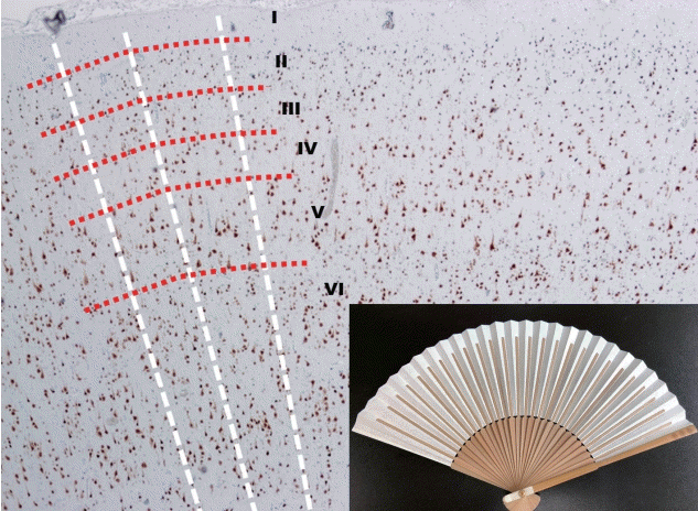 | Fig. 1.The low power view of normal neocortex (NeuN, ×40) and a Korean folding pan (inlet). The imaginary white lines show the direction of radial migrations of neural cells. The lines look like the bones of a folding pan (inlet). The cortical six-layers were seen with red imaginary lines. The red imaginary lines show the tangential composition of neocortex. In the normal neocortex, it is unusual that the neuronal cells exactly align along the white imaginary lines.
|
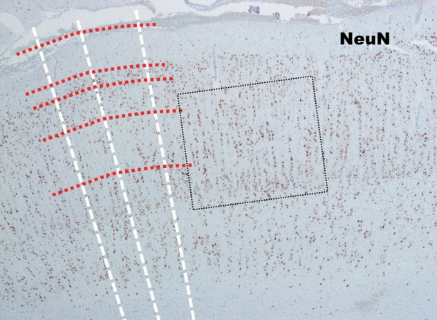

 | Fig. 4.The low power view of FCD type Ib (NeuN, ×40). The cortical sixlayers are deranged (too chaotic) compared to those in normal neocortex (Fig. 1). FCD : focal cortical dysplasia.
|
FCD type II
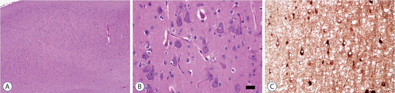 | Fig. 5.The histopathological findings of FCD type IIa. The low power view (A; H-E, ×40) shows cortical dyslamination which means derangement of normal neocortical six-layers. High power view shows many large dysmorphic neurons with coarse Nissl substances. The size of dysmorphic neuron cytoplasm is more than 20 µm (B; H-E, ×400; black bar, 20 µm). Dysmorphic neurons are highlighted by immunostaining with nonphosphorylated neurofilament (C; SMI132, ×200). FCD : focal cortical dysplasia.
|
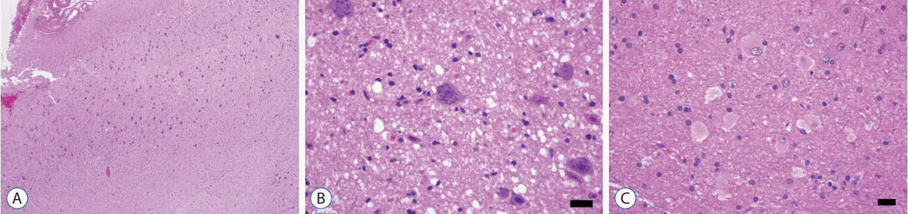 | Fig. 6.The histopathological findings of FCD type IIb. The low power view (A; H-E, ×40) shows cortical dyslamination as FCD type IIa. High power view shows many large dysmorphic neurons with coarse Nissl substances (B; H-E, ×400; black bar, 20 µm) and balloon cells showing eosinophilic homogenous cytoplasm (C; H-E, ×400; black bar, 20 µm). FCD : focal cortical dysplasia.
|
FCD type III
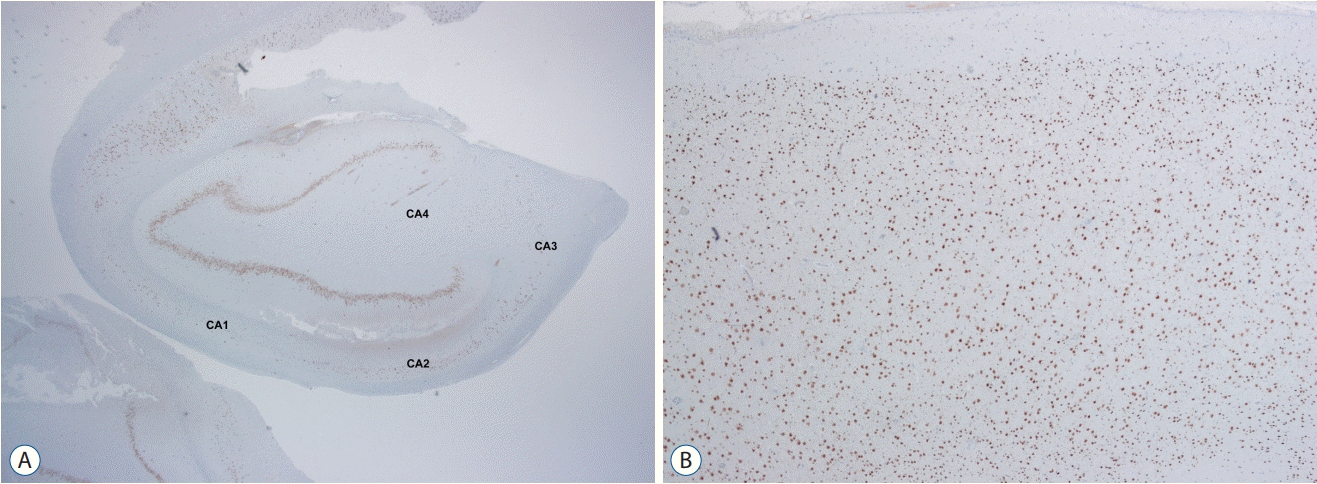 | Fig. 7.FCD type IIIa is a hippocampal sclerosis (ILAE type Ia) showing neuronal loss in CA4, 3, 2 and 1 (A; NeuN, ×12) with associating FCD type Ib which shows a breakdown of cortical tangential composition in overlying cortex (B; NeuN, ×40). FCD : focal cortical dysplasia.
|
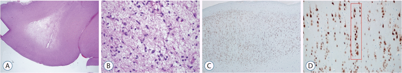 | Fig. 8.FCD type IIIb is combined with a low grade-epilepsy associated tumor (LEAT). Diagnosis of “the associated tumor” in this case is dysembryoplastic neuroepithelial tumor (DNT) (A; H-E, ×12 and B; H-E, ×200). Overlying cortex shows FCD type Ia (C; NeuN, ×40) characterized by microcolumns (D; red box; NeuN, ×200). FCD : focal cortical dysplasia.
|
Table 2.
FCD type III (NOS) : if clinically/radiologically suspected principal lesion is not available for microscopic inspection. Please note that the rare association between FCD type IIa and IIb with hippocampal sclerosis, tumors, or vascular malformations should not be classified as FCD type III variant. ILAE : international league against epilepsy, FCD : focal cortical dysplasia, NOS : not otherwise specified
![]()
Limitations, genetic progress and prospect
Table 3.
![]()
 XML Download