Abstract
We report two rare cases of spontaneously regressed Rathke's cleft cyst (RCC). A 52-year-old woman presented with headache. A pituitary hormone study was normal. Brain magnetic resonance imaging (MRI) showed a 0.45-cm3 cystic sellar lesion. The cyst was hyperintense on T1-weighed imaging and hypointense on T2-weighted imaging without rim enhancement, comparable to a RCC. Six months later, brain MRI showed no change in the cyst size. Without any medical treatments, brain MRI 1 year later revealed a spontaneous decrease in cyst volume to 0.05 cm3. A 34-year-old woman presented with headache and galactorrhea lasting 1 week. At the time of the visit, the patient's headache had disappeared. Her initial serum prolactin level was 81.1 ng/mL, and after 1 week without the cold medicine, the serum prolactin level normalized to 11.28 ng/mL. Brain MRI showed a RCC measuring 0.71 cm3. Without further treatments, brain computed tomography 6 months later showed a spontaneous decrease in cyst volume to 0.07 cm3. Another 6 months later, brain MRI revealed that the cyst had remained the same size. Neither patient experienced neurological symptoms, such as headache or visual disturbance, during the period of cyst reduction. The RCCs in both patients underwent spontaneous regression without any medical treatment during a period of 6 months to 1 year. Although spontaneous regression of a RCC is rare, it is still possible and a sufficient follow-up period should be considered.
Go to : 
A Rathke's cleft cyst (RCC) is a benign cyst in the sellar region that arises from the residual of Rathke's pouch [2]. Most RCCs are small and asymptomatic, however, symptomatic RCCs may cause headache, visual disturbance, and endocrinopathy [4]. Symptomatic RCCs are treated with surgical resection, however, asymptomatic RCCs are treated with conservative treatment and follow-up. The majority of asymptomatic RCCs are unchanged in size at follow-up, but rare cases have been reported to decrease spontaneously in size [2-5,7,9-11,13].
In the present study, we report two rare cases of spontaneously regressed RCCs without medical treatment and review the current literature.
Go to : 
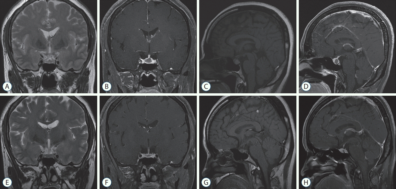
A 52-year-old woman presented with headache due to a common cold. There were no visual or hormonal symptoms. The patient's ophthalmologic examination and hormone study were normal, and her medical history was unremarkable. Brain magnetic resonance imaging (MRI) showed a cystic lesion in the sellar region, measuring 8.6×6.1×12.2 mm with a volume of 0.45 cm3 (Fig. 1). The cyst was hyperintense on T1-weighed imaging and hypointense on T2-weighted imaging without rim enhancement, comparable to a RCC. The enhancement was seen in the pituitary gland surrounding the cyst. The cyst did not compress the optic chiasm. As the patient's cold improved, the headache disappeared. Since there were no other symptoms, she did not take any medication. Six months later, a brain MRI obtained for routine followup showed no change in the size of the cyst. One year later, a subsequent brain MRI revealed the spontaneous decrease in cyst volume to 0.05 cm3. The patient did not experience neurological symptoms, such as headache or visual disturbance, during the period of cyst reduction.
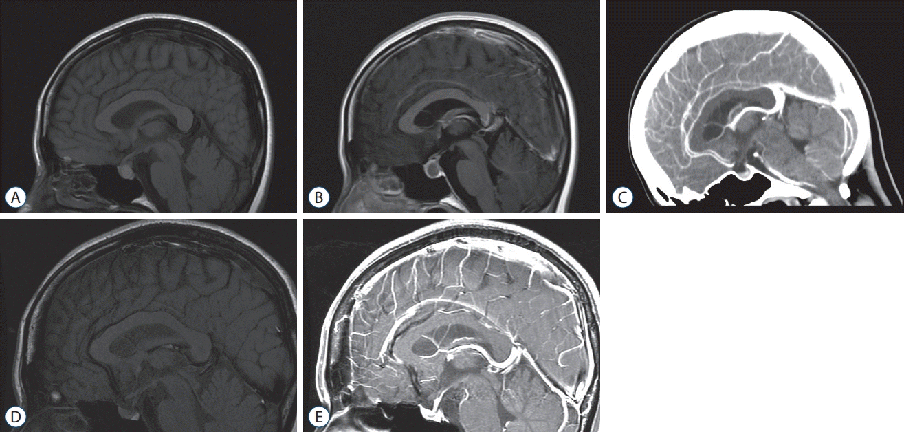
A 34-year-old woman presented with headache due to a common cold, and galactorrhea lasting 1 week. At the time of the visit, her headache had disappeared. There were no visual symptoms and an ophthalmologic examination was normal. A hormone study revealed that her serum prolactin level was 81.1 ng/mL (normal range, 1.9–25.0) and we suspected galactorrhea as a result of the cold medicine. The cold medicine was stopped, and serum prolactin level normalized to 11.28 ng/mL 1 week later. In order to investigate other causes of hyperprolactinemia, a brain MRI was obtained. The brain MRI showed a RCC, measuring 10.5×8.3×15.9 mm, with a volume of 0.71 cm3 , without compression of optic chiasm (Fig. 2). Enhancement was seen in the pituitary gland surrounding the cyst. Without further treatments, a brain computed tomography 6 months later showed the spontaneous decrease in volume to 0.07 cm3. Another 6 months later, a brain MRI revealed that the cyst had remained the same size. Recurrence after spontaneous regression was not observed over a 6-month follow-up period. The patient did not experience neurological symptoms, such as headache or visual disturbance during the period of cyst reduction.
The RCCs in both patients underwent marked spontaneous regression without any treatment during a period of 6 months to 1 year.
Go to : 
RCCs are benign lesions that account for 1.8% of all tumors in the sellar region, and the majority of RCCs remain asymptomatic, requiring no intervention [11]. A series of regular imaging followups are recommend for asymptomatic RCCs, and spontaneous regression occurs rarely [2-5,7,9-11,13]. Simmons and Simmons [13] first described a spontaneous regression of a pituitary cyst in an amenorrhoeic patient in 1999. However, the incidence of spontaneous regression of RCCs has not been well demonstrated [7].
Because of the low rate of pathologic diagnosis by surgical resection, an accurate imaging diagnosis of RCC is important. However, findings on MR imaging are similar to many other pituitary lesions, including cystic pituitary adenoma, and craniopharyngioma. An RCC, which is a well-defined, round, non-calcified lesion within or adjacent to the pituitary, may contain cerebrospinal fluid-like fluid, thick mucoid material, or waxy cellular debris, and show variable densities depending on the intracystic components [10,11,13]. Unlike RCC, calcification, and cyst components that show high intensity on T2-weighted images are common in craniopharyngioma. Additionally, in pituitary adenoma, the cyst wall is irregularly enhanced [2,6].
The mechanism of spontaneous regression of RCCs is not precisely known, and is explained as an unbalanced secretion and absorption of cystic fluid, or cyst rupture [3,10,11]. Nishio et al. [10] suggested that, based on the initial onset of severe headache and MRI findings, cystic lesions in patients may shrink after pituitary apoplexy. Spontaneous regression in our cases, however, is not thought to be caused by pituitary apoplexy. Because the initial headaches were mild and due to the common cold, there was no evidence of pituitary hemorrhage in MR images, and no symptoms related to cyst reduction during follow-up. Saeki et al. [12] reported that RCCs with isointensity to high intensity on T1- weighted images cause clinical symptoms at a smaller size than cysts of low intensity; thus, it is possible that chronic inflammation participates in the development of some cases of pituitary dysfunction. However, in our cases with high intensity on T1- weighted images, no pituitary dysfunction was observed. Maruyama et al. [8] suggested that the inflammatory nature of RCCs may explain the observed short-term size changes in response to glucocorticoid administration. Additionally, Amhaz et al. [3] suggested that spontaneous involution of an RCCs may be more common than prior reports would suggest, because the natural progression of RCCs is poorly understood. There are debates regarding the management of asymptomatic RCCs, and the potential for spontaneous involution supports a conservative approach for patients with symptomatic RCCs presenting solely with headache. Follow-up examinations are important as RCCs often show an unpredictable natural progression and may later become larger, or rarely become smaller, like in our cases. According to previous reports, the time to spontaneous regression of RCCs varies from 0.5 months to 27 months [2,4,8-11]. Our cases showed marked spontaneous regression during a period of 6 months to 1 year. Although it is difficult to determine the exact follow-up period, spontaneous regression is often found in 1-year follow-up MRI.
RCCs are generally considered to be nonexpansile lesions that are stable over time. The effect of medical treatment is not well known, although glucocorticoids may promote cyst size reduction [8]. Our patients experienced spontaneous regression without any medical therapy. If headache, and visual and endocrine symptoms are not well controlled for conservative treatment, surgical treatment should be considered. However, high rates of recurrence after surgical resection have been reported [9]. Aho et al. [1] reported that aggressive surgical resection of the entire cyst wall was associated with higher morbidity. Further studies on the natural progression of symptomatic and asymptomatic RCCs are needed in concordance with the increasing trend of minimally invasive surgery for RCCs. Our report can aid in choosing the appropriate treatment for RCCs.
Go to : 
Notes
INFORMED CONSENT
Informed consent was obtained from all individual participants included in this study.
AUTHOR CONTRIBUTIONS
Data curation : CL
Formal analysis : CL
Funding acquisition : SHP
Methodology : CL
Project administration : SHP
Visualization : CL
Writing - original draft : CL
Writing - review & editing : SHP
Go to : 
References
1. Aho CJ, Liu C, Zelman V, Couldwell WT, Weiss MH. Surgical outcomes in 118 patients with Rathke cleft cysts. J Neursurg. 102:189–193. 2005.

2. Al Safatli D, Kalff R, Waschke A. Spontaneous involution of a presumably Rathke’s cleft cyst in a patient with slight subclinical hypopituitarism: a case report and review of the literature. Case Rep Surg. 2015:971364–971365. 2015.

3. Amhaz HH, Chamoun RB, Waguespack SG, Shah K, McCutcheon IE. Spontaneous involution of Rathke cleft cysts: is it rare or just underreported? J Neurosurg. 112:1327–1332. 2010.

4. Cheng L, Guo P, Jin P, Li H, Fan M, Cai E. Spontaneous involution of a Rathke cleft cyst. J Craniofac Surg. 27:e791–e793. 2016.

5. Hatayama K, Abe T, Endo T, Kobayashi N, Shimazu M, Sasaki K, et al. Pituitary cystic mass with spontaneous disappearance: three cases of equivocal Rathke’s cleft cyst. No To Shinkei. 52:929–933. 2000.
6. Igarashi T, Saeki N, Yamaura A. Long-term magnetic resonance imaging follow-up of asymptomatic sellar tumors. Their natural history and surgical indications. Neurol Med Chir (Tokyo). 39:592–598. 1999.

7. Kim CW, Hwang K, Joo JD, Kim YH, Han JH, Kim CY. Spontaneous involution of Rathke’s cleft cysts without visual symptoms. Brain Tumor Res Treat. 4:58–62. 2016.

8. Maruyama H, Iwasaki Y, Tsugita M, Ogami N, Asaba K, Takao T, et al. Rathke’s cleft cyst with short-term size changes in response to glucocorticoid replacement. Endocr J. 55:425–428. 2008.

9. Munich SA, Leonardo J. Spontaneous involution of a Rathke’s cleft cyst in a patient with normal cortisol secretion. Surg Neurol Int. 3:42. 2012.

10. Nishio S, Morioka T, Suzuki S, Fukui M. Spontaneous regression of a pituitary cyst: report of two cases. Clin Imaging. 25:15–17. 2001.

11. Rasmussen Z, Abode-Iyamah KO, Kirby P, Greenlee JD. Rathke’s cleft cyst: a case report of recurrence and spontaneous involution. J Clin Neurosci. 32:122–125. 2016.

Go to : 


 PDF
PDF Citation
 Print
Print


 XML Download