Journal List > Ann Surg Treat Res > v.102(4) > 1160646

Hong, Lee, Lee, Hong, Suh, Han, Choi, Yi, and Suh: Impact of tumor size on hepatectomy outcomes in hepatocellular carcinoma: a nationwide propensity score matching analysis

Abstract

Purpose

The aim of this study was to compare surgical outcomes after liver resection for hepatocellular carcinoma (HCC) according to tumor size using a large, nationwide cancer registry-based cohort and propensity score matching.

Methods

From 2008 to 2015, a total of 12,139 patients were diagnosed with liver cancer and registered in the Korean Primary Liver Cancer Registry. Patients without distant metastasis who underwent hepatectomy as a primary treatment were selected. We performed 1:1 propensity score matching between the small (<5 cm), large (≥5 cm and <10 cm), and huge (≥10 cm) groups.

Results

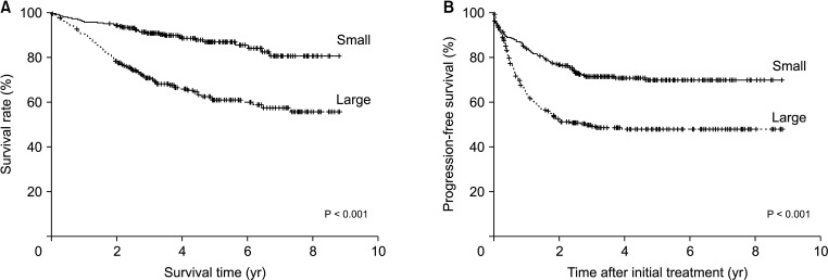
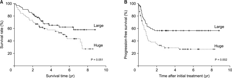
Overall, 265 patients in the small and large groups were compared, and 64 patients each in the large and huge groups were compared. The overall and progression-free survival rates were significantly lower in the large group than in the small group (P < 0.001 and P < 0.001, respectively). Overall survival tended to be poorer in the huge group than in the large group (P = 0.051). The progression-free survival rate was significantly lower in the huge group than in the large group (P = 0.002).

Conclusion

Although primary liver resection can be considered even in patients with huge HCC, greater caution with careful screening for recurrence is needed.

INTRODUCTION

Liver resection is the first-line treatment for patients with single hepatocellular carcinoma (HCC) without cirrhosis, as well as for patients with cirrhosis but with adequate liver functional reserve [123]. The outcome of liver resection has been improved due to recent advances in preoperative examinations and surgical techniques, along with the accumulation of postoperative management [45]. According to recent reports, the 5-year survival rate after liver resection for HCC is 46%–69.5% and the 5-year disease-free survival rate is 23%–56.3% [678]. In general, liver resection is reported to have a good prognosis when performed in 1 or 2 small tumors [47]. Large tumor size, microvascular invasion (MVI), tumor rupture, severity of underlying cirrhosis, and tumor multiplicity are known to be associated with poor prognosis [59101112].
According to the studies reported so far, it is generally considered to be large if the tumor size is larger than 5 cm and huge if it is larger than 10 cm [111314151617]. The American Joint Committee on Cancer staging system for HCC also includes a 5 cm value in determining T3 [18].
However, some recent studies have shown that no MVI was observed in 1/3 of patients with tumors larger than 10 cm, indicating that the size solely itself did not adversely affect the prognosis [1117]. Most of the existing studies are limited in that factors other than tumor size are not properly controlled, the sample size is small, they are not a comparative study, or their study was limited to single-center experience [111217].
This study aimed to assess and compare outcomes after liver resection for HCC according to tumor size (<5 cm vs. ≥5 cm and <10 cm and ≥5 cm and <10 cm vs. ≥10 cm) using a large, nationwide cancer registry-based cohort and propensity score matching to adjust for differences between the groups.

METHODS

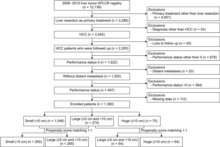
This study was conducted according to the ethical guidelines of the Declaration of Helsinki and exempted from further ethical review by the Institutional Review Board of Seoul National University Hospital (No. 2111-081-1272). The study population included 12,139 patients who were diagnosed with liver cancer and registered in the Korean Primary Liver Cancer Registry (KPLCR) from 2008 to 2015. Fig. 1 shows a schematic of the study. Patients who underwent hepatectomy as the primary treatment for HCC without distant metastasis were included. Patients with an Eastern Cooperative Oncology Group performance status other than 0 or missing were excluded. After exclusion of patients who had missing values for follow-up date, body mass index (BMI), serology, Child-Pugh score, number of tumors, size of tumor, platelet count, PT, or performance status, 1,390 patients were finally included in the study. We then divided the patients according to maximal tumor size, defined using preoperative imaging: small (<5 cm, n = 1,046), large (≥5 cm and <10 cm, n = 274), and huge (≥10 cm, n = 70). Progression-free survival (PFS) was measured from the day of initial treatment to the day of the second or last follow-up.

Statistical analysis

Categorical variables were presented as numbers and percentages, and continuous variables were presented as mean ± standard deviation or median (range). Continuous variables were compared using a Student t-test, whereas categorical variables were compared using the chi-square test or Fisher exact test, as appropriate. Patient overall survival and PFS rates were calculated using the Kaplan-Meier method and compared using the log-rank test. To overcome possible selection bias, 1:1 propensity score matching between small and large and large and huge cohorts was applied using multiple logistic regression and a 1:1 matching requirement via the nearest-neighbor matching method. Factors such as age, sex, BMI, etiology (HBV, HCV, or non-B non-C), platelet count, PT, tumor number, and Child-Pugh score were matched. The absolute standardized differences method was used to diagnose the balance after matching, and all were checked to be less than 0.25. Statistical significance was set at P < 0.05. Statistical analysis was performed using IBM SPSS Statistics ver. 25.0 (IBM Corp., Armonk, NY, USA).

RESULTS

Small (<5 cm) vs. large (≥5 cm and <10 cm)

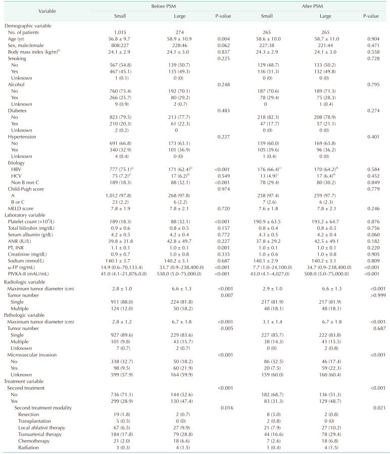
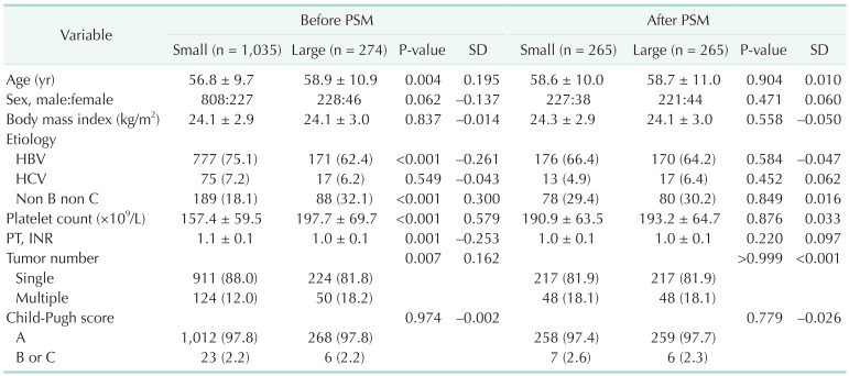
A total of 1,046 patients in the small group and 274 patients in the large group were included (Table 1). After matching, 265 patients were included in each group. Before matching, the large group was older and had a higher proportion of etiology with non-B non-C than the small group. Platelet count was higher and PT was lower in the large group than in the small group. The large group had a higher proportion of patients with multiple tumors than the small group. After matching, there were no significant intergroup differences in relation to the 8 factors encompassing the baseline characteristics of the small and large groups.
The outcomes of demographic, radiologic, pathologic, and treatment variables are summarized in Table 2. Preoperative levels of α-FP (median [range]: 7.7 ng/mL [1.0–24,100.0 ng/mL] vs. 34.7 ng/mL [0.9–238,400.0 ng/mL], P < 0.001), protein induced by vitamin K absence-II (PIVKA-II) (43.0 mAU/mL [4.1–4,027.0 mAU/mL] vs. 508.0 mAU/mL [5.0–75,000.0 mAU/mL], P < 0.001), and the proportion of MVI was higher in the large group than in the small group. A higher proportion of patients experienced a second treatment after resection (31.3% vs. 48.7%, P < 0.001), most of which were non-radical treatments including transarterial therapy, chemotherapy, and radiation, in the large group than in the small group (P = 0.021).
Kaplan-Meier analysis showed that the 1-, 2-, 3-, and 5-year survival rates were 97.0%, 93.9%, 91.5%, and 88.7%, respectively, in the small group; and 90.5%, 78.4%, 71.2%, and 61.5%, respectively, in the large group (P < 0.001) (Fig. 2A). The 1-, 2-, 3-, and 5-year PFS rates were 84.8%, 74.2%, 69.1%, and 66.4%, respectively, in the small group; and 64.0%, 52.1%, 49.8%, and 48.0%, respectively, in the large group (P < 0.001) (Fig. 2B).
When performing subgroup analysis by excluding 96 patients with multiple HCCs, the 1-, 2-, 3-, and 5-year survival rates were 97.7%, 95.4%, 92.9%, and 92.2%, respectively, in the small group (n = 217); and 92.1%, 78.2%, 71.5%, and 62.7%, respectively, in the large group (n = 217) (P < 0.001) (Supplementary Fig. 1A). The 1-, 2-, 3-, and 5-year PFS rates were 87.9%, 77.1%, 71.9%, and 68.8%, respectively, in the small group; and 64.9%, 51.5%, 49.3%, and 48.6%, respectively, in the large group (P < 0.001) (Supplementary Fig. 1B).

Large (≥5 cm and <10 cm) vs. huge (≥10 cm)

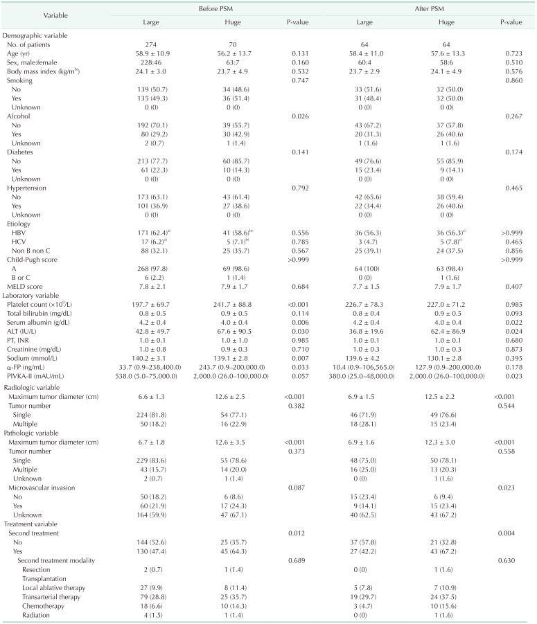
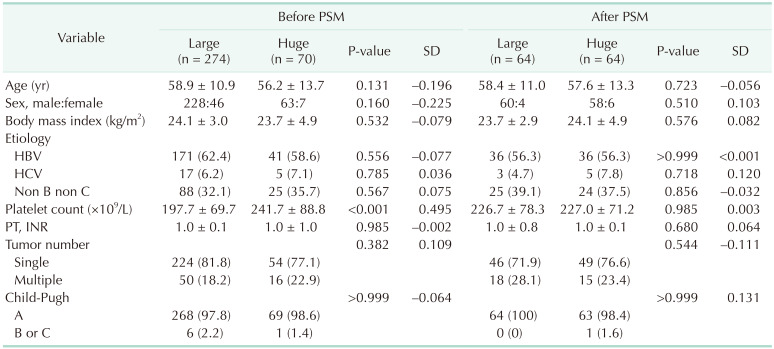
A total of 274 patients in the large group and 70 patients in the huge group were included (Table 3). Before matching, the preoperative platelet count was higher in the huge group than in the large group. After matching, 64 patients were included in each group and they were well matched for age, sex, BMI, etiology, preoperative platelet count, PT, tumor number, and Child-Pugh score.
Table 4 summarizes the outcomes of the demographic, radiologic, pathologic, and treatment variables of the 2 groups. Serum albumin was significantly lower (4.2 vs. 4.0 g/dL, P = 0.022) and ALT was higher in the huge group than in the large group. Preoperative α-FP levels were similar between the 2 groups (P = 0.178), but the preoperative PIVKA-II level was higher in the huge group than in the large group (median [range]: 380.0 mAU/mL [25.0–48,000.0 mAU/mL] vs. 2,000.0 mAU/mL [26.0–100,000.0 mAU/mL], P = 0.023). The rate of MVI was higher in the huge group than in the large group (14.1% vs. 23.4%, P = 0.023).
Kaplan-Meier analysis showed that the 1-, 2-, 3-, and 5-year survival rates were 90.6%, 84.4%, 72.0%, and 62.3%, respectively, in the large group; and 82.8%, 73.4%, 62.0%, and 53.2%, respectively, in the huge group (P = 0.051) (Fig. 3A). The 1-, 2-, 3-, and 5-year PFS rates were 63.0%, 56.5%, 56.5%, and 56.5%, respectively, in the large group; and 43.1%, 36.2%, 28.9%, and 26.7%, respectively, in the huge group (P = 0.002) (Fig. 3B).
When performing subgroup analysis by excluding 33 patients with multiple HCCs, the 1-, 2-, 3-, and 5-year survival rates were 91.3%, 82.6%, 73.1%, and 67.2%, respectively, in the large group (n = 46); and 81.6%, 75.5%, 67.0%, and 59.0%, respectively, in the huge group (n = 49) (P = 0.088) (Supplementary Fig. 2A). The 1-, 2-, 3-, and 5-year PFS rates were 61.5%, 56.9%, 56.9%, and 56.9%, respectively, in the large group, and 48.6%, 41.7%, 34.1%, and 31.0%, respectively, in the huge group (P = 0.032) (Supplementary Fig. 2B).

DISCUSSION

Most previous studies demonstrating the impact of tumor size in HCC patients who underwent hepatectomy are limited by selection bias, small sample size, and/or single-center experience [111217]. The present study has several strengths that can overcome these limitations. First, the patient cohort was sorted from a large nationwide cancer registry database to maintain statistical power. The samples registered in the KPLCR are guaranteed to be representative of all HCCs in Korea, and the statistics of HCC in Korea have been continuously reported using this registry [19]. Second, the propensity score matching method was used after excluding patients with a performance status other than 0, distant metastasis, or missing data to minimize selection bias. Third, the patients were classified into 3 groups (small, large, and huge) and they were serially compared (small vs. large, large vs. huge) to accurately identify the impact of maximum tumor size while maintaining adequate sample size. Comparing the 3 groups at once can lead to the weakness of tailoring to the huge group, which includes only 70 patients before matching. Instead, we serially compared the small group (n = 1,035) with the large group (n = 274) first, and then the large group (n = 274) with the huge group (n = 70).
Our study showed that patients with HCC larger than 10 cm showed a 5-year overall survival rate of 53.2% and a 5-year PFS rate of 26.7%, which are higher than those of previous reports showing recurrence-free survival rates after resection in patients with these huge tumors as 35.5%–42.9% and 9.7%–14.2% [1320]. This can be explained by the inclusion and exclusion criteria of the study design. Patients who underwent hepatectomy as primary treatment were included, and patients with a performance status other than zero and those with distant metastasis were excluded. These inclusion and exclusion criteria may have improved the survival outcomes. As expected, our study showed significantly better overall survival and PFS in the small group than in the large group; and better PFS in the large group than in the huge group. There was also a clear tendency for better overall survival in the large group than in the huge group, but the difference was not statistically significant (P = 0.051). Altogether, this study confirmed once again that overall and PFS worsened as the maximal tumor size increased.
The α-FP and PIVKA-II are widely used serum tumor markers, despite that facts that approximately 40% of patients with HCC have negative α-FP, the positive predictive value ofα-FP is about 9%–32%, and PIVKA-II levels are influenced by several factors other than HCC [2122232425]. Previous studies have reported the correlations between serum α-FP and PIVKA-II levels and the size of HCC [2122232425]. Similarly, the present study showed that serum α-FP and PIVKA-II levels in the large group were significantly higher than those in the small group. Serum PIVKA-II levels in the huge group were higher than those in the large group; however, serum α-FP levels were similar between the 2 groups. As expected, the present study showed that the median values of serum α-FP and PIVKA-II levels serially increased according to the size of HCC.
Although not significant when comparing the small group with the large group, serum albumin was higher and ALT levels were lower in the huge group compared to the large group. Larger HCCs are reported to be associated with lower albumin levels, possibly due to decreased liver function as a result of the original liver disease, liver destruction by extensive HCC growth, and systemic inflammation [2627]. These factors might have been dramatically aggravated as HCC grew to more than 10 cm. Elevation of serum ALT levels can also be explained in a similar context. ALT is a protein that is expressed in various human tissues and organs, with the highest expression observed in the mitochondria of hepatocytes [23]. ALT can be used as a biomarker evaluating hepatocyte destruction. Elevation of ALT level with a mean value of 62.4 IU/L in the huge HCC group may have resulted from liver destruction due to extensive HCC growth. Tarao et al. [27] reported a close correlation between ALT elevation and histological necroinflammation using biopsy specimens. Another explanation can be deduced from previous studies that reported an association between ALT elevation and risk of HCC [282930]. Lin et al. [30] demonstrated that elevated serum levels of ALT were significantly associated with an increased HCC risk in a large cohort of chronic HBV carriers.
It has been reported that as the size of the tumor increases, the frequency of MVI increases and the prognosis is poor [9]. However, according to recent studies, MVI was not observed in approximately one-third of patients with tumors larger than 10 cm [1117]. The present study showed a significant difference in the proportion of MVI between the small and large groups (P < 0.001) and large and huge groups (P = 0.023). There are several missing data on MVI because the registry began to include MVI data since 2013. When performing subgroup analysis including only the period after 2013, the rate of MVI was 18.9% in the small group and 56.2% in the large group. According to the comparison between the large and huge groups, the rate of MVI was 37.5% in the large group and 71.4% in the huge group. Similar to a previous study [11], approximately 30% of patients with huge HCC larger than 10 cm had no MVI.
This study has several limitations. First, it was a retrospective study using a national database registry that relied on the completeness of medical records. Second, Korea is an HBV endemic country, and it is also clear in the present study showing that 56.3%–64.2% of the patients had an etiology of HBV. Thus, our results may not be generalizable to patients with HCC with other etiologies. Third, recurrence-free survival could not be evaluated using this registry. Instead, PFS, which was calculated by the date of initial hepatectomy and the date of the second treatment, was used. Fourth, this study did not compare hepatectomy as an initial treatment with other treatment methods such as transarterial embolization. Despite these limitations, this study has a strength in that it is a large study using a national registry database and it minimized selection bias by excluding patients with certain conditions and using propensity score matching.
In conclusion, this study reports overall survival and PFS, which progressively increased with increasing size of HCC. However, at the same time, it showed a favorable outcome of primary liver resection even in HCCs larger than 10 cm. Primary liver resection should not be excluded based solely on tumor size. Of course, after performing primary liver resection in huge HCC patients, greater caution with careful screening for recurrence is needed.

ACKNOWLEDGEMENTS

The database used in this study was provided by the Korean Central Cancer Registry, Ministry of Health and Welfare, South Korea, and the Korean Liver Cancer Association.

Notes

Fund/Grant Support: None.

Conflict of Interest: No potential conflict of interest relevant to this article was reported.

Author Contribution:

  • Conceptualization, Methodology, Project Administration: SKH, KWL.

  • Formal Analysis: SKH, KWL, SL, SH, SS.

  • Investigation: SKH, KWL, ESH, YC, NJY, KSS.

  • Writing – Original Draft: SKH, KWL.

  • Writing – Review & Editing: All authors.

References

1. Lang H, Sotiropoulos GC, Dömland M, Frühauf NR, Paul A, Hüsing J, et al. Liver resection for hepatocellular carcinoma in non-cirrhotic liver without underlying viral hepatitis. Br J Surg. 2005; 92:198–202. PMID: 15609381.
crossref
2. Capussotti L, Muratore A, Massucco P, Ferrero A, Polastri R, Bouzari H. Major liver resections for hepatocellular carcinoma on cirrhosis: early and long-term outcomes. Liver Transpl. 2004; 10(2 Suppl 1):S64–S68. PMID: 14762842.
crossref
3. Poon RT, Fan ST, Lo CM, Ng IO, Liu CL, Lam CM, et al. Improving survival results after resection of hepatocellular carcinoma: a prospective study of 377 patients over 10 years. Ann Surg. 2001; 234:63–70. PMID: 11420484.
crossref
4. Korean Liver Cancer Association. National Cancer Center. 2018 Korean Liver Cancer Association-National Cancer Center Korea Practice Guidelines for the management of hepatocellular carcinoma. Gut Liver. 2019; 13:227–299. PMID: 31060120.
5. Andreou A, Vauthey JN, Cherqui D, Zimmitti G, Ribero D, Truty MJ, et al. Improved long-term survival after major resection for hepatocellular carcinoma: a multicenter analysis based on a new definition of major hepatectomy. J Gastrointest Surg. 2013; 17:66–77. PMID: 22948836.
crossref
6. Huang J, Zhang Y, Peng Z, Gao H, Xu L, Jiao LR, et al. A modified TNM-7 staging system to better predict the survival in patients with hepatocellular carcinoma after hepatectomy. J Cancer Res Clin Oncol. 2013; 139:1709–1719. PMID: 23982274.
crossref
7. Torzilli G, Belghiti J, Kokudo N, Takayama T, Capussotti L, Nuzzo G, et al. A snapshot of the effective indications and results of surgery for hepatocellular carcinoma in tertiary referral centers: is it adherent to the EASL/AASLD recommendations?: an observational study of the HCC East-West study group. Ann Surg. 2013; 257:929–937. PMID: 23426336.
crossref
8. Lee EC, Kim SH, Park H, Lee SD, Lee SA, Park SJ. Survival analysis after liver resection for hepatocellular carcinoma: a consecutive cohort of 1002 patients. J Gastroenterol Hepatol. 2017; 32:1055–1063. PMID: 27797420.
crossref
9. Goh BK, Chow PK, Teo JY, Wong JS, Chan CY, Cheow PC, et al. Number of nodules, Child-Pugh status, margin positivity, and microvascular invasion, but not tumor size, are prognostic factors of survival after liver resection for multifocal hepatocellular carcinoma. J Gastrointest Surg. 2014; 18:1477–1485. PMID: 24855028.
crossref
10. Minagawa M, Ikai I, Matsuyama Y, Yamaoka Y, Makuuchi M. Staging of hepatocellular carcinoma: assessment of the Japanese TNM and AJCC/UICC TNM systems in a cohort of 13,772 patients in Japan. Ann Surg. 2007; 245:909–922. PMID: 17522517.
11. Hwang S, Lee YJ, Kim KH, Ahn CS, Moon DB, Ha TY, et al. Long-term outcome after resection of huge hepatocellular carcinoma ≥ 10 cm: single-institution experience with 471 patients. World J Surg. 2015; 39:2519–2528. PMID: 26126423.
crossref
12. Zhang H, Yuan SX, Dai SY, Zhang JM, Huang X, Lu CD, et al. Tumor size does not independently affect long-term survival after curative resection of solitary hepatocellular carcinoma without macroscopic vascular invasion. World J Surg. 2014; 38:947–957. PMID: 24258262.
crossref
13. Zhou YM, Li B, Xu DH, Yang JM. Safety and efficacy of partial hepatectomy for huge (≥10 cm) hepatocellular carcinoma: a systematic review. Med Sci Monit. 2011; 17:RA76–RA83. PMID: 21358616.
crossref
14. Wakayama K, Kamiyama T, Yokoo H, Orimo T, Shimada S, Einama T, et al. Huge hepatocellular carcinoma greater than 10 cm in diameter worsens prognosis by causing distant recurrence after curative resection. J Surg Oncol. 2017; 115:324–329. PMID: 28192617.
crossref
15. Ettorre GM, Levi Sandri GB, Colasanti M, Mascianà G, de Werra E, Santoro R, et al. Liver resection for hepatocellular carcinoma ≥5 cm. Transl Gastroenterol Hepatol. 2017; 2:22. PMID: 28447057.
crossref
16. Noh JH, Kim TS, Ahn KS, Kim YH, Kang KJ. Prognostic factors after hepatic resection for the single hepatocellular carcinoma larger than 5 cm. Ann Surg Treat Res. 2016; 91:104–111. PMID: 27617250.
crossref
17. Allemann P, Demartines N, Bouzourene H, Tempia A, Halkic N. Long-term outcome after liver resection for hepatocellular carcinoma larger than 10 cm. World J Surg. 2013; 37:452–458. PMID: 23188527.
crossref
18. AJCC. AJCC cancer staging manual. 8th ed. Updates and corrections [Internet]. New York: Springer;2017. cited 2021 Sep 30. Available from: www.cancerstaging.org .
19. Yoon JS, Lee HA, Kim HY, Sinn DH, Lee DH, Hong SK, et al. Hepatocellular carcinoma in Korea: an analysis of the 2015 Korean Nationwide Cancer Registry. J Liver Cancer. 2021; 21:58–68.
crossref
20. Chen JH, Wei CK, Lee CH, Chang CM, Hsu TW, Yin WY. The safety and adequacy of resection on hepatocellular carcinoma larger than 10 cm: a retrospective study over 10 years. Ann Med Surg (Lond). 2015; 4:193–199. PMID: 26052436.
crossref
21. Toro A, Ardiri A, Mannino M, Arcerito MC, Mannino G, Palermo F, et al. Effect of pre- and post-treatment α-fetoprotein levels and tumor size on survival of patients with hepatocellular carcinoma treated by resection, transarterial chemoembolization or radiofrequency ablation: a retrospective study. BMC Surg. 2014; 14:40. PMID: 24993566.
crossref
22. Bai DS, Zhang C, Chen P, Jin SJ, Jiang GQ. The prognostic correlation of AFP level at diagnosis with pathological grade, progression, and survival of patients with hepatocellular carcinoma. Sci Rep. 2017; 7:12870. PMID: 28993684.
crossref
23. Wang G, Lu X, Du Q, Zhang G, Wang D, Wang Q, et al. Diagnostic value of the γ-glutamyltransferase and alanine transaminase ratio, alpha-fetoprotein, and protein induced by vitamin K absence or antagonist II in hepatitis B virus-related hepatocellular carcinoma. Sci Rep. 2020; 10:13519. PMID: 32782270.
crossref
24. Si YQ, Wang XQ, Fan G, Wang CY, Zheng YW, Song X, et al. Value of AFP and PIVKA-II in diagnosis of HBV-related hepatocellular carcinoma and prediction of vascular invasion and tumor differentiation. Infect Agent Cancer. 2020; 15:70. PMID: 33292429.
crossref
25. Bağırsakçı E, Şahin E, Atabey N, Erdal E, Guerra V, Carr BI. Role of albumin in growth inhibition in hepatocellular carcinoma. Oncology. 2017; 93:136–142. PMID: 28486226.
crossref
26. Carr BI, Guerra V. Serum albumin levels in relation to tumor parameters in hepatocellular carcinoma patients. Int J Biol Markers. 2017; 32:e391–e396. PMID: 28862714.
crossref
27. Tarao K, Rino Y, Ohkawa S, Tamai S, Miyakawa K, Takakura H, et al. Close association between high serum alanine aminotransferase levels and multicentric hepatocarcinogenesis in patients with hepatitis C virus-associated cirrhosis. Cancer. 2002; 94:1787–1795. PMID: 11920542.
crossref
28. Fung J, Cheung KS, Wong DK, Mak LY, To WP, Seto WK, et al. Long-term outcomes and predictive scores for hepatocellular carcinoma and hepatitis B surface antigen seroclearance after hepatitis B e-antigen seroclearance. Hepatology. 2018; 68:462–472. PMID: 29534307.
crossref
29. Chen CF, Lee WC, Yang HI, Chang HC, Jen CL, Iloeje UH, et al. Changes in serum levels of HBV DNA and alanine aminotransferase determine risk for hepatocellular carcinoma. Gastroenterology. 2011; 141:1240–1248. PMID: 21703214.
crossref
30. Lin YJ, Lee MH, Yang HI, Jen CL, You SL, Wang LY, et al. Predictability of liver-related seromarkers for the risk of hepatocellular carcinoma in chronic hepatitis B patients. PLoS One. 2013; 8:e61448. PMID: 23613855.
crossref

SUPPLEMENTARY MATERIALS

Supplementary Figs. 1 and 2 can be found via https://doi.org/10.4174/astr.2022.102.4.193.

Supplementary Fig. 1

Kaplan-Meier analysis of survival comparing small and large groups after excluding patients with multiple hepatocellular carcinomas. (A) Overall survival, (B) progression-free survival.
astr-102-193-s001.pdf

Supplementary Fig. 2

Kaplan-Meier analysis of survival comparing large and huge groups after excluding patients with multiple hepatocellular carcinomas. (A) Overall survival, (B) progression-free survival.
astr-102-193-s002.pdf
Fig. 1

Flowchart of the enrolled patients. KPLCR, Korean Primary Liver Cancer Registry; HCC, hepatocellular carcinoma.

astr-102-193-g001
Fig. 2

Kaplan-Meier analysis of survival comparing small and large groups. (A) Overall survival, (B) progression-free survival.

astr-102-193-g002
Fig. 3

Kaplan-Meier analysis of survival comparing large and huge groups. (A) Overall survival, (B) progression-free survival.

astr-102-193-g003
Table 1

Baseline characteristics of patients with small or large hepatocellular carcinoma before and after propensity score matching

astr-102-193-i001

Values are presented as mean ± standard deviation (SD) or number (%).

PSM, propensity score matching; INR, international normalized ratio.

Table 2

Demographic and clinical characteristics of the included patients

astr-102-193-i002

Values are presented as number only, mean ± standard deviation, or number (%).

PSM, propensity score matching; MELD, model for end-stage liver disease; INR, international normalized ratio; PIVKA-II, protein induced by vitamin K absence-II.

a)Four patients had underlying hepatitis B and C coinfections. b)Two patients had underlying hepatitis B and C coinfections. c)Two patients had underlying hepatitis B and C coinfections. d)Two patients had underlying hepatitis B and C coinfections.

Table 3

Baseline characteristics of patients with large or huge HCC before and after propensity score matching

astr-102-193-i003

Values are presented as mean ± standard deviation (SD) or number (%).

PSM, propensity score matching; SD, standard deviation; INR, international normalized ratio.

Table 4

Demographic and clinical characteristics of the included patients

astr-102-193-i004

Values are presented as number only, mean ± standard deviation, or number (%).

PSM, propensity score matching; MELD, model for end-stage liver disease; INR, international normalized ratio; PIVKA-II, protein induced by vitamin K absence-II.

a)Two patients had underlying hepatitis B and C coinfections. b)One patient had underlying hepatitis B and C coinfections. c)One patient had underlying hepatitis B and C coinfections.

TOOLS
Similar articles