Abstract
An accurate understanding of ultrasound findings of post-operative changes in breast cancer would be one of the most important areas of breast cancer surveillance. This is especially true when the ultrasound is performed by breast surgeons who have in-depth knowledge of oncoplastic breast surgery as it would be easier for them to distinguish between true recurrence of breast cancer and post-operative changes. In this article, the various but typical post-operative changes in breast cancer are described. These findings would be truly helpful to breast surgeons for the detection of early recurrence and treatment of post-operative complications.
유방의 양성 또는 악성결절을 수술한 후의 추적검사의 방법으로서 이학적 검사와 유방촬영술이 중요하지만, 수술 후 변화된 유선의 구조왜곡(architectural distorsion)이나 granulation tissue가 만져지는 경우에는 질환의 재발과 혼동될 수 있어 추가적인 초음파가 많은 도움을 줄 수 있다.(1) 특히, 유방 초음파는 유방질환의 진단에서부터 치료 및 추척관찰에 이르기까지 반드시 필요한 영상검사 기법으로서 민감도와 특이도가 상당히 높은 편이다.(2,3)
유방암의 추적 관찰로서의 초음파의 역할은 종양의 절제부위의 재발을 감시하고 새로운 결절의 발생시 조직검사가 가능하게 하며, 이전 영상과의 비교를 통하여 실제 발생병변인지 수술 후 변화인지 구분이 가능하게 한다.(3) 특히 유방촬영술이나 유방 자기공명영상(magnetic resonsnce imaging)에 비하여 방사선방출이 거의 없어 단기간 내에도 반복적으로 시행할 수 있는 뛰어난 장점을 가진다. 그러나, 객관적인 영상이 얻어지는 유방촬영영상이나 유방 자기공명영상과는 달리 초음파 시술자에 의하여 비교적 주관적인 영상이 얻어지며 반복적인 검사에서 동일하지 않은 영상이 획득될 가능성이 높으며, 경험도에 따라 판단도 달라질 수 있다.(4) 따라서 유방외과의에게 초음파는 유용한 검사임에는 분명하지만, 수술 후 유방의 변화소견을 정확히 숙지함으로서 초음파 영상에 대한 정확도를 높일 수 있을 것이다.
유방암은 다학제적 접근이 표준화된 질환으로서 수술 뿐 아니라 항암화학요법, 방사선 요법, 표적치료 및 호르몬 치료 등 분자학적 특성에 따라 각각 다른 치료가 적용되는 질환이다.(5,6) 심지어 선행항암화학요법이나 선행방사선치료 등은 수술의 적용 없이도 유선의 형태나 부피의 변화를 야기할 수 있으며, 수술 후 시행되는 여러 치료들 역시 유방실질에 많은 영향을 주기 때문에 초음파 추적검사 전 환자가 시행받은 치료가 무엇인지 파악하는 것이 무엇보다 중요하다.(7-9) 또한 수술방법에 따라서 유방의 형태와 반흔이 다르기 때문에 초음파를 보기 전에 환자가 시행받은 수술명이 무엇인지도 확인하여야 한다.(10)
유방보존술은 유방의 부분절제술을 의미하는 것으로 유방암을 중심으로 주변의 정상조직을 안전절제연으로 확보하여 유방의 일부를 절제하는 것을 말한다. 남은 유선조직은 일차적으로 봉합되기도 하지만 결손부위가 큰 경우는 유선의 이동이나 피판술로 채워지기도 한다.
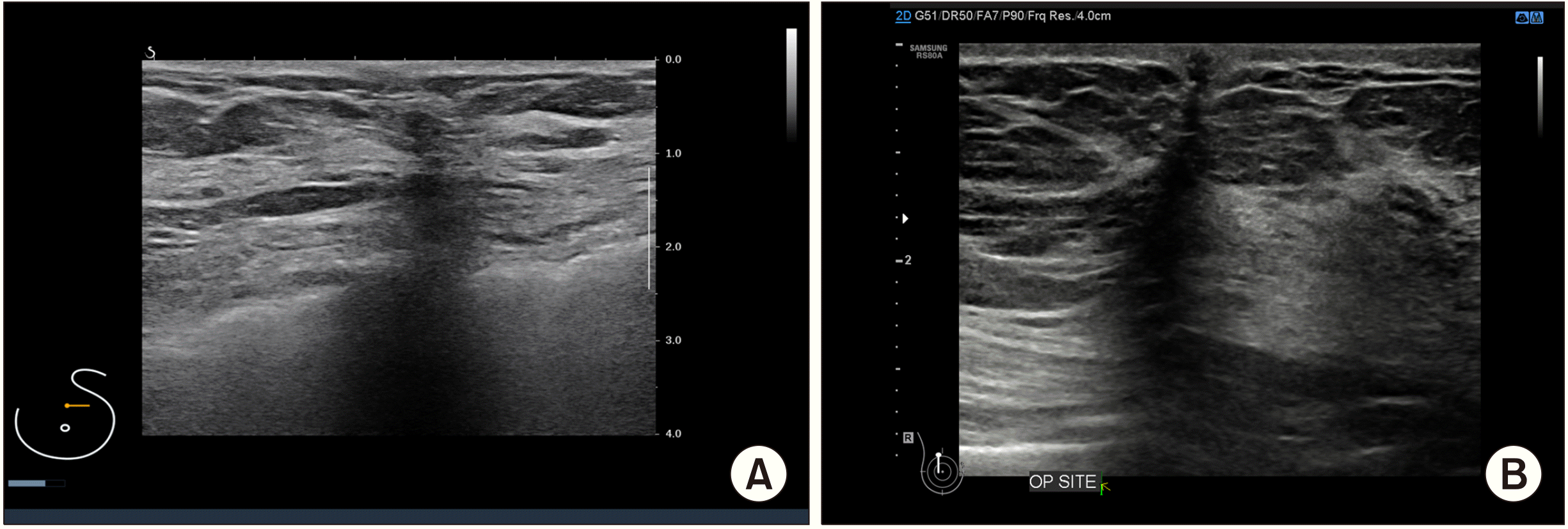
대부분의 악성질환에서 보이는 초음파 소견은 상하로 길이가 긴 (taller than wide) 형태를 보이는데, 누운 자세에서 초음파를 받는 경우 양성결절은 비교적 덜 단단하여 중력에 의한 유선의 압력에 의하여 타원형으로 보이지만 악성질환은 단단하기 때문에 이러한 압력에 영향을 적게 받기 때문이다.(11) 따라서 누운 자세에서 수술을 시행할 때에도 유선을 피부절개선에서부터 대흉근에 이르기까지 원통형으로 일정한 넓이로 절제하는 것이 필요하다. 이러한 수술기법이 향후 초음파 영상에서 보면 절개선에서부터 대흉근에 이르기까지 수직절개선의 형태로 반흔을 남기는 이유라 할 수 있다(Fig. 1).
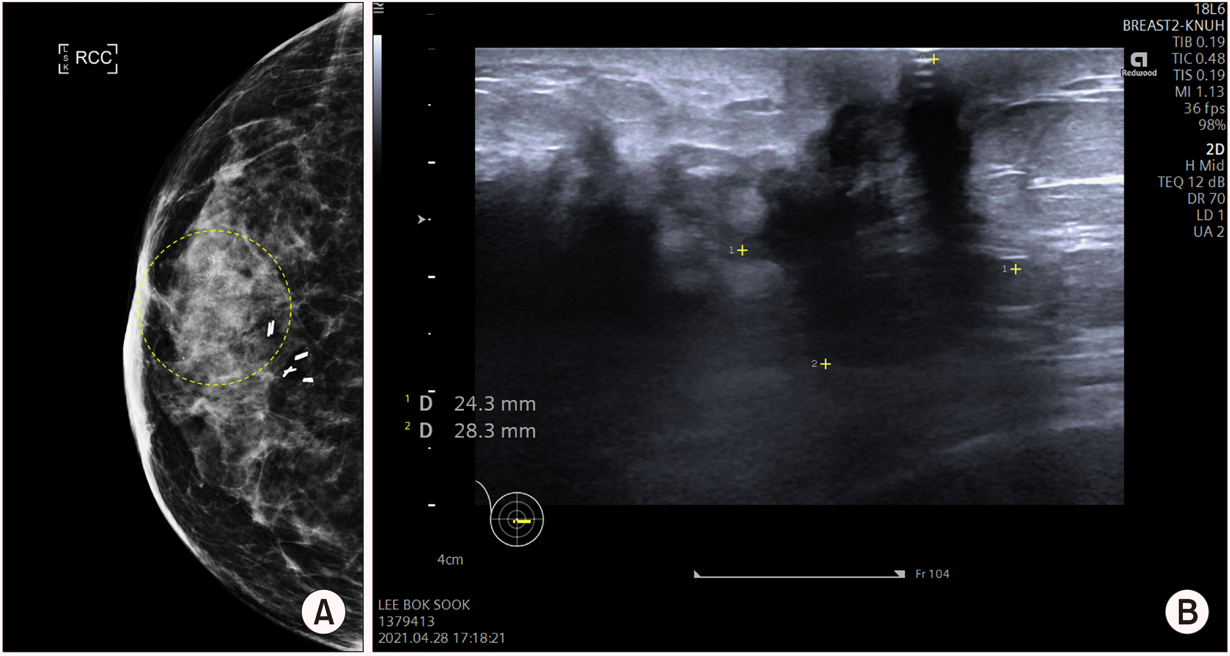
이러한 반흔은 대부분 하나의 선형태로 존재하며, 주변에 결절이나 다발성 반흔의 흔적으로 보인다면 유방암의 재발을 의심해보아야 한다. 특히, 수술 반흔 주변은 암이 존재했던 위치이기 때문에 국소재발의 위험성이 가장 높은 부위이기도 하므로 수술을 시행받은 시기로부터 얼마가 경과하였는지 확인해보고, 수술 직후 발생할 수 있는 혈종이나 염증소견에 의한 결절이 아니라면 반드시 조직검사를 통하여 정확한 진단이 필요할 것이다(Fig. 2). 다른 영상검사소견과 비교해보면 조금 더 정확하게 재발에 대한 가능성을 예측해볼 수 있는데, 유방촬영술의 경우 결절이나 미세석회화를 동반할 수 있고 유방 자기공명영상소견의 경우 조영증강을 보이는 병변이 동일한 위치에서 확인된다면 재발의 가능성이 높다고 판단할 수 있을 것이다(Fig. 3).(12)
만약 이러한 결절이 발생하였지만 수술로부터 불과 1-2개월 미만의 짦은 기간만이 경과하였다면 재발보다는 수술 후 남은 혈종이나 granulation tissue일 가능성이 높으며, 6개월 이상의 시간이 경과한 시점에서 결절이 확인되었더라도 이후 6개월-1년 이상의 기간동안 변화가 없다면 유방암의 재발은 아닐 가능성이 높다. 반대로 조직검사를 통하여 유방암의 재발이 아님이 확인되었더라도 이후 결절의 크기가 증가하거나 개수가 많아지는 경우에는 추가적인 조직검사가 다시 필요하다.
단순 유방전절제술은 흉벽을 덮을 수 있는 최소한의 피부만 보존하고 유두-유륜 복합체를 포함한 나머지 유방피부를 모두 절제하는 방법으로 수술 후에는 피부 바로 아래 얇은 지방층 외에는 남는 구조물은 없다. 따라서 초음파에서도 피부 바로 아래 피부와 평행하게 위치하는 지방층을 관찰할 수 있으며, 새로 생긴 결절이 있는 경우에는 쉽게 만져지기도 하고 초음파에서도 결절을 쉽게 확인할 수 있다. 피하지방층의 아래에는 대흉근과 소흉근이 위치하며 늑골의 단면이나 종면이 순서대로 확인가능하다.(13) 간혹 늑골을 저에코음영의 유방결절로 오인할 수 있으나, 위치상 대흉근이나 소흉근의 아래쪽에 위치하면서 동일한 저에코음영이 반복적으로 관찰된다면 그것은 늑골로 판단할 수 있다(Fig. 4).
간혹 수술 직후 환자가 피부 아래 만져지는 결절을 유방암의 재발로 오인하는 경우도 있으나, 이는 초음파상에서 비교적 타원형의 형태로 실제 절개선의 끝부분에 만든 매듭과 일치하는 위치라면 suture tie에 의한 granuloma로 판단할 수 있다. 피부의 거의 바로 아래 존재하기 때문에 조직검사는 쉽지 않으며, 신경이 많이 쓰인다면 국소마취하에 간단하게 절제가 가능할 것이다. 그러나, 발생하거나 만져지는 결절의 형태가 타원형이 아닌 경계가 모호하거나 불규칙한 변연부를 가지는 경우라면 suture granuloma보다는 유방암 재발의 가능성이 조금더 높을 수 있다. 이 때 도플러신호를 띄워보면 혈류를 공급하는 동맥이 명확히 관찰되거나 주변으로 혈류의 시그널이 높게 관찰될 수 있는데, 이러한 소견을 보인다면 유방암 재발의 가능성은 조금 더 높다고 볼 수 있다.(14) 조직검사를 통하여 쉽게 진단할 수 있지만, 조직검사 후 출혈이나 혈종발생을 예방하기 위하여 혈류를 공급하는 영양동맥(feeding artery)을 피하여 조직검사는 것이 좋다(Fig. 5).
수술 부위 또는 피부 아래 만져지는 결절이 발생하였지만 수술로부터 불과 1-2개월 미만의 짦은 기간만이 경과하였다면 유방보존술의 경우와 비슷하게 재발보다는 수술 후 suture 등에 의한 granulation일 가능성이 높으며, 장시간의 추적관찰 도중 결절이 발생하였더라도 지속적인 변화가 없는 경우라면 유방암의 재발은 아닐 가능성이 높다. 그러나, 결절의 크기가 증가하거나 개수가 많아지는 경우에는 추가적인 조직검사가 필요하다.
REFERENCES
1. Chansakul T, Lai KC, Slanetz PJ. 2012; The postconservation breast: part 1, expected imaging findings. AJR Am J Roentgenol. 198:321–30. DOI: 10.2214/AJR.10.7298. PMID: 22268174.

2. Duijm LE, Guit GL, Zaat JO, Koomen AR, Willebrand D. 1997; Sensitivity, specificity and predictive values of breast imaging in the detection of cancer. Br J Cancer. 76:377–81. DOI: 10.1038/bjc.1997.393. PMID: 9252206. PMCID: PMC2224070.

3. Yoon JH, Kim MJ, Kim EK, Moon HJ. 2015; Imaging surveillance of patients with breast cancer after primary treatment: current recommendations. Korean J Radiol. 16:219–28. DOI: 10.3348/kjr.2015.16.2.219. PMID: 25741186. PMCID: PMC4347260.

4. Chang JF, Huang CS, Chang RF. 2020; Automated whole breast segmentation for hand-held ultrasound with position information: application to breast density estimation. Comput Methods Programs Biomed. 197:105727. DOI: 10.1016/j.cmpb.2020.105727. PMID: 32916544.

5. Houssami N, Sainsbury R. 2006; Breast cancer: multidisciplinary care and clinical outcomes. Eur J Cancer. 2480–91. DOI: 10.1016/j.ejca.2006.05.023. PMID: 16904313.

6. Fragomeni SM, Sciallis A, Jeruss JS. 2018; Molecular subtypes and local-regional control of breast cancer. SurgOncol Clin N Am. 27:95–120. DOI: 10.1016/j.soc.2017.08.005. PMID: 29132568. PMCID: PMC5715810.

7. Chen JH, Nie K, Bahri S, Hsu CC, Hsu FT, Shih HN, et al. 2010; Decrease in breast density in the contralateral normal breast of patients receiving neoadjuvant chemotherapy:MR imaging evaluation. Radiology. 255:44–52. DOI: 10.1148/radiol.09091090. PMID: 20308443. PMCID: PMC2843830.

8. Yi A, Kim HH, Shin HJ, Huh MO, Ahn SD, Seo BK. 2009; Radiation-induced complications after breast cancer radiation therapy: a pictorial review of multimodality imaging findings. Korean J Radiol. 10:496–507. DOI: 10.3348/kjr.2009.10.5.496. PMID: 19721835. PMCID: PMC2731868.

9. Yilmaz ZN, Neal CH, Noroozian M, Klein KA, Sundaram B, Kazerooni EA, et al. 2014; Imaging of breast cancer-related changes after nonsurgical therapy. AJR Am J Roentgenol. 202:675–83. DOI: 10.2214/AJR.13.11518. PMID: 24555607.

10. Margolis NE, Morley C, Lotfi P, Shaylor SD, Palestrant S, Moy L, et al. 2014; Update on imaging of the postsurgical breast. Radiographics. 34:642–60. DOI: 10.1148/rg.343135059. PMID: 24819786.

11. Stavros AT. 2004. Breast ultrasound. Lippincott Williams & Wilkins;Philadelphia:
12. Gutierrez R, Horst KC, Dirbas FM, Ikeda DM. Dirbas F, Scott-Conner C, editors. 2011. Breast imaging following breast conservation therapy. Breast Surgical Techniques and Interdisciplinary Management. Springer;New York: p. 975–95. DOI: 10.1007/978-1-4419-6076-4_81.

13. Destounis SV. 2005; Imaging of the post-surgical breast. Contemp Diagn Radiol. 1–6. DOI: 10.1097/00219246-200512310-00001.

14. Stuhrmann M, Aronius R, Schietzel M. 2000; Tumor vascularity of breast lesions: potentials and limits of contrast-enhanced Doppler sonography. AJR Am J Roentgenol. 175:1585–9. DOI: 10.2214/ajr.175.6.1751585. PMID: 11090380.
Fig. 1
Ultrasonographicfindings after breast-conserving surgery. (A, B) A vertical shape of postoperative scar is observed from the skin to the pectoralis major muscle, which will be remained almost permanently. If any nodule is found around the scar or there is an increasing sign of blood flow, it should be confirmed by tissue biopsy due to possibility of tumor recurrence.

Fig. 2
Doppler imaging of breast ultrasonography in suspected nodule of local recurrence. (A) A 5 mm-sized hypoechoic nodule (yellow square) occurred near the axillary region, and showed increased blood flow on the doppler mode which indicating a proliferative lesion (confirmed as recurrent invasive cancer on histological examination). (B) A 2 cm-sized hypoechoic nodule (green squares) was identified around the postoperative scar. However, there is no evidence of increased blood flow in the doppler mode, suggesting a high possibility of non-proliferative lesions (fat necrosis was confirmed in biopsy).

Fig. 3
Local recurrence of breast cancer. (A) Malignant microcalcifi-cations (yellow dotted circle) on subareolar region were identified on mammography. (B) In ultrasound finding, a hypoechoic nodule with irregular margins were found at the same location. The lesion was dia-gnosed as a local recurrence of breast cancer in core needle biopsy.

Fig. 4
Ultrasound findings of a patient who were received left side of mastectomy for breast cancer. (A) The right side of contralateral breast is a normal breast. All of the breast structures are observed. (B) The left side of ipsilateral breast, which was removed as total mastectomy. Only the skin is remained overlying the pectoralis major muscle.

Fig. 5
Ultrasonographic finding of locally recurred nodule on ipsilateral chest wall. (A) On chest CT, an enhancing nodule was developed under the pectoralis major muscle, suggesting that blood flow was slightly increased. (B) Ultrasound finding of the same nodule. A 3.1 cm-sized nodule was identified under the pectoralis major muscle, showing irregular margins and hypoechoic material inside, which is strongly suspected of local recurrence (it was confirmed as invasive ductal carcinoma, recurred.).


 PDF
PDF Citation
 Print
Print


 XML Download