

 PDF
PDF Citation
 Print
Print


INTRODUCTION
Atherosclerosis is known to be a complex inflammatory process of vascular cells by risk factors of cardiovascular disease such as diabetes or hypertension.(1) The atherosclerotic atheroma thus formed causes rupture and thrombosis leading to acute myocardial infarction or stroke. Atherosclerosis is a major cause of cardiovascular disease, peripheral vascular disease, and cerebrovascular disease.(2)
Using a B-mode (Brightness mode) of the ultrasound imaging system, atherosclerosis and vascular status can be easily confirmed by ultrasonography. Carotid IMT (Intima-media Thickness) is a clinical prognostic factor using an easily accessible and non-invasive ultrasound imaging system. The role of carotid IMT in coronary artery stenosis has been proven in various studies over the last several decades.(3)
However, if atherosclerosis of the femoral artery is expressed more frequently than the carotid artery,(4) the measurement of intimal-medial thickness using only carotid artery may be misdiagnosed as asymptomatic atherosclerosis as a predictor of atherosclerosis or cardiovascular disease.(5) In addition, during carotid ultrasound examination, the internal carotid artery and the external carotid artery were not anatomic in all patients and the femoral artery test was more reproducible.(6) Femoral artery IMT is associated with atherosclerosis of the coronary arteries, as well as carotid IMT. It is also associated with cardiovascular mortality and myocardial infarction and is a predictor of cardiovascular disease.(6) In order to better define the IMT cut-off value of cardiovascular disease, it is recommended that a combination of carotid IMT and femoral artery IMT be measured.(7)
We investigated whether carotid IMT and femoral artery IMT as a screening test were useful factors for the detection of cardiovascular disease by using each factor or its sum and maximum value.
Go to : 
METHODS
From January 2015 to December 2017, we conducted a study at a university hospital. The subjects were healthy adults who visited the health promotion center and ultrasound examination of carotid artery and femoral artery was performed. A total of 39 patients who underwent coronary angiography or coronary artery CT angiogram ere studied. This study was approved by the Institutional Bioethics Committee (INHA IRB2016-07-001).
A total of 39 examinees were identified through questionnaires prepared at the Health Promotion Center and pre interview information. Based on medical records, electronic medical records, and paperwork, the results were as follows: the presence of hypertension, presence of diabetes mellitus, presence of hyperlipidemia, and waist circumference, systolic and diastolic blood pressure, fasting blood glucose, cholesterol, high density lipoprotein (HDL), low density lipoprotein (LDL), triglyceride. Metabolic syndrome is based on electronic medical record. The waist circumference is more than 90 cm for males, more than 85 cm for females, more than 150 mg/dl for triglycerides, less than 40 mg/dl for men and 50 mg/dl for women, more than 100 mg/dl of fasting glucose and we defined the metabolic syndrome as having three or more of these.
The ultrasound imaging device was a single device, Z.ONE (Zonare medical systems Inc., USA). We used a 5–15 MHz linear probe as transducer. The test was performed by a trained One Vascular Technologist (RVT).
In this study, we defined carotid artery stenosis (CAS) and peripheral artery disease (PAD) is defined as increased peak systolic velocity (PSV > 150 cm/s) or diminished PSV (< 45 cm/s) distal to the stenotic lesion with over 50% stenotic lesion. The coronary artery stenosis was defined by stenosis over 50% in 3D CT angiogram. Thirty one of 39 patients underwent coronary artery 3D CTA and 8 patients underwent coronary angiography.
In this study, SPSS statistics ver. 20 (IBM, USA) were used. The patients were classified as total arterial disease of at least one arterial disease among three arterial diseases and no arterial disease group. The chi-square and T test was used to compare the characteristics between two groups. We also performed the correlation analysis between maximum and sum of both IMT with total arterial disease.
RESULTS
There were 25 patients with arterial diseases in 39 enrolled patients. There were 22 patients with coronary artery disease, 8 patients with carotid artery disease and 7 patients with peripheral artery disease. There were older and more metabolic syndromes in arterial disaes group comparing no arterial disease group (Table 1).
Table 1
Characteristic of the Study Population for with or without Total Arterial Disease Separately
| Characteristic | No AD* (n = 14) | AD* (n = 25) | P |
|---|---|---|---|
| Age (mean ± SD, years)† | 47.5 ± 7.0 | 55.0 ± 6.3 | 0.002 |
| Male (n, %) | 13 (93) | 23 (92) | 0.926 |
| Smoking (%) | 11 (79) | 15 (60) | 0.230 |
| Metabolic syndrome (%) | 0 (0) | 5 (20) | 0.022 |
| Waist (mean ± SD, cm)† | 82.1 ± 6.8 | 83.2 ± 7.2 | 0.646 |
| Hypertension (%) | 9 (64) | 21 (84) | 0.210 |
| Systolic blood pressure (mmHg)† | 123.7 ± 12.8 | 128.2 ± 10.2 | 0.234 |
| Diastolic blood pressure (mmHg)† | 74.1 ± 9.7 | 76.5 ± 8.1 | 0.412 |
| Diabetes (%) | 4 (14) | 9 (36) | 0.125 |
| Glucose (mg/dl)† | 88.4 ± 11.0 | 95.8 ± 13.3 | 0.083 |
| Hyperlipidemia (%) | 10 (71) | 14 (56) | 0.355 |
| Cholesterol (mg/dl)† | 195.3 ± 45.7 | 198.9 ± 38.7 | 0.794 |
| HDL cholesterol (mg/dl)†‡ | 57.8 ± 15.3 | 49.6 ± 11.0 | 0.059 |
| LDL cholesterol (mg/dl)†§ | 118.9 ± 49.0 | 117.6 ± 34.5 | 0.928 |
| Triglyceride (mg/dl)† | 135.1 ± 93.6 | 186.8 ± 263.7 | 0.485 |
![]()
There was a significant difference between any arterial disease groups and no arterial groups in the any values of both the carotid artery and femoral artery IMT (Table 2).
Table 2
IMT Analysis of with and without Total Arterial Disease
| IMT* (mm) | No AD† (n = 14) | AD† (n = 25) | t | P |
|---|---|---|---|---|
| Carotid maximum‡ | 0.707 ± 0.121 | 0.824 ± 0.148 | -2.518 | 0.016 |
| Femoral maximum‡ | 0.543 ± 0.041 | 0.624 ± 0.123 | -2.134 | 0.039 |
| Carotid sum‡ | 1.307 ± 0.209 | 1.520 ± 0.275 | -2.509 | 0.017 |
| Femoral sum‡ | 0.979 ± 0.153 | 1.132 ± 0.216 | -2.348 | 0.024 |
| IMT* average‡ | 0.579 ± 0.058 | 0.660 ± 0.100 | -3.220 | 0.003 |
![]()
There was a significant correlation between all value of carotid IMT and femoral artery IMT except the maximum of the carotid IMT and the sum of the femoral artery IMT (Table 3). The maximum values of carotid IMT was correlative with maximum values of the femoral IMT (R = 0.322, P = 0.023) and the sum of carotid IMT was also correlative sum of femoral IMT (R = 0.346, P = 0.015) (Fig. 1).
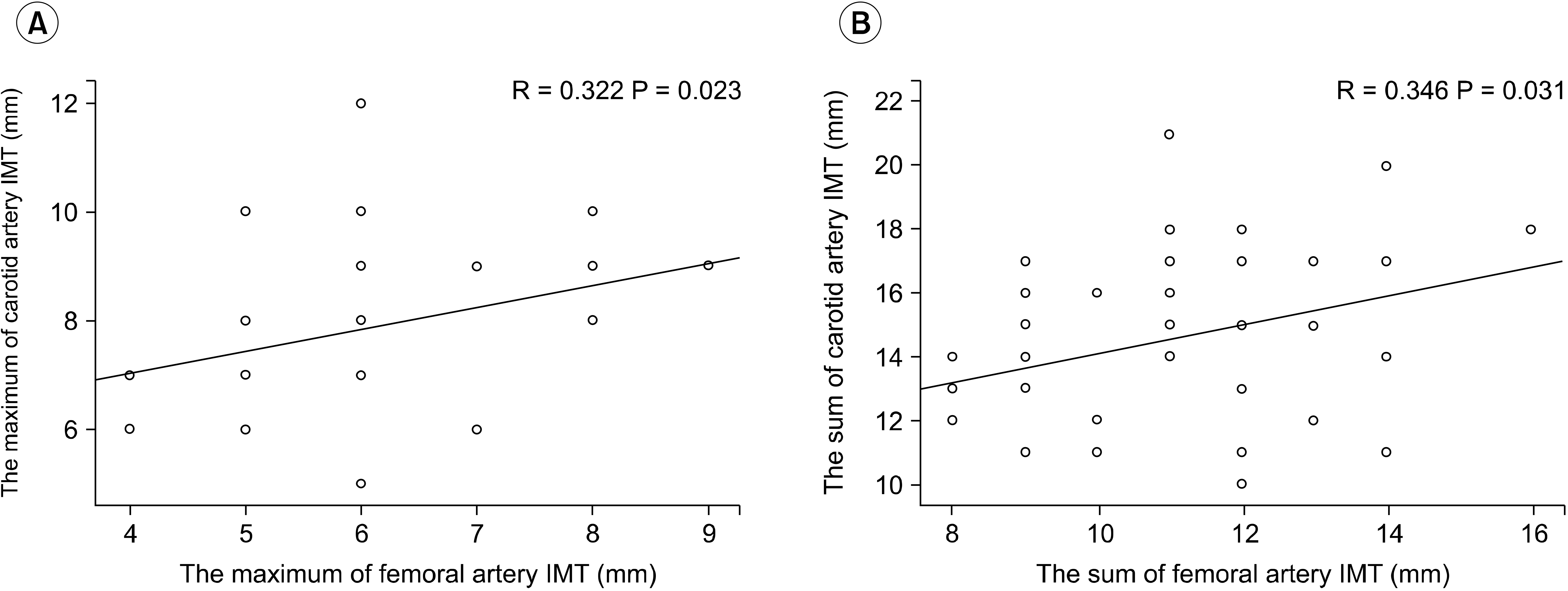 | Fig. 1The relation between femoral artery IMT and carotid artery IMT. The maximum of femoral artery IMT is correlative with that of carotid artery IMT (A, P = 0.023), The sumof femoral artery IMT is correlative with that of carotid artery IMT (B, P = 0.015).
|
Table 3
Correlation Coefficient of Catorid Intima-media Thickness (IMT) and Femoral IMT
| Correlation | Carotid MAX* | Femoral MAX* | Carotid sum | |
|---|---|---|---|---|
| Carotid MAX* | Pearson correlation | 1 | .322 | .928 |
| P (both) | .023 | .000 | ||
| N | 39 | 39 | 39 | |
| Femoral MAX* | Pearson correlation | .322 | 1 | .414 |
| P (both) | .023 | .004 | ||
| N | 39 | 39 | 39 | |
| Carotid sum | Pearson correlation | .928 | .414 | 1 |
| P (both) | .000 | .009 | ||
| N | 39 | 39 | 39 | |
| Femoral sum | Pearson correlation | .252 | .902 | .346 |
| P (both) | .061 | .000 | .015 | |
| N | 39 | 39 | 39 | |
![]()
Go to :
DISCUSSION
The carotid IMT was relatively simple because of the absence of fasting or contrast media, and it was found to be highly correlated with high mortality arterial disease such as cardiovascular disease and cerebrovascular disease.(8) In the past several decades, various studies have confirmed the association between carotid IMT and cardiovascular disease, and carotid IMT has been used as an independent predictor of vascular disease as an independent factor.
However, the internal carotid artery and the external carotid artery are difficult to perform in all patients due to the anatomic structure during carotid ultrasound examination, so that the reproducibility of the test is less accurate than that of the femoral artery.(6) This may lead to errors in the location of the IMT measurement of the carotid artery because the branches cann’t be observed accurately. In addition, the carotid artery may cause vascular bending due to an increase in age, and the IMT may be changed by the examiner. The direction of the femoral artery mainly run straight, and the IMT was measured at the distal portion of the femoral artery, which is the most commonly used puncture site for angiography. For this reason, few studies have reported the use of IMT of the carotid artery and femoral artery, or IMT of the femoral artery alone as predictors of arterial disease.(9) Ultrasound imaging of femoral arteries is cost-effective, non-invasive and reproducible method of assessment comparing carotid arteries but there is some concern regarding the difficulty of the examination of femoral artery, especially in overweight patients.(7)
The correlation between the IMT of the carotid artery and the femoral artery was correlated with Framingham heart study risk values when combined with each other.(10) There was a strong correlation between the femoral artery IMT and moderate CAD expressed in Gensini numbers.(9)
As mentioned above, studies on IMT, atherosclerosis and arterial disease have been studied for many years since the 1980’s, and the research is still underway. Based on these various studies, we analyzed the correlation between carotid artery disease, peripheral arterial disease and total arterial disease as well as coronary artery disease which has undergone study about correlations of IMT of carotid artery and femoral artery.
In our study with patients with arterial disease with at least one of the three arterial diseases, the maximum value and sum of IMT of both carotid arteries and femoral arteries were analyzed. Though this study carried out on a small number of patients that visited health promotion center, there was a significant difference in all IMT between the patients with three arterial disease and those without arterial disease. There is strong evidence suggesting angiographic finding arterial disease with large sample size is correlated with femoral IMT. Furthermore, the authors consider that a follow-up study of the patients could bring more information about the progression of arterial disease and might link the femoral arterial ultrasonographic finding to arterial disease such as carotid arterial plaque.
The maximum and the sum of carotid artery and femoral artery IMT were significant factors in evaluating the early arterial disease.
Go to :
CONCLUSION
The IMT of the carotid artery and the femoral artery were closely correlated with each other. The maximum values and sum of IMT of the carotid artery and femoral artery were significantly correlated with each other. Therefore, it may be necessary to additionally examine the IMT of femoral artery as predictors of early arterial disease like carotid IMT.
Go to :
 XML Download