Abstract
Background
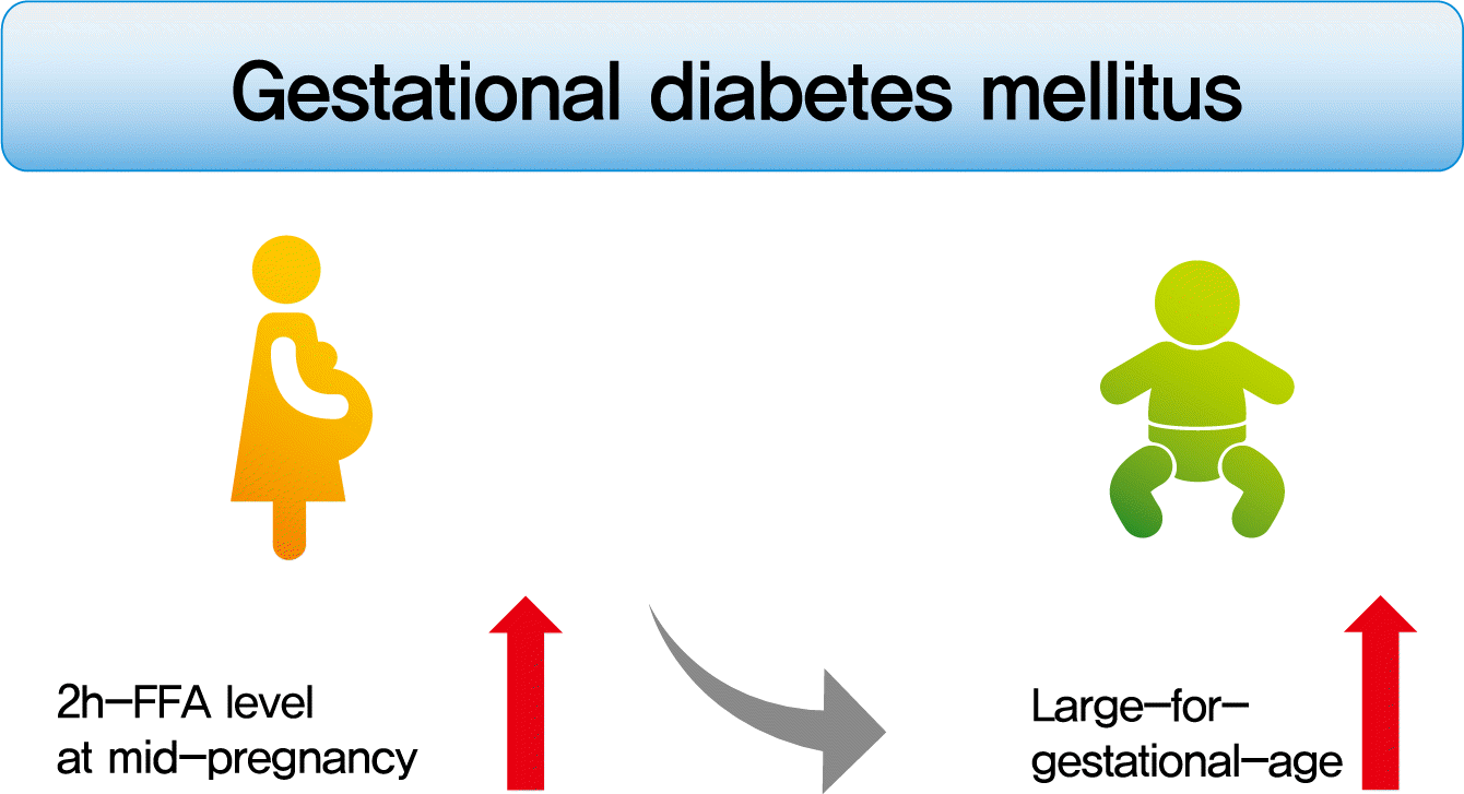
To investigate the association between free fatty acid (FFA) level at mid-pregnancy and large-for-gestational-age (LGA) newborns in women with gestational diabetes mellitus (GDM).
Methods
We enrolled 710 pregnant women diagnosed with GDM from February 2009 to October 2016. GDM was diagnosed by a ‘two-step’ approach with Carpenter and Coustan criteria. We measured plasma lipid profiles including fasting and 2-hour postprandial FFA (2h-FFA) levels at mid-pregnancy. LGA was defined if birthweights of newborns were above the 90th percentile for their gestational age.
Results
Mean age of pregnant women in this study was 33.1 years. Mean pre-pregnancy body mass index (BMI) was 22.4 kg/m2. The prevalence of LGA was 8.3% (n=59). Levels of 2h-FFA were higher in women who delivered LGA newborns than in those who delivered non-LGA newborns (416.7 μEq/L vs. 352.5 μEq/L, P=0.006). However, fasting FFA was not significantly different between the two groups. The prevalence of delivering LGA newborns was increased with increasing tertile of 2h-FFA (T1, 4.3%; T2, 9.8%; T3, 10.7%; P for trend <0.05). After adjustment for maternal age, pre-pregnancy BMI, and fasting plasma glucose, the highest tertile of 2h-FFA was 2.38 times (95% confidence interval, 1.11 to 5.13) more likely to have LGA newborns than the lowest tertile. However, there was no significant difference between groups according to fasting FFA tertiles.
Gestational diabetes mellitus (GDM) is defined as “diabetes diagnosed during pregnancy that is not clearly overt diabetes” [1]. GDM is steadily increasing. It is becoming an emerging burden in global health [2,3]. GDM has a variety of negative implications for mothers and their offspring. For mothers, it is associated with higher rates of preeclampsia, Cesarean deliveries, shoulder dystocia, and type 2 diabetes mellitus (T2DM) in the postpartum period [4-8]. For offspring born to mothers with GDM, they have a higher likelihood of developing obesity, having an impaired glucose tolerance, and developing T2DM in childhood or in early adulthood [9,10].
Large-for-gestational-age (LGA) newborn is an important determinant factor in perinatal morbidity and mortality. Women with GDM are more likely to deliver LGA newborns than those without GDM [11]. In general, maternal serum lipid levels will increase during mid to late gestation. Such increase is believed to be beneficial to mother and fetus in terms of lactation and nutrition [12]. It is already known that maternal fasting serum triglycerides (TG) level at mid-pregnancy can predict LGA newborns independent of maternal pre-pregnancy body mass index (BMI), weight gain during pregnancy, age, and parity in women with GDM [13]. Maternal insulin resistance may link lipid levels and fetal growth in women with GDM. Free fatty acid (FFA) is a fundamental player in insulin resistance. Thus it might be associated with birth weight. However, few studies have examined the association between serum FFA level and LGA newborns in women with GDM. Because LGA newborns have various risks, it is very important to predict LGA during pregnancy. Thus, the aim of this study was to investigate the association between FFA level at mid-pregnancy and LGA newborns in women with GDM.
This observational study was conducted in CHA Bundang Medical Center (Seongnam, Korea) from February 2009 to October 2016, enrolling 710 pregnant women aged ≥20 years who were diagnosed with GDM. The diagnosis of GDM was made by a two-step approach. Patients were initially screened with a 1-hour 50-g glucose challenge test as screening at 24 to 28 weeks of gestation. A 3-hour 100-g oral glucose tolerance test (OGTT) was performed for a subset of women who exceeded glucose threshold value (≥140 mg/dL at 1-hour 50-g glucose challenge test). The diagnosis of GDM was made if at least two of four plasma glucose levels (measured fasting and 1, 2, and 3 hours after the 100-g OGTT) met or exceeded glucose threshold value according to the Carpenter and Coustan criteria [14]. Women who were known to have had type 1 diabetes mellitus or T2DM before pregnancy, thyroid disorders, and twin pregnancy were excluded.
At 24 to 32 weeks of gestation (baseline visit), anthropometric assessments were carried out. We also measured blood pressure, and collected venous blood sample, including fasting glucose, glycosylated hemoglobin (HbA1c), lipid profiles, and FFA for all study participants. FFA and TG were also measured at postprandial 2-hour after eating a standard mixed meal (480 kcal, 60% carbohydrate, 20% protein, and 20% fat) composed of Korean food. We collected baseline clinical and demographic characteristics (including a history of obstetric or medical diseases, family history of diabetes, pre-gestational BMI) from medical records. These women were given medical nutrition therapy during their antenatal follow-up. LGA was defined as birth weight above the 90th percentile for their gestational age based on intrauterine growth percentile curve in Korean 1999 birth cohort [15]. We obtained informed consent from all study participants. This study was approved by the Institutional Review Board of CHA Bundang Medical Center (2017-08-004-001).
Height and weight were measured for all subjects (wearing minimal clothing without shoes). Blood pressure was measured by trained nurses using an automatic sphygmomanometer after participants were seated for 10 minutes. Serum FFAs were measured by enzymatic colorimetry using a Labospect 008AS (Hitachi, Tokyo, Japan). Reagents used to measure FFAs were acyl-coenzyme A (CoA) synthetase, acyl-CoA oxidase, and 3-methyl-N-ethyl-N-(β-hydroxyethyl)-aniline (MEHA). We measured plasma glucose, total cholesterol, TG, and highdensity lipoprotein cholesterol (HDL-C) using a Hitachi 7600 analyzer (Hitachi, Tokyo, Japan). We measured HbA1c using a high-performance liquid chromatography (before May 2013, Bio-Rad Variant II, Hercules, CA, USA; after May 2013, Tosoh G8, San Francisco, CA, USA). We assessed C-peptide and insulin levels by chemiluminescence immunoassay (Roche Diagnostic GmbH, Mannheim, Germany). We carried out homeostasis model assessment of insulin resistance (HOMA-IR) and β-cell function (HOMA-β) according to suggested formulas [16].
Data for categorical factors are reported as percentages. Continuous variables are presented as mean±standard deviation. Characteristics of participants at baseline were compared using Student’s t-test or chi-square test. Subjects were divided into fasting FFA and 2-hour postprandial FFA (2h-FFA) tertiles (T) as follows: fasting FFA T1, ≤603 μEq/L; T2, 604–796 μEq/L; T3, ≥797 μEq/L; 2h-FFA T1, ≤261 μEq/L; T2, 262–400 μEq/L; and T3, ≥401 μEq/L. Pearson’s correlation analysis was performed to analyze the correlation between FFA level and various factors after adjusting for age and pre-pregnancy BMI. Binary logistic regression analyses were performed to determine the odds ratio and 95% confidence intervals (CIs) of delivering LGA newborns after adjusting for covariates. All statistical analyses were performed using SPSS version 26.0 (IBM Co., Armonk, NY, USA). P<0.05 was considered statistically significant.
Table 1 shows baseline characteristics of women with GDM at mid-pregnancy. Mean age was 33.1 years and mean pre-pregnancy BMI was 22.4 kg/m2. Among 710 women with GDM, 59 (8.3%) delivered LGA newborns. Women who delivered LGA newborns had higher pre-pregnancy BMI, fasting plasma glucose (FPG), HbA1c, 2h-FFA than those who delivered non-LGA neonates. There were no significant differences in age, history of GDM, family history of diabetes, parity, gestational weeks, blood pressure, total cholesterol, fasting TG, 2h-TG, fasting FFA, HOMA-IR, HOMA-β, or 100-g OGTT plasma glucose levels between women who delivered LGA and those who delivered non-LGA newborns.
Pregnancy outcomes such as gestational age at delivery, preterm delivery, Cesarean section, and neonate weight were not significantly different among three groups of fasting and 2h-FFA tertile (Table 2). The prevalence of delivering LGA newborns was increased with increasing tertile of 2h-FFA (T1, 4.3%; T2, 9.8%; T3, 10.7%; P for trend <0.05) (Fig. 1). However, there was no significant difference in the prevalence of delivering LGA newborns according to tertiles of fasting FFA. In addition, there was no difference in 2h-FFA between women who managed lifestyle modification and those who treated insulin (356.8 μEq/L vs. 362.2 μEq/L, P=0.740).
2h-FFA, but not fasting FFA, was positively correlated with HbA1c after adjusting for age and pre-pregnancy BMI (r=0.096, P=0.011) (Table 3). FFA (fasting and 2h) was correlated with total cholesterol and TG (fasting and 2h), but not with HDL-C. Interestingly, although it was not significantly associated after adjusting for age and pre-pregnancy BMI, 2h-FFA was positively correlated with HOMA-IR. On the other hand, fasting FFA was negatively associated with HOMA-IR.
The risk of delivering LGA newborns was 2.66 times (95% CI, 1.25 to 5.66) higher in the highest tertile of 2h-FFA compared with that in the lowest tertile (Table 4). Even after adjusting for age, pre-pregnancy BMI, and FPG, the risk of delivering LGA newborns was still 2.38 times (95% CI, 1.11 to 5.13) higher in the highest tertile of 2h-FFA. The cut-off value of 2h-FFA for predicting LGA newborns was 343.5 μEq/L and its sensitivity and specificity were 66.1% and 59.0%, respectively (Supplementary Table 1). However, there was no significant difference for the risk of delivering LGA newborns among tertiles of fasting FFA.
LGA newborns are common in women with GDM. They should be carefully managed because various metabolic disorders can be developed later in life [11]. Many risk factors have been investigated for delivering LGA newborns in women with GDM. However, few studies have evaluated the association between FFA level at mid-pregnancy and LGA newborns. In this study, we found that a high 2h-FFA level, but not fasting FFA, at mid-pregnancy was associated with an increasing risk of delivering LGA newborns in women with GDM.
LGA is a risk factor for complications among both newborns and mothers [17,18]. For infants, LGA has been associated with increased risks of infant mortality and traumatic injuries during delivery [19,20]. These traumatic injuries include shoulder dystocia, clavicle or humerus fractures and brachial or facial paralysis [21,22]. High birth weight is also extended to adverse consequences in later life, including later development of overweight [23]. For mothers, delivering LGA newborns is associated with genital tract injury, prolonged labor, risk of postpartum bleeding, and an increased risk of cesarean delivery [18,20,22]. Because GDM is an important risk factor of delivering LGA, proper screening and medical care for LGA in women with GDM are likely to be important.
To date, many risk factors for delivering LGA newborns have been identified, such as advanced maternal age, obesity, multiparity, gestational weight gain, smoking, and GDM [24-26]. A Swedish population-based cohort study of 1,260,297 women has reported that women with GDM have 3.4 times higher risk of delivering LGA newborns than women without GDM [11]. In general, fetal growth is determined not only by plasma glucose levels, but also by other fuels such as lipids and amino acids [27]. Several studies have found that maternal lipids are associated with fetal growth [28,29]. It is well known that maternal TG concentration is independently associated with LGA newborns [13,30-32]. In this study, however, there was no difference in maternal TG concentration (fasting and 2h) between women who delivered LGA and those who delivered non-LGA newborns, unlike FFA levels. There may be several reasons, such as ethnic characteristics, variability of measurements, etc., but not all of them are certain. Because FFA as fundamental lipid is an essential component of lipid metabolism and insulin resistance, we could assume that FFA might be associated with LGA newborns in women with GDM. We evaluated the association between FFA (fasting and 2h) and LGA newborns and found that only 2h-FFA was associated with elevated risk of LGA newborns in women with GDM. However, as opposed to our expectation, 2h-FFA was not significantly associated with HOMA-IR. This might be because HOMA-IR values were relatively low (mean 1.57) compared with those in Western populations due to low pre-pregnancy BMI (mean 22.4 kg/m2).
FFAs are lipids bound to transport protein, such as albumin. They are also known as non-esterified fatty acids [33]. FFAs play a critical role in the development of T2DM possibly due to lipotoxicity defined as cellular dysfunction caused by elevated level of FFAs [34], thereby reducing glucose uptake by peripheral tissues and stimulation of endogenous glucose production [35]. Several studies have shown higher plasma levels of FFA in T2DM and obesity [36-39]. Hawkins et al. [39] have found that plasma FFA concentrations were higher in individuals with poor glycemic control than in individuals with good glycemic control and nondiabetic individuals during euglycemia (poor glycemic control, 400±95 μmol/L; good glycemic control, 237±28 μmol/L; nondiabetic individuals, 170±28 μmol/L; P<0.01). During hyperglycemia, FFA concentrations were also higher in individuals with poor glycemic control than other two groups (poor glycemic control, 381±75 μmol/L; good glycemic control, 198±19 μmol/L; nondiabetic individuals, 170±28 μmol/L; P<0.01). In addition, plasma FFA levels are commonly increased during late gestation [40]. They are responsible for much of physiologic insulin resistance that occurs in all women during the 2nd half of normal pregnancy [40]. Because fetal birth weight is mainly associated with maternal insulin resistance and lipid metabolism, FFA might be a crucial role for LGA newborns. However, how FFA, especially 2h-FFA, influences fetal growth has not been sufficiently investigated yet.
Inappropriately increased plasma FFA levels could have adverse metabolic effects such as reduced glucose uptake by peripheral tissues and stimulation of endogenous glucose production [35,36]. Some reviews have suggested that detrimental effects of elevated FFAs are likely to be more severe in a postprandial state than in a fasting state [41,42]. Wang et al. [43] have also suggested that higher concentrations of 2h-saturated fatty acid (SFA), not fasting SFA, are independently associated with an increased risk of T2DM. The postprandial state is vital because it not only represents an early metabolic response to glucose, meals, or nutrients, but also accounts for the most time spent in meal sequence during daytime and the usual long duration of certain postprandial metabolites such as the periods for lipids and fatty acids [44]. The ability to regulate postprandial metabolites and decrease blood glucose is apparently a crucial reflection of metabolic efficiency. In addition, previous studies have found that an exacerbated accumulation of 2h-SFA is involved in insulin resistance [45,46]. Therefore, it is likely that increased 2h-FFA levels in women with GDM might have detrimental effects regarding the risk of delivering LGA newborns due to increased insulin resistance. In this study, we found that the prevalence of delivering LGA newborns was increased in women with high 2h-FFA, rather than with high fasting FFA.
Some limitations need to be considered when interpreting our findings. First, 2h-FFA was measured after eating a standard mixed meal, not a 75-g OGTT. Second, as our study participants were confined to Korean women with a singleton pregnancy, we could not generalize these results to other ethnic populations. Third, because we could not get the data of known risk factors for LGA newborns such as smoking history and gestational weight gain, we could not evaluate the association between FFA and LGA newborns after adjustment for aforementioned factors. Finally, we only measured serum FFA once at mid-pregnancy. Thus we could not evaluate effects of dynamic changes of FFA during pregnancy.
In conclusion, a high 2h-FFA level (but not a high fasting FFA) at mid-pregnancy is associated with an increasing risk of delivering LGA newborns in women with GDM. Consequently, we should try to prevent excessive fetal growth if women with GDM have high 2h-FFA levels at mid-pregnancy.
Supplementary Materials
Supplementary materials related to this article can be found online at https://doi.org/10.4093/dmj.2021.0023.
Notes
REFERENCES
1. American Diabetes Association. 2. Classification and diagnosis of diabetes: standards of medical care in diabetes-2020. Diabetes Care. 2020; 43(Suppl 1):S14–31.
2. Kim MH, Kwak SH, Kim SH, Hong JS, Chung HR, Choi SH, et al. Pregnancy outcomes of women additionally diagnosed as gestational diabetes by the International Association of the Diabetes and Pregnancy Study Groups Criteria. Diabetes Metab J. 2019; 43:766–75.
3. Hong S, Lee SM, Kwak SH, Kim BJ, Koo JN, Oh IH, et al. A comparison of predictive performances between old versus new criteria in a risk-based screening strategy for gestational diabetes mellitus. Diabetes Metab J. 2020; 44:726–36.
4. Kang S, Kim MH, Kim MY, Hong JS, Kwak SH, Choi SH, et al. Progression to gestational diabetes mellitus in pregnant women with one abnormal value in repeated oral glucose tolerance tests. Diabetes Metab J. 2019; 43:607–14.
5. Kim KS, Park SW, Cho YW, Kim SK. Vitamin D deficiency at mid-pregnancy is associated with a higher risk of postpartum glucose intolerance in women with gestational diabetes mellitus. Endocrinol Metab (Seoul). 2020; 35:97–105.
6. Oh TJ, Kim YG, Kang S, Moon JH, Kwak SH, Choi SH, et al. Oral glucose tolerance testing allows better prediction of diabetes in women with a history of gestational diabetes mellitus. Diabetes Metab J. 2019; 43:342–9.
7. Falcone V, Kotzaeridi G, Breil MH, Rosicky I, Stopp T, Yerlikaya-Schatten G, et al. Early assessment of the risk for gestational diabetes mellitus: can fasting parameters of glucose metabolism contribute to risk prediction? Diabetes Metab J. 2019; 43:785–93.
8. Morettini M, Castriota C, Gobl C, Kautzky-Willer A, Pacini G, Burattini L, et al. Glucose effectiveness from short insulinmodified IVGTT and its application to the study of women with previous gestational diabetes mellitus. Diabetes Metab J. 2020; 44:286–94.
9. Di Cianni G, Lacaria E, Lencioni C, Resi V. Preventing type 2 diabetes and cardiovascular disease in women with gestational diabetes: the evidence and potential strategies. Diabetes Res Clin Pract. 2018; 145:184–92.
10. Lowe WL Jr, Scholtens DM, Kuang A, Linder B, Lawrence JM, Lebenthal Y, et al. Hyperglycemia and Adverse Pregnancy Outcome Follow-up Study (HAPO FUS): maternal gestational diabetes mellitus and childhood glucose metabolism. Diabetes Care. 2019; 42:372–80.
11. Fadl HE, Ostlund IK, Magnuson AF, Hanson US. Maternal and neonatal outcomes and time trends of gestational diabetes mellitus in Sweden from 1991 to 2003. Diabet Med. 2010; 27:436–41.
12. Herrera E, Munilla MA. Maternal lipid metabolism and its implications for fetal growth. Placental function and fetal nutrition Nestle Nutrition Workshop Series. Available from: https://www.nestlenutrition-institute.org/sites/default/files/documents-library/publications/secured/8a57f88112fde6fe164baf24e1454918.pdf (cited 2021 Jul 8).
13. Son GH, Kwon JY, Kim YH, Park YW. Maternal serum triglycerides as predictive factors for large-for-gestational age newborns in women with gestational diabetes mellitus. Acta Obstet Gynecol Scand. 2010; 89:700–4.
14. Harper LM, Mele L, Landon MB, Carpenter MW, Ramin SM, Reddy UM, et al. Carpenter-Coustan compared with national diabetes data group criteria for diagnosing gestational diabetes. Obstet Gynecol. 2016; 127:893–8.
15. Hong J. A study on mortality level of newborns and birthweight by gestational age: based on intrauterine growth percentile curve in Korean 1999 birth cohort [master’s thesis]. Seoul: Yonsei University;2003.
16. Wallace TM, Levy JC, Matthews DR. Use and abuse of HOMA modeling. Diabetes Care. 2004; 27:1487–95.
17. Wikstrom I, Axelsson O, Bergstrom R. Maternal factors associated with high birth weight. Acta Obstet Gynecol Scand. 1991; 70:55–61.
18. Lazer S, Biale Y, Mazor M, Lewenthal H, Insler V. Complications associated with the macrosomic fetus. J Reprod Med. 1986; 31:501–5.
19. Wikstrom I, Axelsson O, Bergstrom R, Meirik O. Traumatic injury in large-for-date infants. Acta Obstet Gynecol Scand. 1988; 67:259–64.
20. Spellacy WN, Miller S, Winegar A, Peterson PQ. Macrosomia: maternal characteristics and infant complications. Obstet Gynecol. 1985; 66:158–61.
21. Oral E, Cagdas A, Gezer A, Kaleli S, Aydinli K, Ocer F. Perinatal and maternal outcomes of fetal macrosomia. Eur J Obstet Gynecol Reprod Biol. 2001; 99:167–71.
22. Meshari AA, De Silva S, Rahman I. Fetal macrosomia: maternal risks and fetal outcome. Int J Gynaecol Obstet. 1990; 32:215–22.
24. Surkan PJ, Hsieh CC, Johansson AL, Dickman PW, Cnattingius S. Reasons for increasing trends in large for gestational age births. Obstet Gynecol. 2004; 104:720–6.
25. Kramer MS, Morin I, Yang H, Platt RW, Usher R, McNamara H, et al. Why are babies getting bigger?: temporal trends in fetal growth and its determinants. J Pediatr. 2002; 141:538–42.
26. Choi SK, Park IY, Shin JC. The effects of pre-pregnancy body mass index and gestational weight gain on perinatal outcomes in Korean women: a retrospective cohort study. Reprod Biol Endocrinol. 2011; 9:6.
28. Kulkarni SR, Kumaran K, Rao SR, Chougule SD, Deokar TM, Bhalerao AJ, et al. Maternal lipids are as important as glucose for fetal growth: findings from the Pune Maternal Nutrition Study. Diabetes Care. 2013; 36:2706–13.
29. Adank MC, Benschop L, Kors AW, Peterbroers KR, Smak Gregoor AM, Mulder MT, et al. Maternal lipid profile in early pregnancy is associated with foetal growth and the risk of a child born large-for-gestational age: a population-based prospective cohort study: maternal lipid profile in early pregnancy and foetal growth. BMC Med. 2020; 18:276.
30. Schaefer-Graf UM, Pawliczak J, Passow D, Hartmann R, Rossi R, Buhrer C, et al. Birth weight and parental BMI predict overweight in children from mothers with gestational diabetes. Diabetes Care. 2005; 28:1745–50.
31. Wang D, Xu S, Chen H, Zhong L, Wang Z. The associations between triglyceride to high-density lipoprotein cholesterol ratios and the risks of gestational diabetes mellitus and large-for-gestational-age infant. Clin Endocrinol (Oxf). 2015; 83:490–7.
32. Barat S, Ghanbarpour A, Bouzari Z, Batebi Z. Triglyceride to HDL cholesterol ratio and risk for gestational diabetes and birth of a large-for-gestational-age newborn. Caspian J Intern Med. 2018; 9:368–75.
33. Sieber J, Jehle AW. Free fatty acids and their metabolism affect function and survival of podocytes. Front Endocrinol (Lausanne). 2014; 5:186.
34. Roden M, Krssak M, Stingl H, Gruber S, Hofer A, Furnsinn C, et al. Rapid impairment of skeletal muscle glucose transport/phosphorylation by free fatty acids in humans. Diabetes. 1999; 48:358–64.
35. Lam TK, Carpentier A, Lewis GF, van de Werve G, Fantus IG, Giacca A. Mechanisms of the free fatty acid-induced increase in hepatic glucose production. Am J Physiol Endocrinol Metab. 2003; 284:E863–73.
36. Jensen MD, Haymond MW, Rizza RA, Cryer PE, Miles JM. Influence of body fat distribution on free fatty acid metabolism in obesity. J Clin Invest. 1989; 83:1168–73.
37. Boden G. Obesity, insulin resistance, type 2 diabetes and free fatty acids. Expert Rev Endocrinol Metab. 2006; 1:499–505.
38. Boden G. Free fatty acids, insulin resistance, and type 2 diabetes mellitus. Proc Assoc Am Physicians. 1999; 111:241–8.
39. Hawkins M, Tonelli J, Kishore P, Stein D, Ragucci E, Gitig A, et al. Contribution of elevated free fatty acid levels to the lack of glucose effectiveness in type 2 diabetes. Diabetes. 2003; 52:2748–58.
40. Sivan E, Boden G. Free fatty acids, insulin resistance, and pregnancy. Curr Diab Rep. 2003; 3:319–22.
41. Frayn KN, Summers LK, Fielding BA. Regulation of the plasma non-esterified fatty acid concentration in the postprandial state. Proc Nutr Soc. 1997; 56:713–21.
42. Frayn KN, Williams CM, Arner P. Are increased plasma nonesterified fatty acid concentrations a risk marker for coronary heart disease and other chronic diseases? Clin Sci (Lond). 1996; 90:243–53.
43. Wang Y, Meng X, Deng X, Okekunle AP, Wang P, Zhang Q, et al. Postprandial saturated fatty acids increase the risk of type 2 diabetes: a cohort study in a Chinese population. J Clin Endocrinol Metab. 2018; 103:1438–46.
44. Lairon D, Lopez-Miranda J, Williams C. Methodology for studying postprandial lipid metabolism. Eur J Clin Nutr. 2007; 61:1145–61.
Table 1.
Values are presented as number (%) or mean±standard deviation.
LGA, large-for-gestational-age; BMI, body mass index; GDM, gestational diabetes mellitus; HbA1c, glycosylated hemoglobin; HDL-C, high-density lipoprotein cholesterol; HOMA-IR, homeostasis model assessment of insulin resistance; HOMA-β, homeostasis model assessment of β-cell function; OGTT, oral glucose tolerance test.
Table 2.
Variable | T1 | T2 | T3 | P value |
---|---|---|---|---|
Fasting FFA | ||||
Gestational age at delivery, wk | 38.4±1.8 | 38.5±1.7 | 38.4±1.7 | 0.897 |
Preterm deliverya | 31 (13.4) | 27 (11.2) | 26 (11.0) | 0.678 |
Cesarean section | 89 (49.2) | 97 (54.2) | 87 (47.8) | 0.443 |
Treatment of GDM | 0.698 | |||
Diet and exercise | 186 (80.2) | 199 (82.2) | 187 (79.2) | |
Insulin | 46 (19.8) | 43 (17.8) | 49 (20.8) | |
Neonate weight, g | 3,105.4±534.9 | 3,125.1±479.6 | 3,110.5±486.6 | 0.906 |
2-hour postprandial FFA | ||||
Gestational age at delivery, wk | 38.5±1.7 | 38.4±1.6 | 38.4±1.9 | 0.280 |
Preterm deliverya | 26 (11.2) | 31 (12.7) | 27 (11.5) | 0.867 |
Cesarean section | 99 (56.3) | 89 (49.2) | 85 (45.9) | 0.136 |
Treatment of GDM | 0.877 | |||
Diet and exercise | 185 (79.7) | 199 (81.6) | 188 (80.3) | |
Insulin | 47 (20.3) | 45 (18.4) | 46 (19.7) | |
Neonate weight, g | 3,094.3±507.9 | 3,117.4±478.0 | 3,129.4±515.6 | 0.944 |