

 PDF
PDF Citation
 Print
Print


INTRODUCTION
Knee pain secondary to osteoarthritis (OA) is a common problem in elderly patients. Symptomatic disease is found in 10% of men and in 13% of women more than 60 years of age [1,2]. In India, this disease is the most common cause of chronic pain with a prevalence of 23%–39% [3]. Various treatment modalities include conservative treatment and interventional procedures. Conservative therapy consists of nonpharmacologic modalities in the form of physiotherapy, orthoses, weight reduction, and use of pharmacologic options like acetaminophen, oral nonsteroidal anti-inflammatory drugs (NSAIDs), topical NSAIDs, and opioids [4,5]. Interventional procedures include non-surgical methods like intraarticular steroids, nerve blocks, etc. [6,7]. However, no modality has been proven to be superior in treating OA knee pain.
The knee joint is mainly supplied by four genicular nerves (GN): the superior medial genicular nerve (SMGN), the inferior medial genicular nerve (IMGN), the superior lateral genicular nerve (SLGN), and the inferior lateral genicular nerve (ILGN) [8]. These can be blocked with a combination of corticosteroid and local anesthetic (LA) or a radiofrequency (RF) treatment. RF can be done using conventional radiofrequency (CRF) or pulsed radiofrequency (PRF) [9]. In the CRF technique, tissue temperature reaches up to 60°C–80°C which causes lysis of the nerves with chances of motor fiber damage and deafferentation. On the other hand, in PRF, tissue temperature reaches up to a maximum of 42°C, and thus, there is no irreversible damage to the nerve, and less risk of deafferentation [10–12].
CRF of the GN for the management of OA knee pain has resulted in significant pain reduction and functional improvement in randomised controlled trials [13,14]. On the other hand, PRF of the GN has been evaluated in a case series of 9 patients with 69% of patients reporting a > 50% reduction of their visual analogue scale (VAS) scores at 12 weeks [15].
GN block with CRF or local anesthetic steroid (LAS) block has been known as an effective interventional technique for patients with knee OA with or without total knee arthroplasty (TKA) in previous studies [16–18]. However, head-to-head comparison of CRF to LAS has been done in only one study [18]. There is a knowledge gap in the comparison of the efficacy of PRF and LAS.
Hence, we decided to conduct this study to compare the effectiveness of PRF and LAS block of the targeted GN under ultrasonography (USG) guidance in OA knee patients not responding to conservative therapy for 12 weeks.
Go to : 
MATERIALS AND METHODS
After the approval of the Institute Ethics Committee (approval number: NK/7153) and registration with Clinical Trials Registry - India (registration number: 014956), this study was conducted at a tertiary care center in northern India (Post Graduate Institute of Medical Education and Research, Chandigarh, India) from July 2017 to September 2018. After obtaining written informed consent, adult patients of either gender more than 18 years of age with knee pain were examined to ascertain their eligibility. Eligible patients with chronic knee pain of moderate/greater intensity, that is, a verbal numeric rating scale (VNRS) pain score > 5 for more than 3 months, a KL (Kellgren and Lawrence) Grade of 2 or more (Grade I: unlikely narrowing of the joint space, possible osteophytes, Grade II: small osteophytes, possible narrowing of the joint, Grade III: multiple, moderately sized osteophytes, definite joint space narrowing, and Grade IV: multiple large osteophytes, severe joint space narrowing), which does not respond to other treatment modalities were enrolled in this randomized controlled double-blinded study. Patients with acute knee pain, prior knee surgery, uncontrolled hypertension and diabetes, connective tissue disorders, neurological or psychiatric disorders, patients receiving intra-articular knee injection with steroid or hyaluronic acid within three months, and those with a history of bleeding disorder were excluded.
1. Pre-procedure evaluation and preparation
Consenting patients were evaluated pre-procedure to assess them for the proposed procedure. Patients were informed about probable risks and benefits as well as the importance of maintaining a pain diary. The patient was also informed about the VNRS for pain (0–10, 0 represents no pain and 10 represents the worst imaginable pain) and Western Ontario McMaster Universities Osteoarthritis Index (WOMAC) score. Demographic data including age, gender, body mass index, and duration of illness were recorded.
The WOMAC score has three categories: pain (five questions, possible subscale score 0–20), stiffness (two questions, 0–8), and physical functioning (17 questions, 0–68), with a minimum score of 0, and a maximum score of 96 [19].
2. Randomization and blinding
Patients were randomly allocated into two groups (the PRF group and the LAS group) using computer-generated random numbers, which were kept in sealed, opaque envelopes, numbered sequentially, and opened just before the procedure. In the PRF group, PRF of the targeted nerve was performed. In the LAS group, targeted nerves were blocked using bupivacaine and methyl prednisolone. The physician, who performed the procedure (BG/JKM), as well as the patient, were unaware of the group assigned. A blinding screen was placed between the patient’s head and the procedure shelf. An RF cannula was used for localization of the GN in both the groups. Sensory and motor stimulation was done in both the groups. In the PRF group, a RF probe was connected to a RF generator and PRF at the target nerves was performed for 3 cycles of 2 minutes each. In the LAS group, the start button was not initiated and the investigator waited for the same duration. Light music was played in both the groups to mask the RF machine alarm sound. Thereafter, patients in the PRF group received 2 mL of saline while in the LAS group patients received 2 mL of drug solution. Both the syringes were covered with silver foil. Further follow-up of the patients was carried out in the pain clinic by another investigator blinded to the group assigned (MK).
3. Localization of the GN
All procedures were performed in a pain operating theater under sterile conditions in the supine position. Appropriate monitoring (electrocardiography, pulse oximetry, and non-invasive blood pressure) was attached. Intravenous access was secured. To avoid discomfort to the patient, support was maintained under the popliteal fossa. Knees were scanned using a 5–12 MHz linear transducer (M-Turbo; FUJIFILM Sonosite Inc., Bothwell, WA). The SMGN, IMGN, and SLGN were located. The SMNG curves around the shaft of the femur and passes in between the adductor magnus tendon and femoral medial epicondyle then descends 1 cm anteriorly to the adductor tubercle. The IMGN is located horizontally around the tibial medial epicondyle and the insertion of the medial collateral ligament on the tibia.
The SLGN consistently travels in the superolateral side of the popliteal fossa in the horizontal course of the sciatic nerve to reach the medial aspect of the biceps femoris tendon and approximately 2.6 cm proximal to the lateral femoral epicondyle tip. During the procedure, the linear USG probe was kept sagittal to the medial side of the partially flexed knee and then the anatomic landmarks were imaged. For the SMGN, the transducer was placed sagittally over the femoral lateral epicondyle with the corresponding arterial pulsation of the superior lateral genicular artery. SLGN was targeted around 2.6 cm proximal to the lateral femoral epicondyle tip. The ILGN was not targeted due to its proximity to the common peroneal nerve, which might have resulted in the risk of motor blockade [20]. Before proceeding, a LA was infiltrated at each of the marked points using 25-gauge insulin needles, loaded with 1 mL of 2% lidocaine.
4. The PRF group
After localization of the SMGN, IMGN, and SLGN using USG guidance, a 22-gauge, 10 cm RF cannula (Cosman CannulaTM; Cosman Medical Inc., Burlington, MA) with a 10 mm active tip was progressively advanced at the corresponding target point described using an in-plane approach. An RF electrode was introduced into each cannula and was connected to an RF generator (Cosman G 4 generator; Cosman Medical Inc.). Sensory stimulation (50 Hz) of up to 1.0 V was performed at each targeted nerve separately. Similarly, the motor response was checked by application of motor stimulation (2 Hz) at 2 V to each cannula. PRF of the target nerves at 42°C and 45 V was performed for 3 cycles of 2 minutes each at all three GN. Two cubic centimeters of normal saline was then injected in a syringe covered with silver foil.
5. The LAS group
Following GN localization using an RF needle, the probe was introduced. Sensory and motor stimulation was applied. The operator waited for the same duration without activating the RF generator and 2 mL of a solution consisting of 1.5 mL of 0.5% bupivacaine and 0.5 mL (20 mg) of methylprednisolone (1 mL = 40 mg, DEPO-MEDROLTM injection; Pfizer products India Pvt. Ltd, Mumbai, India) was injected in a syringe covered with silver foil.
Patients were assessed for any adverse event during the procedure such as intravascular injection, paresthesia as well as sensory and motor deficits.
6. Follow up
Patients were sent home with analgesics (aceclofenac 100 mg twice daily) for three days. All patients were advised to do stretches and strengthening exercises initially, and endurance training later, by a physical therapist. After 3 days, a fixed-dose combination of tramadol (37.5) + paracetamol (375 mg) was used as rescue analgesia. Patients were advised to maintain a pain diary and rescue analgesia diary. Patients were followed up for a period of 12 weeks at intervals of 2-, 4-, 8-, and 12-weeks post-procedure for VNRS and WOMAC scores. Any adverse events including deafferentation pain was screened via history taking and examination at follow up visits.
7. Primary and secondary objectives
The primary objective of the study was the difference in VNRS scores at 12 weeks as compared to the baseline. Secondary outcome variables were VNRS and WOMAC scores at 2, 4, and 8 weeks, proportion of patients achieving effective pain relief (at least a 50% reduction in VNRS scores at 12 weeks), rescue analgesia, and any adverse event.
8. Statistical analysis
1) Sample size calculation
Thirteen patients per group were required to detect a mean difference of 1.7 with a standard deviation (SD) of 1.73, i.e., a variance of 3 (square of the SD) at a confidence interval of 95% and a power of 80%. We enrolled 15 patients per group taking into account possible dropouts [18].
The distribution of the variables was tested with Kolmogorov–Smirnov tests for normality. Continuous data that were not normally distributed were reported as median and interquartile range (IQR). Categorical variables were reported as counts and percentages. Proportions were compared using the chi-square test or Fisher’s exact test.
For normally distributed data, a Student’s t-test was applied to compare 2 groups. For skewed data, Mann–Whitney U-test was applied. A related-samples Friedman’s two-way analysis of variance was used to analyse the VNRS and WOMAC scores over time. When the difference was identified, the Bonferroni’s post-test was performed. A P value < 0.05 was considered statistically significant. Analysis was conducted using SPSS statistics (version 22.0; IBM Co., Armonk, NY).
Go to :
RESULTS
The flow of participants is represented in Fig. 1. Sixty patients visiting a pain clinic with a chief complaint of knee pain were screened for eligibility. Of these, 40 patients fulfilled the eligibility criteria. Eight patients did not provide consent for the study after a detailed explanation of the procedure. Thirty-two patients were enrolled to receive treatment as per the study protocol. There was loss to follow up in one patient. Protocol violation occurred in one patient. Therefore, the data were analysed for 30 patients. Demographic data was comparable between the two groups (Table 1).
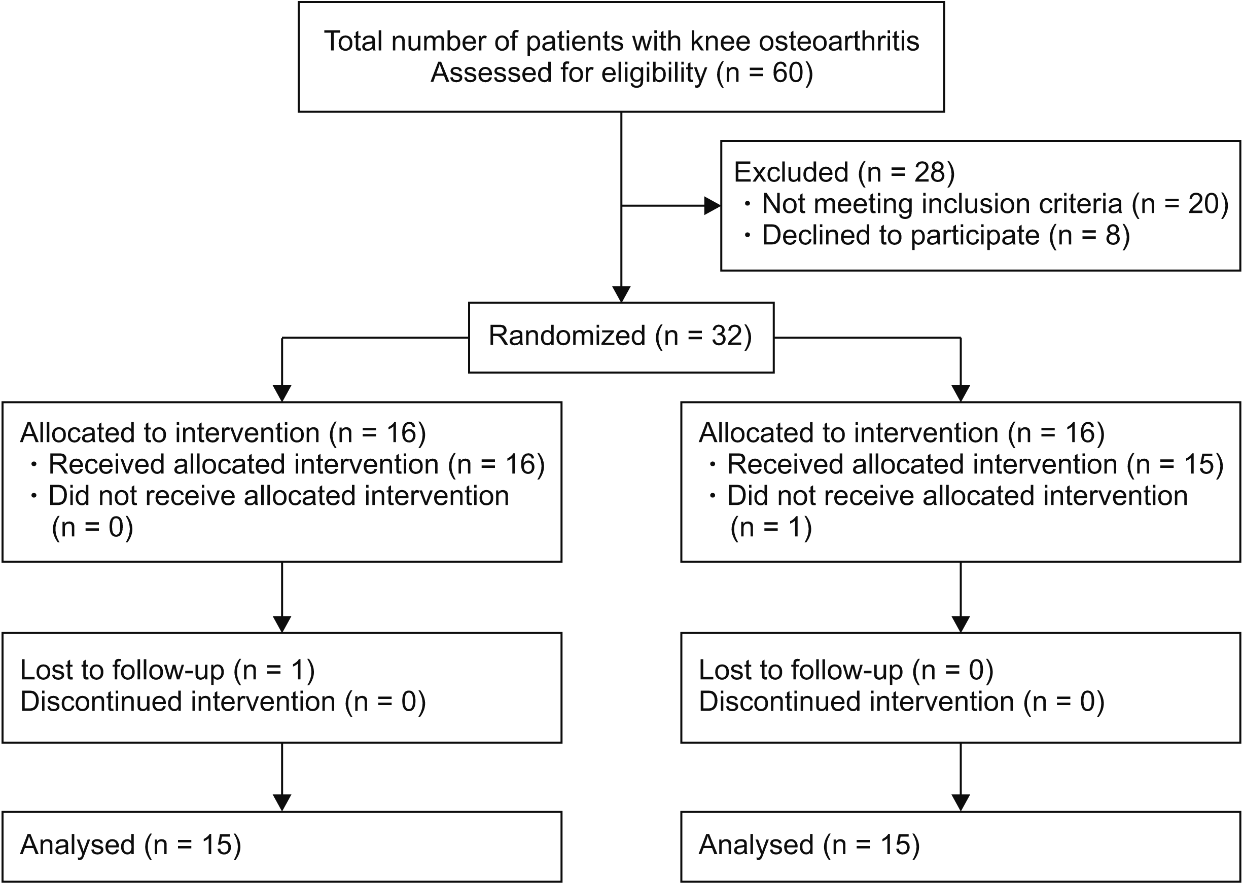
Table 1
Demographic characteristics
| Variable |
PRF group (n = 15) |
LAS group (n = 15) |
P value |
|---|---|---|---|
| Age (yr) | 60.8 ± 12.7 | 57.4 ± 9.7 | 0.419 |
| Gendera | 0.427 | ||
| Man | 6 (40.0) | 3 (20.0) | |
| Woman | 9 (60.0) | 12 (80.0) | |
| Body mass index (kg/m2) | 26.9 ± 4.0 | 26.6 ± 5.2 | 0.851 |
| Side of interventiona | 0.215 | ||
| Right | 13 (86.7) | 10 (66.7) | |
| Left | 2 (13.3) | 5 (33.3) | |
| VNRS score | 8.0 (8-9) | 8.0 (8-9) | 0.909 |
| WOMAC score | 53.0 (47-63) | 50 (43-54) | 0.405 |
| Comorbidities | |||
| Controlled hypertension | 6 (40.0) | 5 (33.3) | 0.724 |
| Controlled hypothyroidism | 2 (13.3) | 1 (6.7) | 0.612 |
| Duration of pain (mo) | 17 ± 7.9 | 19.3 ± 7.9 | 0.432 |
| Kellgren–Lawrence gradinga | |||
| 2 | 2 (13.3) | 2 (13.3) | > 0.999 |
| 3 | 13 (86.7) | 13 (86.7) |
![]()
The baseline VNRS score (median IQR) of group PRF was 8.0 (8–9) and for group LAS was 8.0 (8–9) before treatment. At 12 weeks, no difference was found in the VNRS scores between the two groups (P = 0.724). VNRS scores improved significantly over time in both groups as compared to baseline (group PRF 4 [3–4], P < 0.001; group LAS 4.0 [3–5]; P < 0.001). The baseline WOMAC scores (median IQR) of group PRF was 53.0 (47–63) and group LAS was 50.0 (43–54) before treatment. There was no difference in WOMAC scores between the two groups at 12 weeks (P = 0.983). WOMAC scores improved significantly over time in both groups as compared to baseline (group PRF 34.0 [21–37], P = 0.001; group LAS 32.0 [22–43]; P = 0.001).
The results of Friedman’s two ways analysis of variance revealed a significant difference between the VNRS scores measured at baseline, 2 weeks, 4 weeks, 8 weeks, and 12 weeks (degree of freedom [df] [4] = 35.34, P < 0.001 for group PRF and df [4] = 38.17, P < 0.001 for group LAS). Within the groups pairwise analysis revealed that VNRS scores decreased at all-time intervals compared with baseline in both groups (Fig. 2). Similarly, within-group pairwise analysis revealed that WOMAC scores significantly decreased at all-time intervals compared with baseline in both groups (Fig. 3).
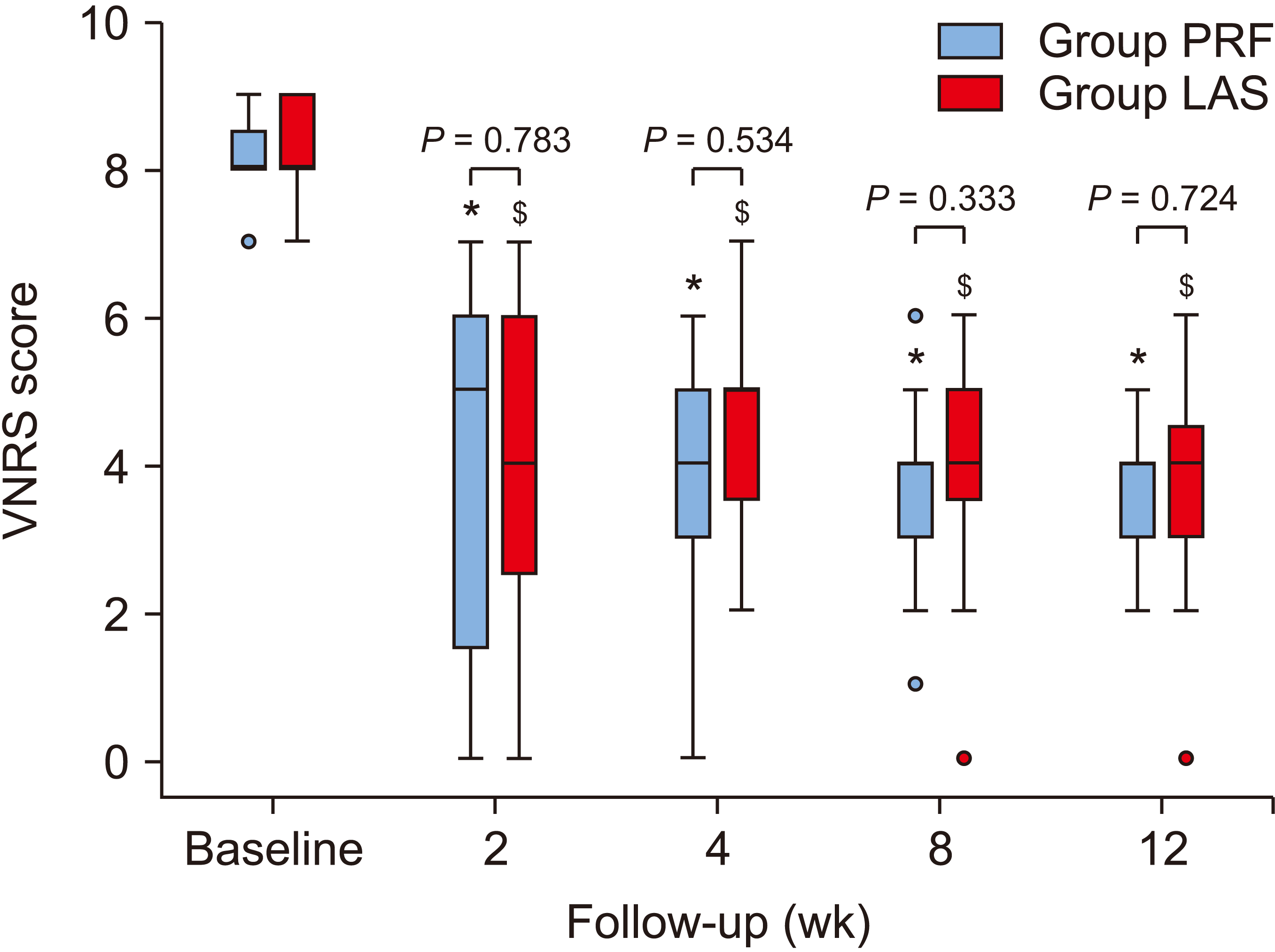 | Fig. 2Box plot analysis of VNRS scores at various time intervals. Boxplots show the median, interquartile range and outliers. Box edge indicates 25th and 75th percentiles, Whiskers indicate 5th and 95th percentiles. Red/blue dots represent outliers. Within group comparison, VNRS score were significant in both the PRF group and LAS group at various time interval of follow-up. Error bars indicate 95% confidence interval. VNRS: verbal numeric rating scale, PRF: pulsed radiofrequency, LAS: local anesthetic steroid. * indicates P < 0.001 compared to baseline in PRF group. $ indicates P < 0.001 compared to baseline in LAS group.
|
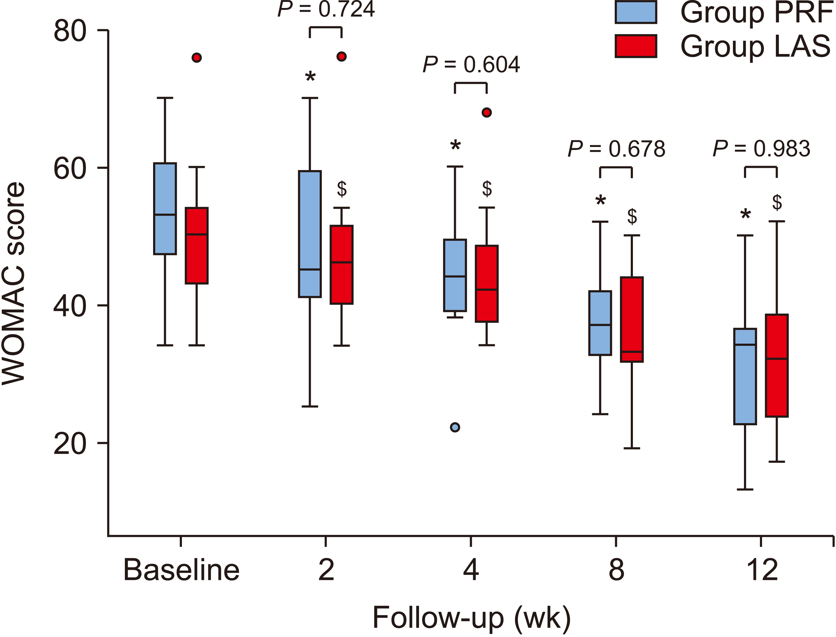 | Fig. 3Box plot analysis of WOMAC score at various time intervals. Boxplots show the median, interquartile range and outliers. Box edge indicates 25th and 75th percentiles, Whiskers indicate 5th and 95th percentiles. Red/blue dots represent outliers. Within group comparison, WOMAC score were significant in both the PRF group and LAS group at various time interval of follow-up. Error bars indicate 95% confidence interval. WOMAC: Western Ontario and McMaster Universities Arthritis Index, PRF: pulsed radiofrequency, LAS: local anesthetic steroid. * indicates P < 0.05 compared to baseline in PRF group. $ indicates P < 0.05 compared to baseline in LAS group.
|
The proportion of patients achieving effective pain relief (whose VNRS value decreased by at least 50%) at 12 weeks was comparable (70% in the PRF group and 66% in the LAS group, P > 0.999). Six patients required rescue analgesia in the PRF group as compared to four patients in the LAS group (P = 0.700, chi-square test). We did not encounter any bleeding, localized swelling, motor weakness, sensory deficit, deafferentation pain, or any other complication in any of our patients immediately after the procedure or during the follow up period.
Go to :
DISCUSSION
We conducted this study to compare ultrasound-guided PRF of the GN with GN block using LAS for management of OA knee pain. The VNRS score decreased significantly in both groups at 12 weeks and other follow up time intervals as compared to baseline. There was also a significant improvement in the WOMAC score in both groups. There was no difference in VNRS and WOMAC scores between the two groups. Eleven patients (73%) in group PRF and ten patients (66%) in group LAS had effective pain relief (≥ 50% reduction in pain) at 12 weeks. We did not come across any adverse effects during the procedure or follow-up.
RF neurotomy was first used in the treatment of chronic pain patients who failed to respond to conservative treatment modalities. Following good results with RF neurotomy in trigeminal neuralgia, it was applied to the sacroiliac and facet joints for chronic pain [21,22]. The technique was later applied to the cervical facet, discogenic pain, and cancer pain treatment [23]. In the management of OA knee pain, CRF ablation was applied for the first time by Choi et al. [14]. Thirty eight patients with positive diagnostic GN block were randomised into a CRF or control group. Ten patients (59%) in the RF group achieved a primary outcome of at least 50% knee pain relief at 12 weeks. In a similar study conducted by Pineda et al. [24] to reproduce the effects of RF genicular neurotomy described by Choi et al. [14], the authors reported an 88% reduction in short term VAS scores, which decreased to 32% at the end of one year. In another study by Qudsi-Sinclair et al. [18], CRF was used, and its efficacy was compared with LAS in patients experiencing pain more than six months after TKA. Twenty-eight patients were followed up over a period of one year. Significant pain relief and knee function improvement was observed with similar results in both the groups. The best results were obtained during the first 6 months, worsening afterwards [18]. Authors recommended that patients of knee OA should undergo regular follow ups to evaluate need for timely repetition of procedure.
In the CRF technique, tissue temperature reaches up to 60°C–80°C which causes lysis of the nerves with a chance of motor fiber damage and deafferentation. With the purpose of finding an equally effective and less destructive technique, PRF was invented. In PRF, the pulse generator creates pulses with an amplitude of 45 V lasting 20 milliseconds followed by a 480-millisecond silent phase. This allows heat to spread and tissue temperature does not exceed a maximum of 42°C. As temperature does not reach the damage threshold (45ºC–50ºC), no irreversible tissue damage occurs [10,25]. The basic principle of PRF is a neuromodulation effect at the target tissue caused by stopping nociceptive (A-δ and C-fibers) pain input from the periphery to the central nervous system without destroying the motor or sensory (A-β) fibers, whereas high-temperature ablation may result in neuromas and deafferentation [25].
Studies on use of PRF in GN block are limited to only two case series [15,26]. Results obtained are promising and support the outcome of our study. In a preliminary case series by Kesikburun et al. [15], the efficacy of PRF was evaluated using USG guidance in 9 patients. PRF was performed only to the SMGN and IMGN in patients who experienced pain on the medial side of the knee. The study revealed significant pain reduction in 66% (6/9) of patients and knee function improvement throughout the study period of 12 weeks. However, the study was limited by the lack of a control group and short follow-up [15]. Further, only patients with pain on the medial side of the knee were enrolled. In another case series of 10 patients, USG-guided PRF of the composite nerves of the knee (saphenous, femoral, common peroneal, and tibial nerves along with the plexus of the popliteal, sub sartorial, and peripatellar nerves) was performed in OA knee patients. The authors reported sustained pain relief and muscle relaxation that enabled patients to optimize physiotherapy, possibly due to reduced peripheral and central sensitization [26]. With the purpose of elucidating the efficacy of the technique further, the authors conducted this study. The results showed that 73% (11/15) of patients had effective pain relief in the PRF group and 63% (10/15) in the LAS group at the 12 weeks follow-up. Our study had an active control group in the form of an LAS group, thus eliminating the risk of bias. We also found a significant reduction in VNRS and WOMAC scores with the use of PRF in OA knee pain management.
The decision to administer a corticosteroid in addition to LA for peripheral nerve block is limited by local or systemic side effects associated with the use of steroids and with their short-term effects [17,27]. Adding a steroid to LA may prolong the duration of the LA effect and also inhibits the transmission of afferent c-fibers that are mainly involved in the pain pathway [17]. Steroids alter the nociceptive and neuropathic brain pathway mechanism responsible for the integration of pain.
Previous studies have reported contradictory results on the effectiveness of LAS for GN block [17,18,27]. Authors found that a GN block administered with LAS is as effective as CRF of the GN in post-knee arthroplasty patients [18]. In a case series of two brain-injured patients who developed heterotropic ossification in the knee joint, USG-guided GN block administered with lignocaine and betamethasone led to a decrease in VAS scores at the 3rd month of follow-up [17]. On the other hand, Kim et al. [27], compared USG-guided GN block with LAS vs LA alone and found no added advantage from adding a steroid to the local anaesthetic, as clinically significant improvement in both the VAS and Oxford knee score was observed only up to 2 weeks and 1 week respectively in both groups. The authors used 20 mg of triamcinolone acetonide mixed with 6 mL of lignocaine in 3 divided doses at the target site. It is possible that the total dose of steroid used was less, resulting in subclinical pain relief. In the presence of contradictory literature, the authors decided to evaluate the effect of PRF with LAS in the management of pain for OA knee patients. They used 60 mg of methylprednisolone acetate in 3 divided at the target site.
GN block can be performed using ultrasound or fluoroscopic guidance. USG allows the direct visualization of soft-tissue structures such as the nerve bundles and blood vessels. USG also has the advantage of causing no exposure to ionizing radiation, as with the fluoroscopic technique [28]. Application of USG for GN RF was found to be easier and safer than fluoroscopy [28]. Sari et al. [28] studied fluoroscopy and USG as an imaging modality for the application of CRF on the GN. They found a similar reduction in VNRS and WOMAC scores at three months with a shorter procedure time with USG (20.2 ± 6.4 minutes) as compared to fluoroscopy (25.0 ± 4.8 minutes) [28]. In the present study, we didn’t encounter any complications during either the procedure or follow-up at various time intervals.
Our study has a few limitations. Firstly, we did not give any diagnostic blocks. At this point, there is no evidence-based recommendations for performing a diagnostic nerve block to ensure a proper selection of patients for RF treatment [29]. McCormick et al. [29] randomized 29 participants (36 knees) to receive a prognostic GN block or no block prior to cooled RF treatment and reported ≥ 50% pain relief in 64% of patients in the no block group at six months. Secondly, follow-up was done until 12 weeks. This was due to logistic constrains with many patients coming from remote areas. Long term follow-up would have allowed documentation of the duration of the interventions and time period after which repeated procedures might be required. Thirdly, the study is limited by a small sample size. Further studies with larger sample sizes are warranted.
To conclude, both USG-guided PRF of the GN and LAS block of the GN provided a comparable reduction in pain and improvement in WOMAC scores up to 12 weeks of follow-up in patients with OA knee pain without any complication. We cannot recommend one treatment option over the other, but PRF can avoid corticosteroid usage and possible complications, especially in older patients. However, PRF of the GN is a procedure that takes more time and equipment than the GN block using LAS.
Go to :
 XML Download