Abstract
Background
Subjects with frozen shoulder (FS) might not be comfortable with vigorous physical therapy. Clinical trials assessing the effect of graded motor imagery (GMI) in FS are lacking. The aim of this study was to determine the effect of GMI as an adjunct to conventional physiotherapy in individuals with painful FS.
Methods
Twenty subjects aged 40–65 years having stage I and II of FS were randomly divided into two study groups. The conventional physiotherapy group (n = 10) received electrotherapy and exercises while the GMI group (n = 10) received GMI along with the conventional physiotherapy thrice a week for 3 weeks. Pre- (Session 1) and post- (Session 9) intervention analysis for flexion, abduction, and external rotation range of motion (ROM) using a universal goniometer, fear of movement using the fear avoidance belief questionnaire (FABQ), pain with the visual analogue scale, and functional disability using the shoulder pain and disability index (SPADI) was done by a blinded assessor.
Results
Statistically significant difference was seen within both the groups for all the outcomes. In terms of increasing abduction ROM as well as reducing fear of movement, pain, and functional disability, the GMI group was significantly better than control group. However, both groups were equally effective for improving flexion and external rotation ROM.
Adhesive capsulitis or frozen shoulder (FS), can be defined as a “condition of uncertain etiology, characterized by a significant restriction of active and passive shoulder motions which occurs in the absence of known intrinsic shoulder disorder” [1]. It is a common shoulder pathology which is characterized by limited and painful shoulder range of motion (ROM) [1–3]. It commonly affects individuals between 40 and 65 years of age. The literature suggests around 2% to 5% of general population is affected by FS [4]. Although it is more common in females (approx. 70%), males exhibit higher risk of slower recovery and more disability [5]. In patients with diabetes mellitus or thyroid diseases the occurrence increases to 18% to 38%. Adhesive capsulitis or FS is believed to be a self-controlling condition, lasting 18–30 months [4–6]. A primary or idiopathic FS occurs when there is no cause that may be associated with another systemic illness which most commonly includes diabetes mellitus [3,7]. Secondary FS can be traumatic or nontraumatic. Dislocation, fractures or soft tissue injuries around the shoulder joint incorporate traumatic causes. Nontraumatic pathologies include osteoarthritis (OA), rotator cuff tendinopathy, and calcific tendinitis etc. [3].
FS can be divided into four stages: the pre-adhesive, freezing, frozen, and thawing stage. The pre-adhesive stage may last up to three months. Subjects might experience aching pain at rest as well as severe pain at the end of the ROM. The freezing phase persists from 3 to 9 months. Subjects experience slow but progressive loss of ROM in all directions. The frozen stage persists from 9 to 14 months, whereas the thawing stage lasts from 15 to 24 months [8–10].
Physiotherapy management of FS includes therapeutic modalities such as heat and cold, interferential therapy, transcutaneous electrical nerve stimulation, etc. and exercises such as active/passive ROM, capsular stretching, proprioceptive neuromuscular facilitation stretching, and strengthening also can be prescribed [2,9]. Manual therapy treatment for FS includes various joint mobilization and manipulation techniques. But, in some chronic musculoskeletal impairments, the central nervous system can become hypersensitive, which is referred to as central sensitization (CS) [11]. Evidence suggests that CS may be present in acute FS. Hence, subjects having FS might experience loss of pain inhibition along with non-noxious stimuli like simple movements being interpreted as threatening and painful. This results in developing fear of movement and kinesiophobia in subjects with FS. Hence, subjects might not be comfortable with vigorous physical therapy [12].
Graded motor imagery (GMI) is a therapeutic approach to treat a hypersensitive nervous system which targets activation of different neural webs in a graded way. The brain is the target organ for GMI, which includes three sequential stages [11–14]. GMI has been found to reduce pain and hypersensitivity along with improved functions and mobility in conditions with suspected CS, such as phantom limb pain, complex regional pain syndrome, and chronic low back pain [14–17]. Mirror therapy alone has also been proven to be effective in subjects with shoulder pain [11]. But data showing the effectiveness of all the 3 components of GMI on various measures in cases of FS is scarce. Also, clinical trials are lacking comparing the effects of GMI with conventional physiotherapy.
Therefore, the need arises to conduct a preliminary study to find out if sequential 3 stage GMI will prove to be an effective alternative therapy in the treatment of FS. Hence, the aim of this study was to study and compare the effects of GMI as an adjunct to conventional physiotherapy in subjects with FS.
This was a pilot randomized controlled trial in which subjects diagnosed with FS were included. Ethical clearance was obtained from the Institutional and research ethics committee, KAHER Institute of Physiotherapy, Belagavi (SL No. 445). Also, the study was registered in the Clinical Trials Registry – India (CTRI/2020/05/025201). The study was designed as an assessor-blinded study, as the outcome assessor was blinded to the subjects’ randomization into the groups. After obtaining ethical clearance, subjects who were willing to participate signed the written informed consent to publish the photographs of participants with concealed identity.
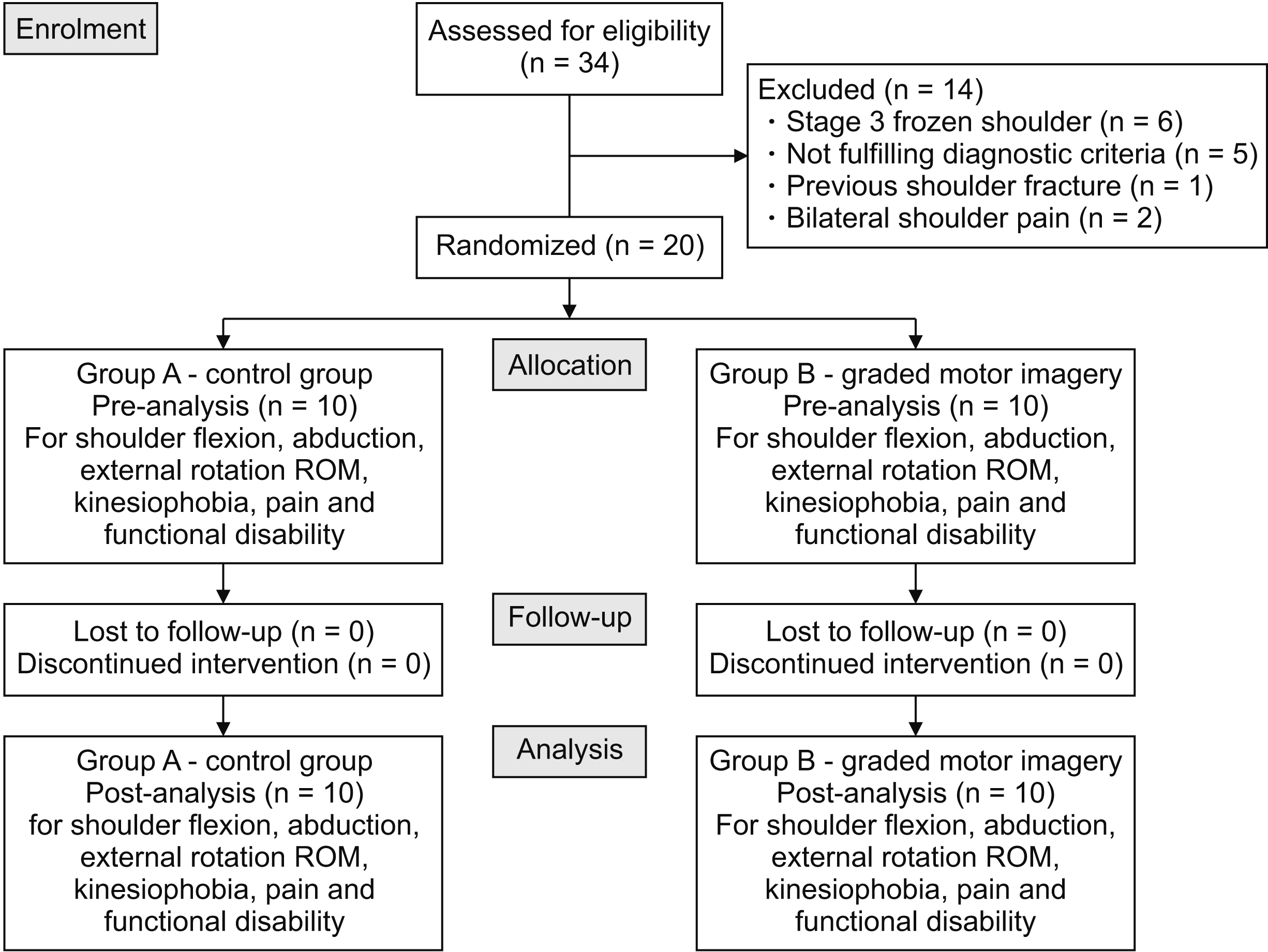
As the study design was a pilot study, the sample size was kept open-ended as suggested by the statistician. Thirty-four subjects visiting a tertiary care hospital from May 2020 to November 2020 with primary diagnosis of FS were screened for inclusion and exclusion criteria. Fourteen subjects who didn’t fulfill the criteria were excluded. A total of 20 subjects (10 in each group) were randomly allocated into two groups by random allocation using a lottery method by primary the investigator. There were no dropouts, and all the included participants completed the study protocol (Fig. 1).
The inclusion criteria were: 1) subjects fulfilling the clinical practice guideline criteria [8] for analysis of FS which includes (1) age between 40–65 years, (2) limited and painful shoulder passive ROM (mostly external rotation), and (3) gradual onset and progressive worsening of pain and stiffness; 2) stage I and II of FS; 3) subjects with or without diabetes mellitus; and 4) both male and female subjects being willing to participate. The exclusion criteria were visually impaired subjects, subjects having pain and incomplete ROM in both shoulders, previous neck or shoulder surgery, subjects having subacromial pain syndrome or painful arc syndrome, a positive apprehension test, subjects having any history of shoulder fracture or dislocation, and/or having signs of neurological involvement. Subjects who were on oral analgesics or any pain-relieving medications were excluded.
After collecting a small amount of demographic data from the subjects, pre-treatment ROM, fear of movement, pain, and functional disability outcomes were assessed by a blinded outcome assessor. Primary outcome measures were shoulder flexion, abduction, and external rotation ROM using a universal goniometer and fear avoidance belief questionnaire (FABQ) score, whereas secondary outcomes were visual analogue scale (VAS) and shoulder pain and disability index (SPADI) scores.
Fair to good reliability was noted for assessment of shoulder flexion, abduction, and external rotation ROM using a goniometer (Inter-rater = 0.64–0.69; intra-rater = 0.53–0.65) [18].
ROM was assessed in a supine position with hips flexed, knees flexed, and arms kept in the anatomical position. To assess shoulder flexion ROM, the goniometer axis was placed at the lateral side of the head of the humerus. The stationary arm was kept parallel to the trunk whereas the movable arm was placed laterally pointing the lateral epicondyle and the patient was told to flex the shoulder in the sagittal plane. To assess abduction ROM, the goniometer axis was placed at the front part of the shoulder inferior to the coracoid process. The stationary arm was kept parallel to the mid-axillary line and the movable arm was placed in line with the humerus. Patient was then asked to abduct the shoulder in frontal plane. To measure shoulder external rotation ROM, the arm was positioned as the shoulder was abducted to 90° and the elbow flexed to 90°. The forearm was placed halfway between pronation and supination. The goniometer axis was kept at the olecranon process and the arm was placed perpendicular to the floor. The movable arm was pointed towards the ulna styloid process and kept parallel to the longitudinal axis of the ulnar side of forearm. Subjects were then asked to perform an external rotation movement [19].
The FABQ is used to check fear related to movement. The FABQ has a total of 16 questions about physical activity (FABQ-PA) and work (FABQ-W). Each item is scored from 0 to 6. Lower scores indicate less fear of movement. Since we were studying FS, the term back from the questionnaire was replaced with shoulder. As the questionnaire is originally in English language, it was translated in the most understandable language for each subject. This questionnaire has fair reliability in patients having shoulder pain (intraclass correlation coefficient [ICC] = 0.88) [12].
The VAS permits a person to visually score pain along a 10 cm line where 0 represents no pain and 10 represents maximum pain. The procedure was explained to each subject in simple language and then they were asked to put a mark on the VAS line at the point that represented their pain intensity. Using a ruler, the score is calculated by determining the distance. It has fair reliability in both literate (r = –0.94, P = 0.001) and illiterate (r = –0.71, P = 0.001) subjects [20,21].
SPADI contains 13 items, with 5 items measuring pain and 8 items measuring disability. Each item of the scale was explained to the subjects in a language they understand, and they were then asked to score each item from 0 to 10 where 0 represents no pain and 10 represents the worst imaginable pain. The total score out of 100 was calculated. SPADI has good test-retest reliability (ICC = 0.89) in subjects with FS [22,23].
Subjects in this group received conventional physiotherapy treatment which is strongly recommended by various authors for improving ROM and decreasing pain in subjects with FS [10]. It included a hot moist pack applied in supine or sitting position for 15 minutes, interferential therapy (vectristim-100; Technomed Electronics, Chennai, India) for 20 minutes at 4,000 Hz carrier frequency with an amplitude modulated frequency of 0 to 250 Hz [24]. Along with the physical agents, exercise like Codman’s pendulum exercises in flexion-extension, abduction-adduction, and circular motion, active assisted ROM exercises with a wand, capsular stretching, wall ladder, and shoulder wheel exercises were also prescribed [2,4,10,25].
The treatment was provided 3 times a week for 3 weeks by a qualified orthopedic physiotherapist in an outpatient department of physiotherapy. Subjects were also asked to perform same exercises at home along with application of a hot moist pack twice a day for 3 weeks. Hence, a total of 9 sessions of conventional physiotherapy were given face to face along with the home exercise program. A diary was provided to each subject to maintain their record of daily exercises. The intensity and repetitions of the exercises were increased from week 1 to week 3 according to the subject’s tolerance and response to the treatment.
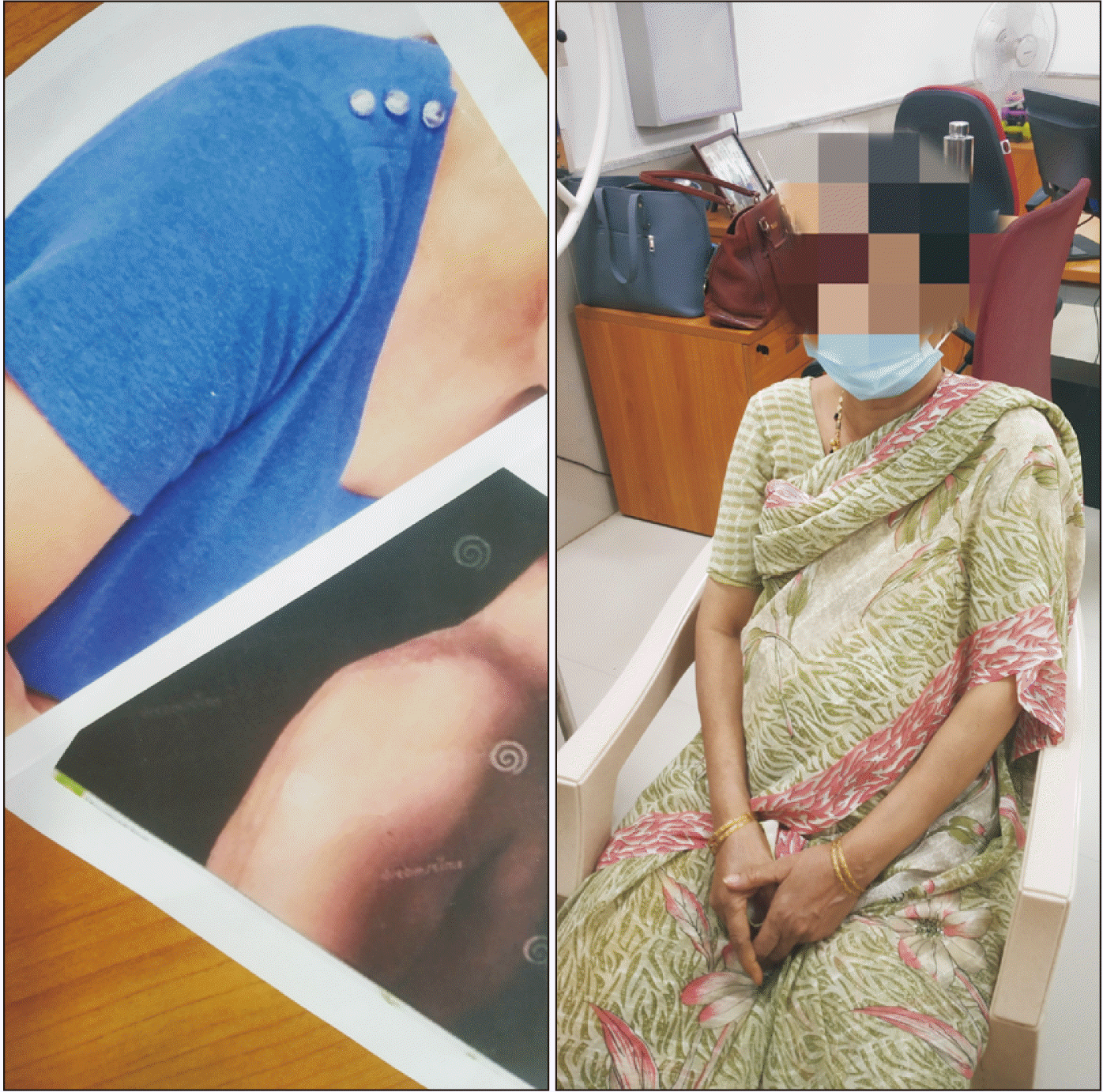
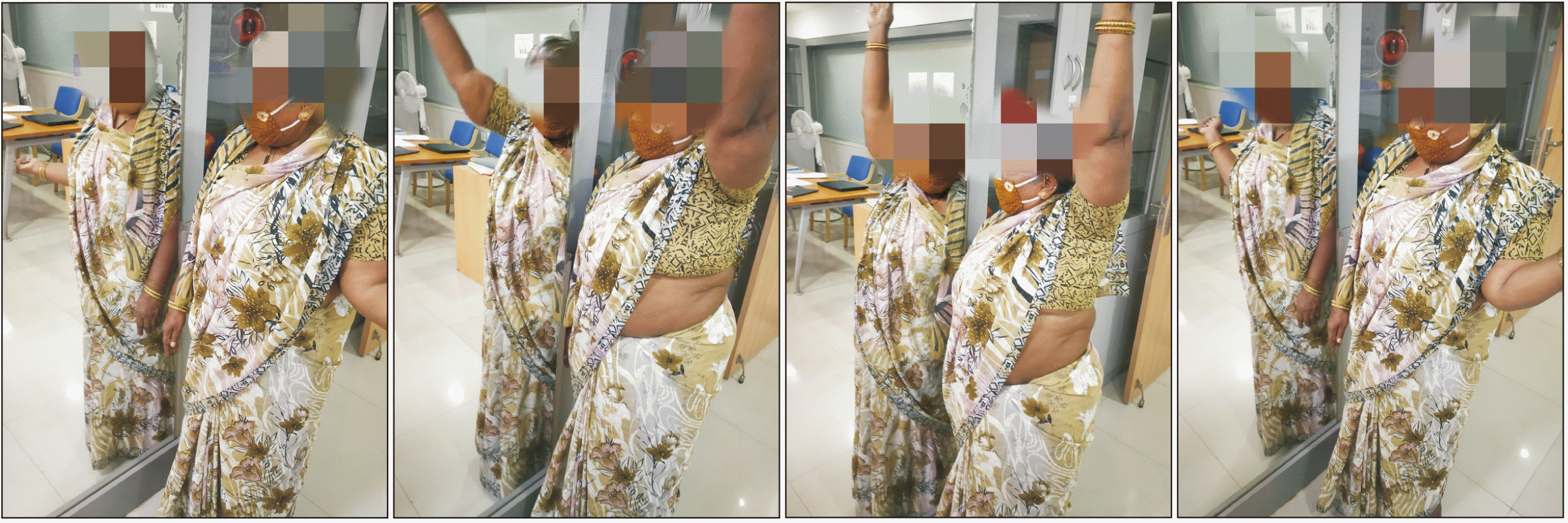
Subjects received GMI along with the conventional physiotherapy. Each stage of GMI was prescribed for 1 week. In the 1st week, laterality recognition was taught to the subjects. It includes left/right judgments of photographs that depict the affected area. For the shoulder this involves viewing an image of a shoulder and judging whether that image is a left or a right shoulder. Numerous flash cards showing images of left and right shoulder were provided to the patient and they were asked to recognize the left and right shoulders (Fig. 2). Also, they were asked to see peoples’ shoulders and judge whether they were left or right. In the 2nd week movement visualizations were performed. These included visualizing the affected area without actually moving the area. Images of both the shoulders were randomly given and subjects were advised to imagine the movements shown in the picture. The subjects were asked to imagine pain free, smooth and full range movements (Fig. 3). The final step of GMI was mirror therapy, which included observation of the movements of unaffected body part in the mirror. This gives an illusion of a moving, but pain free, affected limb, thus providing positive feedback that the movement can be pain free (Fig. 4).
Subjects preformed each stage of GMI three times a week in the presence of a qualified physiotherapist in the outpatient Physiotherapy department, along with the conventional treatment as mentioned above. Hence, a total of 9 sessions of GMI (3 sessions of each stage) were provided face to face. Subjects were also asked to perform the same interventions at home for 10 minutes every 2 hourly every day. A diary was provided to maintain records of this [12,13,15].
SPSS (ver. 20.0; IBM Co., Armonk, NY) software and Excel 2013 (Microsoft, Redmond, WA) were used during data analysis. The Kolmogorov–Smirnov test was used to determine the normal distribution. For within-group comparisons, the paired t-test or Wilcoxon singed rank test were used. For between-group comparisons, the Mann–Whitney test, independent t-test, or Welch’s t-test were used. A P value of < 0.05 was considered statistically significant.
Baseline characteristics were compared and no statistical difference was found between the two groups (Table 1). The results showed there were statistically significant differences within both the control group as well as the GMI group in terms of primary outcomes, i.e., flexion, abduction, and external rotation ROM using a universal goniometer and fear of movement with the FABQ as well as in terms of secondary outcomes, i.e., pain using VAS scores and functional disability with SPADI scores (Table 2).
There was significant difference between the groups in terms of improving abduction ROM as well as reducing fear of movement, pain, and functional disability. When the pre- and post-intervention mean differences were compared for both the groups, it was observed that the GMI group showed significantly better improvement in abduction ROM and reduction in fear of movement, pain, and functional disability. However, when compared between the groups, both groups showed equal improvement and there was no statistically significant difference found between the control and GMI groups in terms of flexion and external rotation ROM (Table 3).
The present randomized controlled trial was aimed at assessing and comparing the effects of GMI and conventional physiotherapy in people with FS. The comparison was done in terms of three objective measures (flexion, abduction, and external rotation ROM using a universal goniometer) and subjective measures (fear of movement using the FABQ, pain using the VAS, and functional disability using the SPADI).
The present study showed a significant reduction in fear of movement after 3 weeks of GMI intervention. Similar studies have been done before to assess the effect of GMI on fear avoidance behavior. Evidence supports that in chronic pain there is different primary sensorimotor cortex representation. This rearrangement of cortical structures causes alterations in the body map representation of a subject. These affected representations are responsible for CS. GMI comprises cortical remapping techniques such as left-right judgments, visualization of a body part, and mirror therapy. GMI desensitizes the hypersensitive nervous system and thus helps in reducing fear of movement [11].
In a study, the effect of GMI on kinesiophobia in subjects with knee OA was assessed and in conclusion, the authors suggested the use of GMI to reduce kinesiophobia in subjects with knee OA [26]. Sawyer et al. [12] did a case study on the effect of GMI on a subject with FS and found similar results. The authors proved that for a patient with fear of movement, using a ‘top down’ approach was found to be very effective.
In the present study, the shoulder pain was also significantly reduced post intervention in the GMI group. Moseley [14] assessed the effect of GMI on pathologic pain. This study had included 51 subjects with phantom limb pain and chronic regional pain syndrome. The author concluded that GMI is effective in reducing pain in these conditions. In a systemic review it was observed that the individual stages of GMI as a treatment to reduce pain doesn’t produce a significant effect, but using the 3 sequential components of GMI intervention has shown better alleviation of pain [15]. The reason behind shoulder pain reduction might be the presence of CS in FS. Evidence has shown that pain is associated with CNS. Advanced imaging and cortical mapping techniques have shown that there is positive correlation between sensorimotor cortex disorganization and pain intensity. GMI is a phenomenon which helps in cortical remapping which reduced fear avoidance behavior, thus reducing the pain [27].
Studies have proved that there is positive correlation between fear of movement and shoulder dysfunction. Hence, as the fear of movement increases, the SPADI score increases [28]. In our study it was found that fear of movement was better reduced using GMI intervention. Also, decrease in pain helps in improving shoulder joint function. This is why, in the present study, a significant reduction in the functional disability of the shoulder was seen after 3 weeks of GMI intervention.
In this study, GMI along with conventional physiotherapy showed slightly better outcomes in terms of improving abduction ROM. Evidence has shown that GMI is a “hands off, top down” approach which helps in reducing fear of movement and improves joint ROM. In a pilot study, subjects with knee OA were assessed to compare the effects of GMI and progressive muscle relaxation. It was concluded that both the interventions were effective, but GMI was more effective in subjects with knee OA [26]. Sawyer et al. [12] stated that GMI is believed to be useful in re-arranging the brain maps (homunculus), thus reducing pain and improving ROM [11,12]. But, in a systemic review, it was observed that in acute musculoskeletal conditions, GMI was not useful in improving ROM [29]. As we had included subjects with only stage 1 and stage 2 (acute stages) of FS, there was equal improvement in ROM in both the groups except in regard to abduction.
The present study had certain limitations. Carryover effects of GMI were not assessed, as the follow up was not performed after 3 weeks of intervention. The duration of the GMI intervention was short compared to most of the studies, as there was less subject compliance because of COVID-19. Also, manual therapy was not given to any of the groups. In future studies, a comparison of the effect of GMI versus manual therapy can be made, assessing the long-term effects.
In conclusion, it can be stated that GMI as an adjunct to conventional physiotherapy can be a better approach of treatment in terms of reducing pain, fear of movement, and in improving shoulder function in subjects with acute FS.
ACKNOWLEDGMENTS
With due respect, I extend my special thanks and gratitude towards the statistician who helped me with the statistical analysis. Also, I wholeheartedly thank the subjects who participated in the study.
REFERENCES
1. Chan HBY, Pua PY, How CH. 2017; Physical therapy in the management of frozen shoulder. Singapore Med J. 58:685–9. DOI: 10.11622/smedj.2017107. PMID: 29242941. PMCID: PMC5917053.
2. Uppal HS, Evans JP, Smith C. 2015; Frozen shoulder: a systematic review of therapeutic options. World J Orthop. 6:263–8. DOI: 10.5312/wjo.v6.i2.263. PMID: 25793166. PMCID: PMC4363808.
3. Cho CH, Bae KC, Kim DH. 2019; Treatment strategy for frozen shoulder. Clin Orthop Surg. 11:249–57. DOI: 10.4055/cios.2019.11.3.249. PMID: 31475043. PMCID: PMC6695331.
4. Kelley MJ, McClure PW, Leggin BG. 2009; Frozen shoulder: evidence and a proposed model guiding rehabilitation. J Orthop Sports Phys Ther. 39:135–48. DOI: 10.2519/jospt.2009.2916. PMID: 19194024.
5. Brue S, Valentin A, Forssblad M, Werner S, Mikkelsen C, Cerulli G. 2007; Idiopathic adhesive capsulitis of the shoulder: a review. Knee Surg Sports Traumatol Arthrosc. 15:1048–54. DOI: 10.1007/s00167-007-0291-2. PMID: 17333122.
6. Page P, Labbe A. 2010; Adhesive capsulitis: use the evidence to integrate your interventions. N Am J Sports Phys Ther. 5:266–73. PMID: 21655385. PMCID: PMC3096148.
7. Dias R, Cutts S, Massoud S. 2005; Frozen shoulder. BMJ. 331:1453–6. DOI: 10.1136/bmj.331.7530.1453. PMID: 16356983. PMCID: PMC1315655.
8. Kelley MJ, Shaffer MA, Kuhn JE, Michener LA, Seitz AL, Uhl TL, et al. 2013; Shoulder pain and mobility deficits: adhesive capsulitis. J Orthop Sports Phys Ther. 43:A1–31. DOI: 10.2519/jospt.2013.0302. PMID: 23636125.
9. Hannafin JA, Chiaia TA. 2000; Adhesive capsulitis. A treatment approach. Clin Orthop Relat Res. 372:95–109. DOI: 10.1097/00003086-200003000-00012. PMID: 10738419.
10. Jain TK, Sharma NK. 2014; The effectiveness of physiotherapeutic interventions in treatment of frozen shoulder/adhesive capsulitis: a systematic review. J Back Musculoskelet Rehabil. 27:247–73. DOI: 10.3233/BMR-130443. PMID: 24284277.
11. Louw A, Puentedura EJ, Reese D, Parker P, Miller T, Mintken PE. 2017; Immediate effects of mirror therapy in patients with shoulder pain and decreased range of motion. Arch Phys Med Rehabil. 98:1941–7. DOI: 10.1016/j.apmr.2017.03.031. PMID: 28483657.
12. Sawyer EE, McDevitt AW, Louw A, Puentedura EJ, Mintken PE. 2018; Use of pain neuroscience education, tactile discrimination, and graded motor imagery in an individual with frozen shoulder. J Orthop Sports Phys Ther. 48:174–84. DOI: 10.2519/jospt.2018.7716. PMID: 29257926.
13. Manisha U, Kumar SP, Manu G. 2014; Graded motor imagery program - a review. Indian J Med Health Sci. 1:59–66. https://rfppl.co.in/view_abstract.php?jid=41&art_id=1811.
14. Moseley GL. 2006; Graded motor imagery for pathologic pain: a randomized controlled trial. Neurology. 67:2129–34. DOI: 10.1212/01.wnl.0000249112.56935.32. PMID: 17082465.
15. Bowering KJ, O'Connell NE, Tabor A, Catley MJ, Leake HB, Moseley GL, et al. 2013; The effects of graded motor imagery and its components on chronic pain: a systematic review and meta-analysis. J Pain. 14:3–13. DOI: 10.1016/j.jpain.2012.09.007. PMID: 23158879.
16. Dilek B, Ayhan C, Yagci G, Yakut Y. 2018; Effectiveness of the graded motor imagery to improve hand function in patients with distal radius fracture: a randomized controlled trial. J Hand Ther. 31:2–9.e1. DOI: 10.1016/j.jht.2017.09.004. PMID: 29122370.
17. Anderson B, Meyster V. 2018; Treatment of a patient with central pain sensitization using graded motor imagery principles: a case report. J Chiropr Med. 17:264–7. DOI: 10.1016/j.jcm.2018.05.004. PMID: 30846919. PMCID: PMC6391225.
18. Hayes K, Walton JR, Szomor ZR, Murrell GA. 2001; Reliability of five methods for assessing shoulder range of motion. Aust J Physiother. 47:289–94. DOI: 10.1016/S0004-9514(14)60274-9. PMID: 11722295.
19. Norkin CC, White DJ. 2016. Measurement of joint motion: a guide to goniometry. 5th ed. FA Davis;Philadelphia: p. 45–56. https://www.worldcat.org/title/measurement-of-joint-motion-a-guide-to-goniometry/oclc/965327541?referer=br&ht=edition.
20. Hawker GA, Mian S, Kendzerska T, French M. 2011; Measures of adult pain: Visual Analog Scale for Pain (VAS Pain), Numeric Rating Scale for Pain (NRS Pain), McGill Pain Questionnaire (MPQ), Short-Form McGill Pain Questionnaire (SF-MPQ), Chronic Pain Grade Scale (CPGS), Short Form-36 Bodily Pain Scale (SF-36 BPS), and Measure of Intermittent and Constant Osteoarthritis Pain (ICOAP). Arthritis Care Res (Hoboken). 63 Suppl 11:S240–52. DOI: 10.1002/acr.20543. PMID: 22588748.
21. Boonstra AM, Schiphorst Preuper HR, Reneman MF, Posthumus JB, Stewart RE. 2008; Reliability and validity of the visual analogue scale for disability in patients with chronic musculoskeletal pain. Int J Rehabil Res. 31:165–9. DOI: 10.1097/MRR.0b013e3282fc0f93. PMID: 18467932.
22. Tveitå EK, Ekeberg OM, Juel NG, Bautz-Holter E. 2008; Responsiveness of the shoulder pain and disability index in patients with adhesive capsulitis. BMC Musculoskelet Disord. 9:161. DOI: 10.1186/1471-2474-9-161. PMID: 19055757. PMCID: PMC2633286.
23. Roach KE, Budiman-Mak E, Songsiridej N, Lertratanakul Y. 1991; Development of a shoulder pain and disability index. Arthritis Care Res. 4:143–9. DOI: 10.1002/art.1790040403. PMID: 11188601.
24. Panchal DN, Eapen C. 2015; Effectiveness of end-range mobilization and interferential current or stretching exercise and moist heat in treatment of frozen shoulder- a randomized clinical trial. Int J Curr Res Rev. 7:21–6. http://eprints.manipal.edu/143690/.
25. Kumar A, Kumar S, Aggarwal A, Kumar R, Das PG. 2012; Effectiveness of Maitland techniques in idiopathic shoulder adhesive capsulitis. ISRN Rehabil. 2012:710235. https://www.hindawi.com/journals/isrn/2012/710235/. DOI: 10.5402/2012/710235.
26. Gurudut P, Jaiswal R. 2020; Comparative effect of graded motor imagery and progressive muscle relaxation on mobility and function in patients with knee osteoarthritis: a pilot study. Altern Ther Health Med. AT6436. PMID: 33128533.
27. Priganc VW, Stralka SW. 2011; Graded motor imagery. J Hand Ther. 24:164–8. DOI: 10.1016/j.jht.2010.11.002. PMID: 21306870.
28. Luque-Suarez A, Martinez-Calderon J, Navarro-Ledesma S, Morales-Asencio JM, Meeus M, Struyf F. 2020; Kinesiophobia is associated with pain intensity and disability in chronic shoulder pain: a cross-sectional study. J Manipulative Physiol Ther. 43:791–8. DOI: 10.1016/j.jmpt.2019.12.009. PMID: 32829946.
29. Yap BWD, Lim ECW. 2019; The effects of motor imagery on pain and range of motion in musculoskeletal disorders: a systematic review using meta-analysis. Clin J Pain. 35:87–99. DOI: 10.1097/AJP.0000000000000648. PMID: 30222613.
Table 1
Table 2
Table 3
Outcome measure | Mean difference from baseline | Between group analysis | ||
---|---|---|---|---|
Group A (Control group) | Group B (GMI group) | P value | ||
Primary outcome measure | ||||
ROM | ||||
Flexion | 27.5 | 38.5 | 0.690 | |
Abduction | 23.5 | 45 | 0.014* | |
External rotation | 10 | 24 | 0.060 | |
FABQ score | 7.3 | 24.3 | 0.004* | |
Secondary outcome measure | ||||
VAS score | 1.4 | 4.6 | 0.001* | |
SPADI score | 14.19 | 28.31 | 0.009* |