

 PDF
PDF Citation
 Print
Print


INTRODUCTION
Monoclonal gammopathy is a condition in which the clonal immunoglobulins, fragments, or free light chains of immunoglobulin (M protein) are detected in the blood or urine. In most cases, the presence of M protein is considered as an indicator of plasma cell or B cell malignancies [1]. However, monoclonal gammopathy can be benign. Here, we report a rare case of monoclonal gammopathy associated with aplastic anemia (AA) and provide considerations for its diagnosis.
Go to : 
CASE
A 59-year-old man was referred to our hospital due to condition of post-iñuenza pneumonia and pancytopenia. At the time of admission, he had a fever, but there was no recent reports of weight loss or night sweats. Complete blood cell count at the time of admission was: hemoglobin 9.1 g/dL (reference range 13.0–18.0 g/dL), white blood cells 1.07×10⁹/L (reference range 4.0–10.0×10⁹/L), absolute neutrophils 0.35×10⁹/L (reference range 1.4–7.5×10⁹/L), platelets 12×109/L (reference range 150–400×109/L), and absolute reticulocytes 9.1×10⁹/L (reference range 24–84×109/L). His total protein level was 6.3 g/dL (reference range 5.8–8.1 g/dL), albumin level was 3.7 g/dL (reference range 3.1–5.2 g/dL), and albumin/globulin ratio was 1.4 (reference range 1.2–2.1), which were all normal. However, his C-reactive protein level was elevated (210.01 mg/L, reference range 0–5.0 mg/L). Proteinuria (542.34 mg/day) was detected in urine analysis. M protein was also detected in the serum (0.08 g/dL, Ig G lambda type, Fig. 1), but not in the urine in capillary protein electrophoresis (EP) and capillary immunofixation electrophoresis (IFE) analyses. His serum and urine free light chain concentrations were as follows: free kappa in the serum 35.93 mg/dL (reference range 3.30–19.40 mg/dL) and urine 586.01 mg/dL (reference range 0.78–13.48 mg/dL), and free lambda in the serum 41.66 mg/dL (reference range 5.71–26.30 mg/dL) and in the urine 217.28 mg/dL (reference range 2.22–5.90 mg/dL). The kappa/lambda free light chain ratios were all within the normal limits (0.86 for serum and 2.70 for urine). There was no splenomegaly or enlarged lymph nodes observed in the chest and abdomen computed tomography scans. Bone marrow (BM) examination was performed to evaluate the cause of presence of M protein and pancytopenia, which revealed hypocellular marrow (5% of cellularity, Fig. 2A) with an increase in plasma cell proportion (27.4%, Fig. 2A and B). The immunohistochemical (IHC) staining for CD138, kappa, lambda, and CD56 did not reveal monoclonal or aberrant plasma cells in the BM biopsy (Fig. 2B–D). The ˜ow cytometric (FCM) immunophenotyping with BM aspirate was performed using a myeloma panel consisting of CD19, CD28, CD38, CD45, CD56, CD117, CD138, kappa, and lambda light chain. CD38 and CD138 positive plasma cells were also found to be positive for CD19 and negative for CD56, CD28, and CD117. So, a single population of normal plasma cells without light chain restriction was confirmed (Fig. 2E and F). The diagnosis of severe AA was confirmed and follow-up serum protein EP and IFE tests were recommended. The patient was subjected to the empirical pneumonia antibiotic therapy. However, he died due to the aggravation of pneumonia and multiorgan failure on day 19 of his hospital stay.
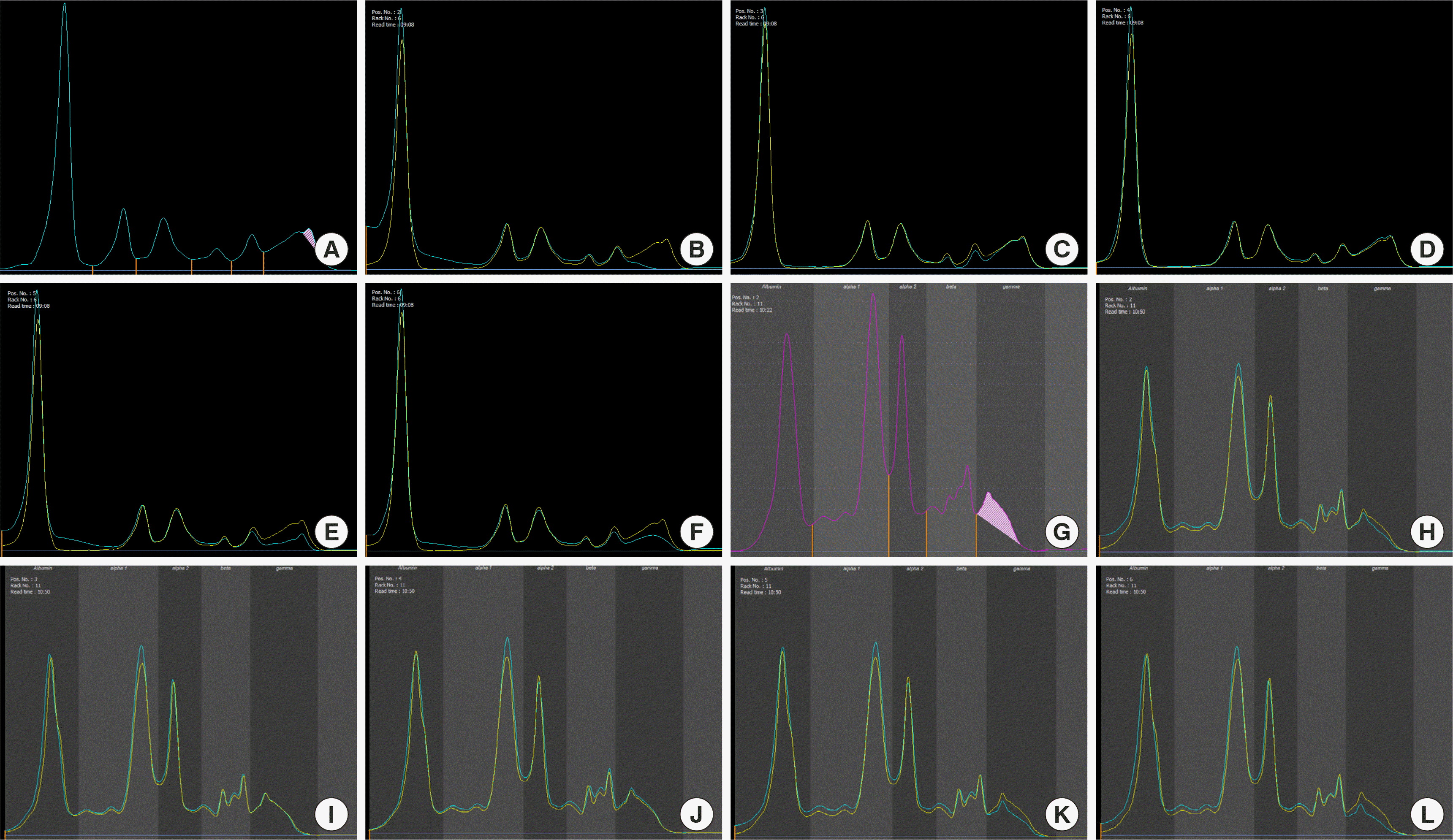 | Fig. 1Capillary electrophoresis (EP) and capillary immunofixation electrophoresis (IFE) of the serum and urine samples. (A) Serum EP. (B-F) Serum IFE: (B) IgG, (C) IgA, (D) IgM, (E) Kappa light chain, and (F) Lambda light chain. (G) Urine EP. (H-L) Urine IFE: (H) IgG, (I) IgA, (J) IgM, (K) Kappa light chain, and (L) Lambda light chain.
|
 | Fig. 2Immunohistochemical (IHC) staining of the bone marrow (BM) biopsy specimen and multicolor flow cytometric (FCM) immunophenotyping of the plasma cells. (A) Hematoxylin and eosin staining (×200). (B-D) IHC staining of (B) CD 138, (C) kappa light chain, and (D) lambda light chain (×400). (E, F) FCM immunophenotyping of plasma cells in BM aspirate. (G) Result of the lymphocyte subset analysis of BM aspirate using FCM panel corresponding to plasma cell myeloma.
|
Go to :
DISCUSSION
In our case, two hypotheses on the origin of M protein were proposed. The first possibility is benign monoclonal gammopathy; it was associated with autoimmune diseases, post-allogenic hematopoietic stem cell transplantation state, and infections [2-5]. So far, there is only one study on monoclonal gammopathy in pediatric patients with severe AA who have undergone immunosuppressive therapy [2]. However, there is only one study that has reported benign monoclonal gammopathy in a AA patient who have not undergone immunosuppressive treatment [6]. The mechanism of benign monoclonal gammopathy is related to T-cell impairment, a mediator of AA, and it could be transient [2, 3]. Generally, cytotoxic T cells which mediate BM destruction are typically activated in AA [7]. Conversely, in our patient, we confirmed an increase in CD19+ B cells over CD28+ T cells in the BM (Fig. 2G), which might be due to a reactive response to viral infection. Therefore, we assumed that the presence of M protein could be an indicator of viral infection-induced B cell activation. Particularly, in a case like this, a careful diagnosis should be performed to avoid misdiagnosis with malignant monoclonal gammopathy; the increased proportion of plasma cells may be due to a decrease in the number of hematopoietic cells in the BM.
Second, M protein could act as a marker of early stage monoclonal gammopathy of undetermined significance (MGUS). To diagnose MGUS, a monoclonal population of plasma cells with an aberrant phenotype must be identified [8]. In our patient, there was no aberrant plasma cell populations identified using both IHC staining and FCM immunophenotyping, and therefore, the possibility of MGUS was ruled out. However, occasionally, aberrant clones could be very small in size and below the detection limit. In such a case, MGUS should be considered when the clonal plasma cells are newly observed or found to be increased over time in BM, and M protein is consistently observed in EP and IFE analyses. Therefore, serial follow-up with serum protein EP and IFE analyses were essential to determine the exact cause of monoclonal gammopathy in our case. However, further tests were not possible since the patient died soon after the diagnosis of severe AA.
Conclusively, establishment of the cause of monoclonal gammopathy may be challenging in cases where it is associated with AA because benign monoclonal gammopathy due to B cell activation over T cell is rare in AA and the plasma cell proportion is found to be relatively increased in hypocellular marrow. Therefore, extensive diagnostic analysis and follow-up tests including IHC staining, FCM immunophenotyping, and lymphocyte subset analysis must be performed for its accurate diagnosis.
Go to :
 XML Download