Journal List > J Rheum Dis > v.29(2) > 1160486

Yeo, Baek, Song, and Lee: Evaluation of Serum Matrix Metalloproteinase-3 as an Objective Indicator for the Disease Activity in Rheumatoid Arthritis Patients Treated With Methotrexate Versus Tocilizumab: 24-week Results From a Prospective Randomized Controlled Study

Abstract

Objective

This study aims to evaluate the change in serum metalloproteinase-3 (MMP-3) following the management of active rheumatoid arthritis (RA) and define the relationships between MMP-3 and disease activity indices.

Methods

Data from a previously reported a 24-week, randomized controlled trial to investigate efficacy of tocilizumab in active RA refractory to methotrexate were analyzed. The serum level of MMP-3 were measured at week 0, 12, 20, and 24. The changes in MMP-3, and the relationship between MMP-3 and clinical parameters was assessed based on treatment group, methotrexate with or without tocilizumab.

Results

A total of 95 patients were included in this study. The serum MMP-3 significantly decreased and showed similar pattern with other disease activity indices during treatment period in both treatment groups (p<0.001). The MMP-3 was positively correlated with ESR, CRP, DAS28, SDAI, and CDAI for 302 visits throughout 24 weeks (p<0.001). In another correlation analysis to evaluate the treatment effect at 24 week time point, methotrexate group showed significant correlation between serum markers MMP-3 (r=0.321, p=0.043); ESR (r=0.450, p=0.002); and CRP (r=0.536, p<0.001), with DAS28, but tocilizumab group didn’t show meaningful correlation between serum markers and DAS28 (p>0.05).

Conclusion

Serum MMP-3 showed positive correlation with disease activity indices in active RA patients. Furthermore, serum MMP-3 significantly decreased from baseline to week 20. As there is no single serum marker that can represent the disease activity particularly in tocilizumab treatment, MMP-3 might be a useful adjunct indicator to evaluate the treatment response in active RA patients.

INTRODUCTION

Rheumatoid arthritis (RA) is a prolonged immune-mediated disease characterized by erosive polyarthritis and joint destruction with systemic complications [1]. It affects a wide range of physical function, well-being, and productivity, in addition to increased demand for health care resources. A strategic approach is required for better outcome in case of high disease activity [2]. Recently, the introduction of disease-modifying antirheumatic drugs (DMARDs) has led to an evolution of therapeutic algorithms and strategies for RA, dramatically improving patient outcomes [3]. Developing better outcome measures is one of the major issues in the treatment of RA [4]. Remission, defined by composite indices such as 28-joint Disease Activity Score (DAS28) and Simplified Disease Activity Index (SDAI), is the therapeutic goal for patients with RA, and it is also regarded as a primary or secondary endpoint for clinical trials [5]. However, most of these measures, including patient or physician global assessment, use swollen or tender joint count, a subjective approach to disease activity. As observer (rheumatologists) variation is not neglected, standardization of these measures is demanding to perform in daily clinical practice.
The determination of serum markers may be one of the objective approaches for estimating disease activity. Erythrocyte sedimentation rate (ESR) and C-reactive protein (CRP) are the most frequently used for assessing disease activity in RA patients. However, those are markers of non-specific inflammation, not specific inflammation in rheumatoid synovium [6]. Unlike other biologic agents, tocilizumab has the mechanism of action that involves inhibition of interleukin (IL)-6 receptors, resulting in CRP levels decreased to negative values. Therefore, outcome measures that depend on CRP levels may not reflect disease activity under the tocilizumab treatment [7,8].
Matrix metalloproteinase-3 (MMP-3) is produced by synoviocytes and responsible for the degradation of collagen in cartilage, thus having pivotal roles in the destruction of cartilage in patients with RA [9]. With respect to its diagnostic and prognostic potential in RA patients, elevated serum concentrations of MMP-3 have been studied to correlate with disease activity indices [10,11]. However, serial MMP-3 levels and the correlation between MMP-3 and clinical parameters at different time points are rare, and comparing the change of MMP-3 levels in methotrexate with tocilizumab therapy have not yet been established in patients with RA.
This study aims to define the relationship between MMP-3 and several disease activity indices, and to compare the change of MMP-3 levels during 24-week treatment of methotrexate with tocilizumab in active RA patients data from prospective randomized controlled study [12].

MATERIALS AND METHODS

Participants

The data of this study were collected from previously reported multicenter randomized clinical trial to investigate the efficacy and safety of tocilizumab in Korean patients with active RA failed to respond to methotrexate with or without other conventional DMARDs [12]. From October 2009 to October 2010, patients with RA who fulfilled the 1987 American College of Rheumatology (ACR) criteria were enrolled [13]. Active RA was defined by 6 or more swollen joints based on 66-joint count; 8 or more tender joints based on 68-joint count; serum CRP 1 mg/dL or higher; or ESR 28 mm/hr or higher. Stable dose of conventional DMARDs was required within 8 weeks prior to the baseline. Concurrent oral glucocorticoids (≤10 mg/day prednisone or equivalents) and non-steroidal anti-inflammatory drugs were allowed. Key exclusion criteria included concurrent active infection or malignancy, or previous hypersensitivity or contraindication to treatment with tocilizumab.

Study design

The main study of previous trial was a 24-week, phase III, randomized double-blind, placebo-controlled study. A total of 99 patients were randomized in a 1:1 ratio to receive either placebo (n=51) or tocilizumab (n=48) stratified according to methotrexate alone or in combination with other conventional DMARDs. During the trial, four patients were excluded from the main study due to Good Clinical Practice (GCP) or protocol violations: 3 from the placebo group and 1 from the tocilizumab group. In this study, we newly defined these two groups as methotrexate group (methotrexate therapy; n=48) and tocilizumab group (methotrexate with tocilizumab therapy; n=47). Concurrent conventional DMARDs was continued during the study. The detailed protocol of main study was provided in Supplementary Data. The study was approved by the Institutional Review Board of Seoul National University Hospital (IRB #1302-028-463 and #1701-018-820) and written informed was obtained from each patients. This study was conducted in accordance with the principles of the Declaration of Helsinki and GCP guidelines.

Data collection

The clinical data included age, sex, disease duration, 66/68-joint count, patient’s pain and global assessment (0~100 scale), physician’s global assessment (0~100 scale), Health Assessment Questionnaire Disability Index (HAQ-DI, 0~3), DAS28, SDAI and Clinical Disease Activity Index (CDAI). DAS28 scores were calculated by using the 66/68 joint count. Therefore, an involvement of over 28 joints was considered as the involvement of 28 joints. Patients were divided into two groups by disease activity at 24 weeks: high disease activity group (HDA at 24 weeks; DAS28>5.1) or remission, low and moderate disease activity group (non HDA at 24 weeks; DAS28≤5.1).
Laboratory data included rheumatoid factor (U/mL), ESR (mm/h), CRP (mg/dL), IL-6 (pg/mL), and soluble IL-6 receptor (sIL-6R) (ng/mL). The levels of IL‐6 were measured by high‐sensitivity Enzyme-Linked Immunosorbent Assay (ELISA) (BMS213HS; Bender MedSystems, Vienna, Austria) and sIL-6R by ELISA (BMS214; Bender MedSystems) according to the manufacturer’s instructions. Serum was collected every 4 weeks.

Determination of serum MMP-3 levels

MMP-3 levels (ng/mL) were analyzed at baseline and 12, 20, and 24 weeks after baseline. The normal range of serum MMP-3 concentrations was from 36.9 ng/mL to 121.0 ng/mL for male or from 17.3 ng/mL to 59.7 ng/mL for female. The values were determined using Nanopia MMP-3, a latex turbidimetric immunoassay (SEKISUI MEDICAL CO., LTD., Tokyo, Japan).

Statistical analysis

Continuous variables were summarized as means with standard deviations (SD) and ranges. Categorical variables were summarized as absolute numbers and percentages. A linear mixed model (LMM) was used to investigate the changes in MMP-3 and continuous variables for disease activities with time between methotrexate group and tocilizumab group adjusted for age, sex, and treatment. The relationship between the various clinical and laboratory parameters was tested using Pearson correlation analysis. p-values of ≤0.05 were considered statistically significant. Furthermore, patients were divided into two groups based on disease activity at 24 weeks: non high disease activity group including patients with remission, low and moderate disease activity (non HDA at 24 weeks; DAS28≤5.1); and high disease activity group (HDA at 24 weeks; DAS28>5.1). We also compared serum markers with time in two different disease activity groups by using LMM adjusted for age, sex, and treatment. All statistical analyses were performed by using SPSS (version 26.0; IBM Corp., Armonk, NY, USA), and data were visualized by Prism (version 8.0.1; GraphPad software, San Diego, CA, USA) for Window 10.

RESULTS

Baseline characteristics of the participants

Data from 95 Korean patients with active RA were analyzed. The patient baseline demographics and clinical characteristics are described in Table 1. In brief, the mean±SD age of the patients was 52.3±11.3 and 88.4% were female. The serum level of MMP-3 at baseline was 290.0±206.4 ng/mL. All patients showed moderate or high disease activity; their mean DAS28-ESR, DAS28-CRP, SDAI, and CDAI were 6.9±0.9, 6.2±1.0, 45.3±14.7, and 42.4±13.6, respectively. There was no statistically difference between methotrexate group and tocilizumab group in baseline disease activity indices including serum markers (ESR and CRP), DAS28-ESR, DAS28-CRP, SDAI, and CDAI (all p>0.05). Patients receiving methotrexate alone were 56.8%, methotrexate with hydroxychloroquine were 33.7%, and methotrexate with sulfasalazine were 9.5% at baseline.

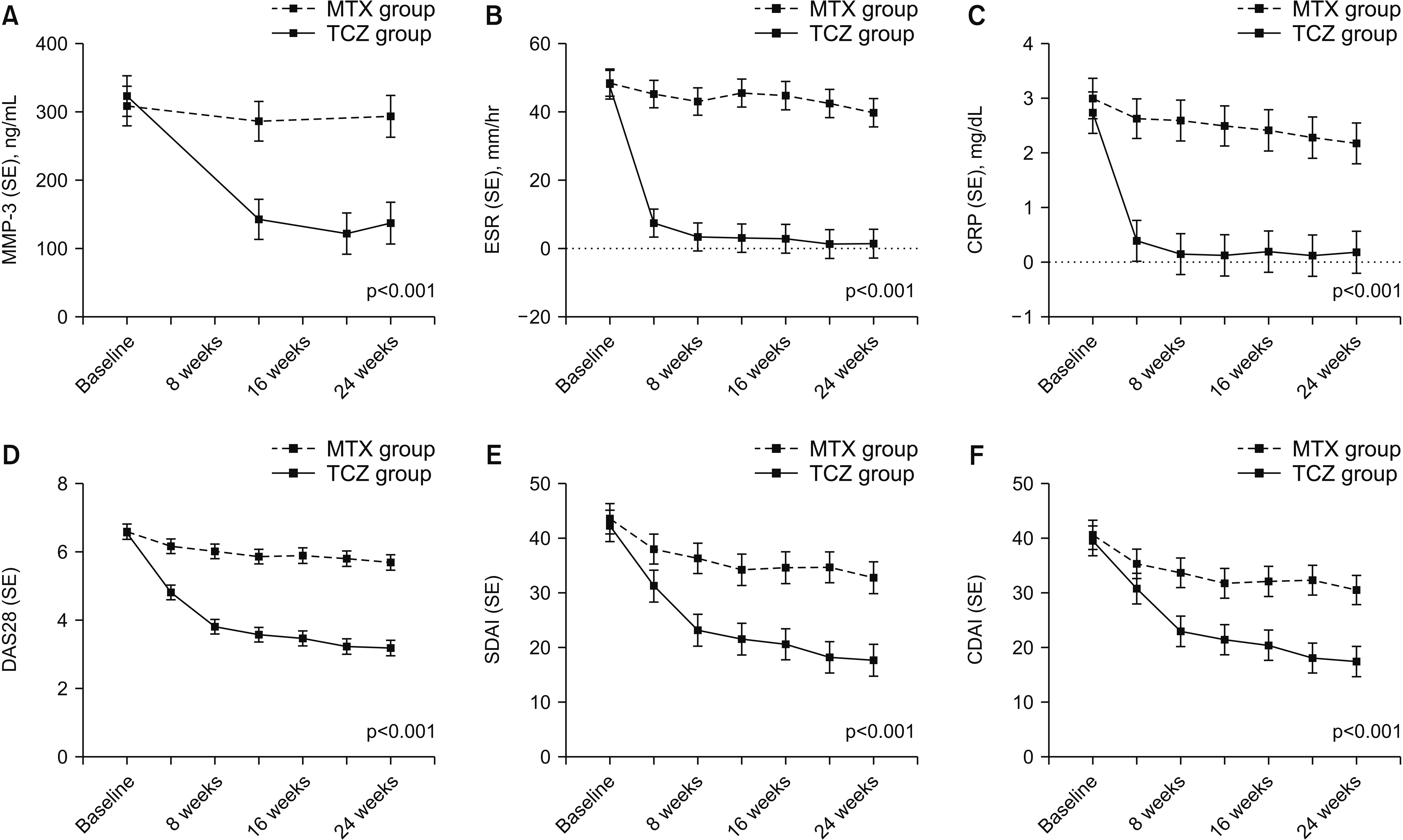
Changes in serum MMP-3 levels and disease activity indices in RA patients during the 24 weeks

The changes in level of serum MMP-3 and disease activity indices were demonstrated in Figure 1. All disease activity indices including MMP-3, ESR, CRP, DAS28, SDAI, and CDAI were significantly higher in methotrexate group than in tocilizumab group throughout the course of treatment (all p<0.001). The mean MMP-3 (standard error) in tocilizumab group largely decreased from baseline to 20 weeks, and then slightly rebounded at 24 weeks: 323.0 (29.8) ng/mL at baseline, 143.4 (29.6) ng/mL at 12 weeks, 122.2 (29.9) ng/mL at 20 weeks, and 137.6 (30.7) ng/mL at 24 weeks (p<0.001, Figure 1A). The changes in other disease activity indices showed similar patterns to MMP-3 but decrements continued to 24 weeks (Figure 1B~F).

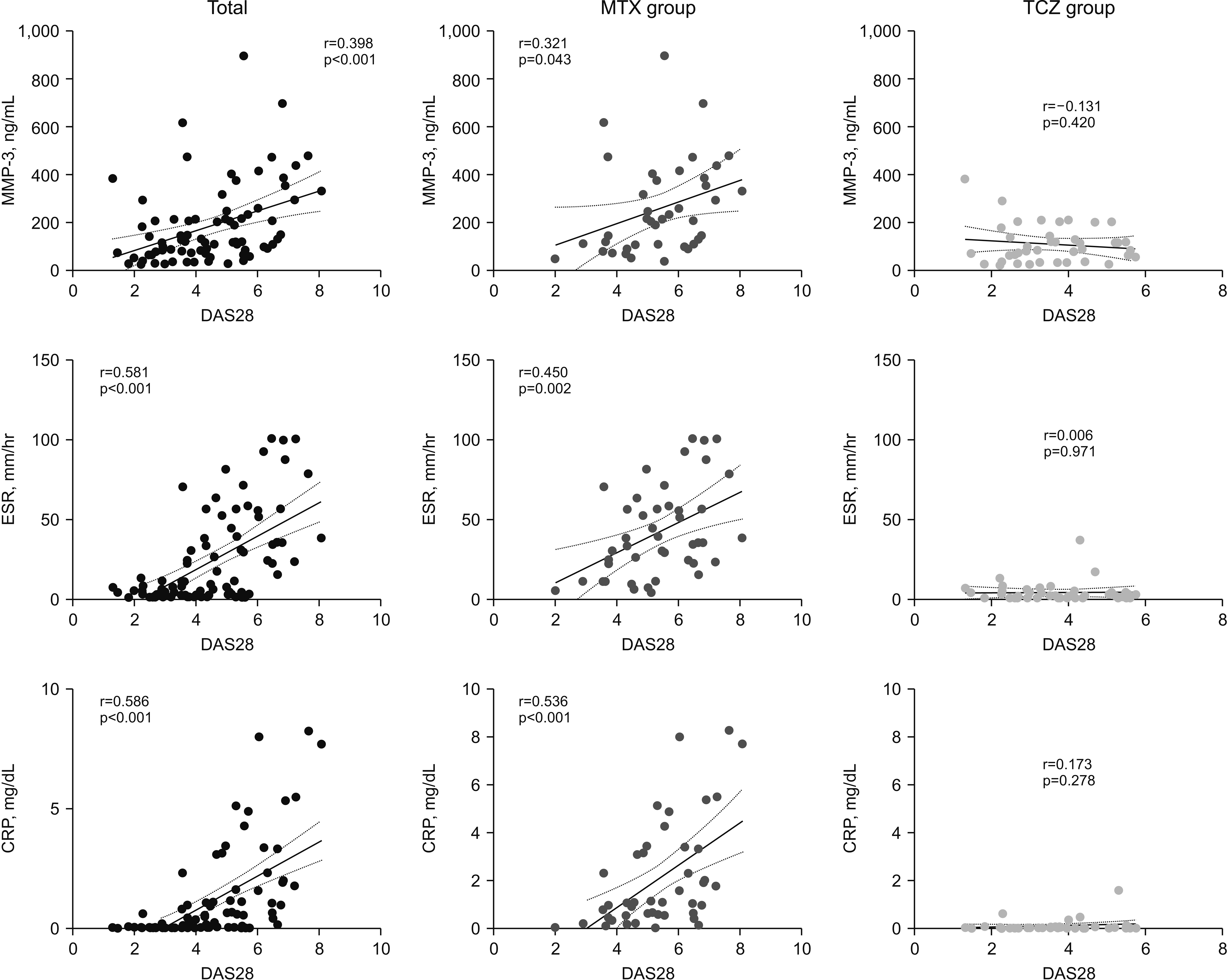
Correlations between MMP-3 and other biomarkers

The serum MMP-3 levels were positively correlated with ESR (r=0.557, p<0.001), CRP (r=0.564, p<0.001), DAS28 (r=0.448, p<0.001), SDAI (r=0.339, p<0.001) and CDAI (r=0.291, p<0.001) for 302 visits throughout 24 weeks (Table 2). IL-6 showed no significant correlation with MMP-3 (r=0.112, p=0.149), sIL-6R showed a weakly negative correlation with MMP-3 levels (r=-0.190, p=0.015). Meanwhile, CRP levels were significantly correlated with IL-6 (r=0.263, p<0.001) and sIL-6R (r=0.266, p<0.001; Supplementary Table 1). We also analyzed the correlation of each serum marker with DAS28 after 24-week treatment. Significantly positive correlation was showed between serum markers and DAS28 (MMP-3, r=0.398; ESR, r=0.581; and CRP, 0.586 with all p<0.001; Figure 2, left panel). Based on treatment group, while methotrexate group showed significantly good correlation between all serum markers and DAS28 (Figure 2, middle panel), tocilizumab group did not show significant correlation of all serum markers with DAS28 at 24 weeks (Figure 2, right panel).

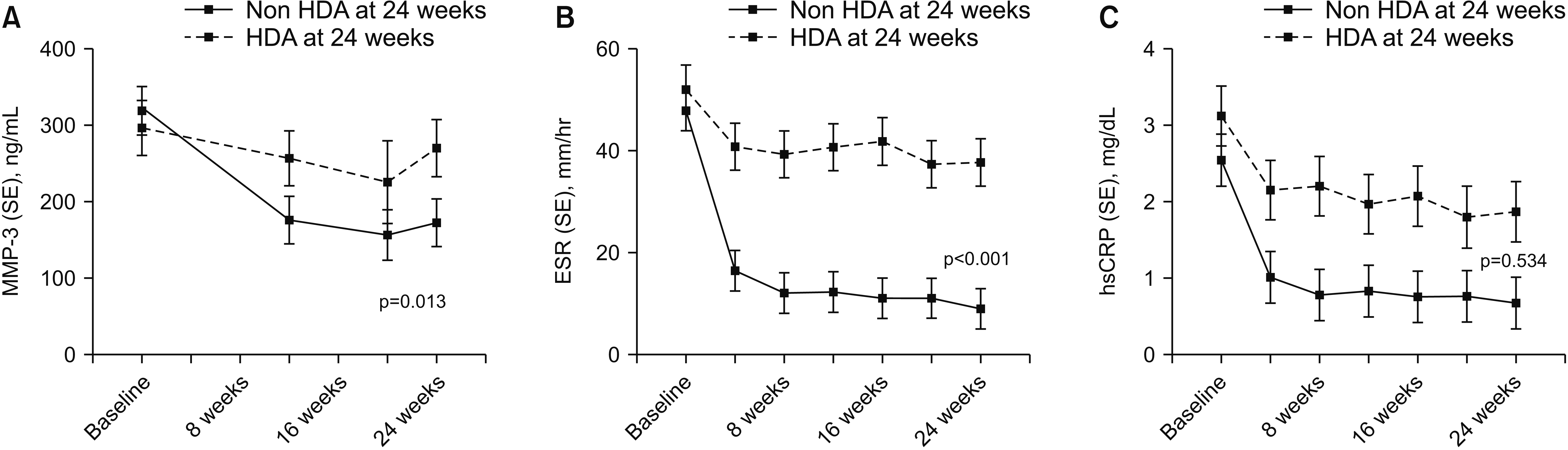
Comparison of changes in serum levels of MMP-3 and acute phase reactants during the treatment period according to disease activity at 24 weeks

When grouped by the DAS28 scores at 24 weeks, 47 (54.7%) were in non HDA group (DAS28≤5.1), and 39 (45.3%) were in HDA group (DAS28>5.1). The mean change of MMP-3 and acute phase reactants levels from baseline to 24 weeks in each group is presented in Figure 3. The mean decrement in the levels of MMP-3 at 20 weeks from baseline was significantly greater in non HDA group than HDA group (156.1 ng/mL and −50.9% from baseline in non HDA group; and 225.4 ng/mL and −23.9% from baseline in HDA group). After 20-week treatment, the MMP-3 levels rebounded greater in HDA group than non HDA group at 24 weeks (172.6 ng/mL in non HDA group; and 269.7 ng/mL in HDA group, p=0.013; Figure 3A). The change of ESR levels was rapidly decreased at 4 weeks from baseline especially in non HDA group and plateaued (16.5 mm/hr and −65.4% from baseline in non HDA group; and 40.8 mm/hr and −21.4% from baseline in HDA group; p<0.001; Figure 3B). While the CRP levels showed similar change to ESR, it did not show the significant difference of change patterns throughout treatment period between non HDA and HDA groups (1.02 mg/dL and −60.0% from baseline in non HDA group; and 3.12 mg/dL and −30.9% from baseline in HDA group; p=0.534; Figure 3C). Unlike MMP-3, there were no significant rebound pattern in the level of ESR and CRP after 20 weeks.

DISCUSSION

This study was performed to evaluate the serial relationships between MMP-3 and several disease activity indices using the data and serum samples obtained in the previous randomized controlled trial in active RA patients treated with methotrexate with or without tocilizumab for 24 weeks [12]. We found that the MMP-3 levels robustly decreased from baseline to 12 weeks, and this decrement continued into week 20 during 24-week treatment period. Furthermore, the MMP-3 was positively correlated with ESR, CRP, DAS28, SDAI, and CDAI for 302 visits throughout 24 weeks.
The evaluation of disease activity by symptoms and sign is important in RA patients; in this regard, composite scores of indices such as DAS28, SDAI, or CDAI have been developed. However, these indices are subjective and require training for the assessors to reduce variability. In fact, standardization of these measures is demanding in clinical practice. Therefore, serum markers in RA are valuable for decision-making as adjunctive measures in clinical situations.
In this study, all enrolled patients were moderate or high disease activity by DAS28, and had elevated MMP-3 levels (290.0 ng/mL) at baseline, consistent with the previous studies [14,15]. Zhou et al. [16] reported serum MMP-3 levels in each group of patients with active RA: 54.25 ng/mL in mild RA, 105.10 ng/mL in moderate RA, and 363.11 ng/mL in severe RA. We also found that the serum MMP-3 levels significantly decreased following the treatment, especially with tocilizumab (122.2 ng/mL at 20 weeks).
Given the tocilizumab acts by different mechanism from other DMARDs, there are some reports that levels of serum markers such as ESR and CRP remain negative values during tocilizumab treatment [14,17]. Tocilizumab is a monoclonal antibodies acting as IL-6 receptor blocker and fully inhibits the production of acute-phase reactants in particular CRP [18]. Therefore, negative values of CRP frequently observed even in some patients do not achieve to remission or lower disease activity under tocilizumab treatment. We demonstrated similar results that ESR and CRP levels in tocilizumab group showed negative values during 24 weeks follow up (Figure 1). Furthermore, our analysis of the correlation between serum markers and DAS28 after 24-week treatment showed that methotrexate group but not tocilizumab group showed significantly positive correlation of serum markers including MMP-3, ESR, and CRP with DAS28 (Figure 2). These results indicate that serum markers are affected by the mechanism of tocilizumab. Although we failed to show the significant correlation between MMP-3 and DAS28 after 24-week tocilizumab treatment, all 8 patients with high disease activity in tocilizumab group showed elevated MMP-3 levels but not ESR and CRP levels (data not shown).
We performed further subgroup analysis to investigate the changes of serum markers based on DAS28 at 24 weeks: non HDA and HDA groups. Interestingly, the levels of MMP-3 and ESR showed significant decrease with treatment during 24 weeks in both two different disease activity groups, while CRP did not show meaningful change in these groups (Figure 3). These results suggest that MMP-3 and ESR rather than CRP might be a good predictors of treatment response in subgroup based on disease activity. In addition, we found that decrement of MMP-3 levels rebounded at 24 weeks. Although we didn’t obtain the information of radiographic damage, it may be attributed to radiographic progression. In fact, we cannot provide the clear relationship of re-elevated MMP-3 levels and radiographic progression. However, several prior studies demonstrated that MMP-3 is potential marker of radiographic damage [15,19,20].
The utility of serum MMP-3 has been studied but little known in longitudinal analysis. Shinozaki et al. reported the relationship of serum MMP-3 and increment in HAQ over 4 years [21]. However, they demonstrated the usefulness of positivity of MMP-3 by qualitative analysis. In other previous studies, longitudinal analysis was done in a small number of patients with RA, or serial MMP-3 levels were not presented [14,16,22]. Compared to these reports, we included 95 patients with active RA from a prospective trial, and performed a quantitative analysis of MMP-3 and an evaluation of the change in MMP-3 levels grouped by methotrexate or tocilizumab treatment for 24 weeks.
This study had several limitations. Firstly, given that we used a cohort of RA patients from a previous randomized controlled trial where tocilizumab was compared with conventional DMARDs in patients with high disease activity, we could not rule out the effects of other biologics, including tumor necrosis factor inhibitor, on MMP-3 levels. Secondly, although previous studies reported serum MMP-3 level as a useful marker for predicting joint damage progression, we could not obtain any information about the radiographic change in our patients. Thirdly, when calculating the disease activity scores, we used 66/68 joint count, which may have overestimated the RA disease activity compared to 28 joint count data. However, our data was based on the prospective controlled trial and could provide the most reliable evidence on the effectiveness of treatment on MMP-3 levels.

CONCLUSION

In conclusion, this study showed the meaningful relationships between MMP-3 and ESR, CRP, DAS28, SDAI, and CDAI in RA patients. Furthermore, the MMP-3 levels largely and significantly decreased from baseline to 20 weeks over the 24-week treatment period.
There is no single serum marker to represent the disease activity particularly in tocilizumab treatment. Thus, our results suggest that MMP-3 might be a useful adjunct indicator to evaluate the treatment response in active RA patients.

SUPPLEMENTARY DATA

Supplementary data can be found with this article online at https://doi.org/10.4078/jrd.2022.29.2.89

ACKNOWLEDGMENTS

We would like to thank JW Bioscience Co. Ltd and SEKISUI MEDICAL CO., LTD for technical help.

Notes

FUNDING

This work was supported by JW Pharmaceutical Co. Ltd (SNUH 0620171030 to E.Y.L) and this work was partly supported by a grant from the Ministry of Science, ICT, and Future Planning (NRF-2020M3E5E2037430 to Y.W.S).

CONFLICT OF INTEREST

No potential conflict of interest relevant to this article was reported.

AUTHOR CONTRIBUTIONS

Conceptualization and supervision: E.Y.L. Acquisition of data: J.Y., Y.W.S., and E.Y.L. Analysis and interpretation: J.Y., H.J.B., and E.Y.L. Writing manuscript: J.Y. All authors approved the manuscript.

REFERENCES

1. McInnes IB, Schett G. 2011; The pathogenesis of rheumatoid arthritis. N Engl J Med. 365:2205–19. DOI: 10.1056/NEJMra1004965. PMID: 22150039.
crossref
2. Choy EH, Panayi GS. 2001; Cytokine pathways and joint inflammation in rheumatoid arthritis. N Engl J Med. 344:907–16. DOI: 10.1056/NEJM200103223441207. PMID: 11259725.
crossref
3. Aletaha D, Smolen JS. 2018; Diagnosis and management of rheumatoid arthritis: a review. JAMA. 320:1360–72. DOI: 10.1001/jama.2018.13103. PMID: 30285183.
4. Kim SH, Jeong HJ, Kim JM, Jun JB, Son CN. 2020; Clinical significance of elevated serum immunoglobulin G4 levels in patients with rheumatoid arthritis. J Rheum Dis. 27:96–9. DOI: 10.4078/jrd.2020.27.2.96.
crossref
5. Felson DT, Smolen JS, Wells G, Zhang B, van Tuyl LH, Funovits J, et al. 2011; American College of Rheumatology/European League Against Rheumatism provisional definition of remission in rheumatoid arthritis for clinical trials. Arthritis Rheum. 63:573–86. DOI: 10.1002/art.30129. PMID: 21294106. PMCID: PMC3115717.
6. Wells G, Becker JC, Teng J, Dougados M, Schiff M, Smolen J, et al. 2009; Validation of the 28-joint Disease Activity Score (DAS28) and European League Against Rheumatism response criteria based on C-reactive protein against disease progression in patients with rheumatoid arthritis, and comparison with the DAS28 based on erythrocyte sedimentation rate. Ann Rheum Dis. 68:954–60. DOI: 10.1136/ard.2007.084459. PMID: 18490431. PMCID: PMC2674547.
crossref
7. Smolen JS, Beaulieu A, Rubbert-Roth A, Ramos-Remus C, Rovensky J, Alecock E, et al. 2008; Effect of interleukin-6 receptor inhibition with tocilizumab in patients with rheumatoid arthritis (OPTION study): a double-blind, placebo-controlled, randomised trial. Lancet. 371:987–97. DOI: 10.1016/S0140-6736(08)60453-5.
crossref
8. Genovese MC, McKay JD, Nasonov EL, Mysler EF, da Silva NA, Alecock E, et al. 2008; Interleukin-6 receptor inhibition with tocilizumab reduces disease activity in rheumatoid arthritis with inadequate response to disease-modifying antirheumatic drugs: the tocilizumab in combination with traditional disease-modifying antirheumatic drug therapy study. Arthritis Rheum. 58:2968–80. DOI: 10.1002/art.23940. PMID: 18821691.
crossref
9. Burrage PS, Mix KS, Brinckerhoff CE. 2006; Matrix metalloproteinases: role in arthritis. Front Biosci. 11:529–43. DOI: 10.2741/1817. PMID: 16146751.
crossref
10. Kobayashi A, Naito S, Enomoto H, Shiomoi T, Kimura T, Obata K, et al. 2007; Serum levels of matrix metalloproteinase 3 (stromelysin 1) for monitoring synovitis in rheumatoid arthritis. Arch Pathol Lab Med. 131:563–70. DOI: 10.5858/2007-131-563-SLOMMS. PMID: 17425385.
crossref
11. Ribbens C, Andre B, Jaspar JM, Kaye O, Kaiser MJ, De Groote D, et al. 2000; Matrix metalloproteinase-3 serum levels are correlated with disease activity and predict clinical response in rheumatoid arthritis. J Rheumatol. 27:888–93.
12. Baek HJ, Lim MJ, Park W, Park SH, Shim SC, Yoo DH, et al. 2019; Efficacy and safety of tocilizumab in Korean patients with active rheumatoid arthritis. Korean J Intern Med. 34:917–31. DOI: 10.3904/kjim.2017.159. PMID: 29334721. PMCID: PMC6610180.
crossref
13. Arnett FC, Edworthy SM, Bloch DA, McShane DJ, Fries JF, Cooper NS, et al. 1988; The American Rheumatism Association 1987 revised criteria for the classification of rheumatoid arthritis. Arthritis Rheum. 31:315–24. DOI: 10.1002/art.1780310302. PMID: 3358796.
crossref
14. Funahashi K, Koyano S, Miura T, Hagiwara T, Okuda K, Matsubara T. 2009; Efficacy of tocilizumab and evaluation of clinical remission as determined by CDAI and MMP-3 level. Mod Rheumatol. 19:507–12. DOI: 10.3109/s10165-009-0203-z. PMID: 19609487.
crossref
15. Ally MM, Hodkinson B, Meyer PW, Musenge E, Tikly M, Anderson R. 2013; Serum matrix metalloproteinase-3 in comparison with acute phase proteins as a marker of disease activity and radiographic damage in early rheumatoid arthritis. Mediators Inflamm. 2013:183653. DOI: 10.1155/2013/183653. PMID: 23690656. PMCID: PMC3649689.
crossref
16. Zhou L, Wang G, Liu X, Song J, Chen L, Xu H. 2017; Matrix metalloproteinase-3 and the 7-joint ultrasound score in the assessment of disease activity and therapeutic efficacy in patients with moderate to severe rheumatoid arthritis. Arthritis Res Ther. 19:250. DOI: 10.1186/s13075-017-1449-z. PMID: 29141665. PMCID: PMC5688630.
crossref
17. Nakajima A, Terayama K, Sonobe M, Aoki Y, Takahashi H, Akatsu Y, et al. 2019; Serum levels of reactive oxygen metabolites at 12 weeks during tocilizumab therapy are predictive of 52 weeks-disease activity score-remission in patients with rheumatoid arthritis. BMC Rheumatol. 3:48. DOI: 10.1186/s41927-019-0096-1. PMID: 31891116. PMCID: PMC6912997.
18. Nishimoto N, Terao K, Mima T, Nakahara H, Takagi N, Kakehi T. 2008; Mechanisms and pathologic significances in increase in serum interleukin-6 (IL-6) and soluble IL-6 receptor after administration of an anti-IL-6 receptor antibody, tocilizumab, in patients with rheumatoid arthritis and Castleman disease. Blood. 112:3959–64. DOI: 10.1182/blood-2008-05-155846. PMID: 18784373.
crossref
19. Ma JD, Wei XN, Zheng DH, Mo YQ, Chen LF, Zhang X, et al. 2015; Continuously elevated serum matrix metalloproteinase-3 for 3 ~ 6 months predict one-year radiographic progression in rheumatoid arthritis: a prospective cohort study. Arthritis Res Ther. 17:289. DOI: 10.1186/s13075-015-0803-2. PMID: 26467222. PMCID: PMC4606896.
20. Tchetverikov I, Lard LR, DeGroot J, Verzijl N, TeKoppele JM, Breedveld FC, et al. 2003; Matrix metalloproteinases-3, -8, -9 as markers of disease activity and joint damage progression in early rheumatoid arthritis. Ann Rheum Dis. 62:1094–9. DOI: 10.1136/ard.62.11.1094. PMID: 14583574. PMCID: PMC1754368.
crossref
21. Shinozaki M, Inoue E, Nakajima A, Hara M, Tomatsu T, Kamatani N, et al. 2007; Elevation of serum matrix metalloproteinase-3 as a predictive marker for the long-term disability of rheumatoid arthritis patients in a prospective observational cohort IORRA. Mod Rheumatol. 17:403–8. DOI: 10.3109/s10165-007-0608-5. PMID: 17929133.
crossref
22. Hattori Y, Kojima T, Kaneko A, Kida D, Hirano Y, Fujibayashi T, et al. 2018; High rate of improvement in serum matrix metalloproteinase-3 levels at 4 weeks predicts remission at 52 weeks in RA patients treated with adalimumab. Mod Rheumatol. 28:119–25. DOI: 10.1080/14397595.2017.1317320. PMID: 28463029.
crossref

Fig. 1
Changes in level of serum MMP-3 (A) and disease activity indices (B~F) between two treatment groups during 24-week follow up. Statistical analysis was performed by linear mixed model adjusted for age, sex, and treatment. Data were expressed as mean (SE) value. CDAI: Clinical Disease Activity Index, CRP: C-reactive protein, DAS28: 28-joint Disease Activity Score, ESR: erythrocyte sedimentation rate, MMP-3: metalloproteinase-3, MTX: methotrexate, SDAI: Simplified Disease Activity Index, SE: standard error, TCZ: tocilizumab.
jrd-29-2-89-f1.tif
Fig. 2
Correlation of each serum marker with DAS28 at 24 weeks. Pearson’s correlation coefficient (r) and p-value levels are decribed. DAS28: 28-joint Disease Activity Score, ESR: erythrocyte sedimentation rate, MMP-3: metalloproteinase-3, MTX: methotrexate, TCZ: tocilizumab.
jrd-29-2-89-f2.tif
Fig. 3
Changes in serum MMP-3 (A), ESR (B), and CRP (C) levels across the 24 weeks according to two disease activity groups. Statistical analysis was performed by performed by linear mixed model adjusted for age, sex, and treatment. Data were expressed as mean (SE) value. Non HDA at 24 weeks; non high disease activity group including patients with remission, low and moderate disease activity at 24 weeks, DAS28≤5.1. HDA at 24 weeks; high disease activity group at 24 weeks, DAS28>5.1. DAS28: 28-joint Disease Activity Score, CRP: C-reactive protein, ESR: erythrocyte sedimentation rate, MMP-3: metalloproteinase-3, RA: rheumatoid arthritis, SE: standard error.
jrd-29-2-89-f3.tif
Table 1
Baseline demographic and clinical characteristics of enrolled RA patients
Characteristics Total
(n=95)
Methotrexate group (n=48) Tocilizumab group (n=47) p-value
Age (yr) 52.3±11.3 52.0±12.24 52.6±10.4 0.806
Female 84 (88.4) 42 (87.5) 42 (89.4) 0.777
Disease duration (yr) 9.87±7.5 8.9±7.2 10.8±7.8 0.220
Smoking history 5 (5.3) 3 (6.3) 2 (4.3) 1.000
Weight (kg) 56.46±9.2 57.7±10.2 55.2±8.0 0.172
TJC (66 assessed) 22.3±13.1 22.8±14.0 21.8±12.1 0.722
SJC (68 assessed) 11.1±7.9 11.9±10.0 10.3±4.9 0.319
Patient’s pain VAS (0~100) 59.7±22.4 60.7±21.8 58.6±23.1 0.654
Patient’s global VAS (0~100) 65.0±22.2 65.9±20.0 64.0±24.4 0.673
Physician’s global VAS (0~100) 63.2±16.9 63.0±18.1 63.4±15.7 0.917
HAQ-DI 1.32±0.67 1.4±0.6 1.3±0.7 0.418
Laboratory findings
RF positivity 58 (61.1) 24 (50.0) 34 (72.3) 0.0256
RF titer (U/mL) (n=54)* 102.27±156.37 164±217 162±191 0.246
ESR (mm/hr) 52.3±26.2 52.5±27.1 52.2±25.6 0.958
CRP (mg/dL) 2.9±2.6 3.0±2.7 2.7±2.5 0.656
MMP-3 (ng/mL) (n=90) 290.0±206.4 277.9±189.7 302.1±223.4 0.581
IL-6 (pg/mL) 8.7±5.1 8.9±4.9 8.6±5.4 0.818
sIL-6R (ng/mL) 130.0±68.7 127.4±68.9 132.7±69.1 0.708
DAS28-ESR 6.9±0.9 6.8±1.0 6.9±0.9 0.972
Low disease activity 0 (0.0)
Moderate disease activity 0 (0.0)
High disease activity 95 (100.0)
DAS28-CRP (choice one) 6.2±1.0 6.2±1.0 6.2±1.0 0.762
Low disease activity 0 (0.0) 0 (0.0) 0 (0.0)
Moderate disease activity 13 (13.7) 6 (12.5) 7 (14.9) 0.734
High disease activity 82 (86.3) 42 (87.5) 40 (85.1)
SDAI 45.3±14.7 45.7±15.6 44.8±13.9 0.754
CDAI 42.4±13.6 42.8±15.0 42.0±12.3 0.800
DMARDs
MTX alone 54 (56.8) 26 (54.2) 28 (59.6) 0.595
MTX dose (mg/wk) 14.18±3.21 13.6±3.4 14.5±3.0 0.314
Hydroxychloroquine 32 (33.7) 19 (39.6) 13 (27.7) 0.219
Sulfasalazine 9 (9.5) 3 (6.3) 6 (12.8) 0.278
Oral glucocorticoid 84 (88.4) 45 (93.8) 39 (83.0) 0.101

Values are presented as mean±standard deviation or number (%). CDAI: Clinical Disease Activity Index, CRP: C-reactive protein, DAS28: 28-joint Disease Activity Score, DMARD: disease-modifying antirheumatic drug, ESR: erythrocyte sedimentation rate, HAQ-DI: Health Assessment Questionnaire disability index, IL-6: interleukin-6, MMP-3: metalloproteinase-3, MTX: methotrexate, RF: rheumatoid factor, SDAI: Simplified Disease Activity Index, sIL-6R: soluble IL-6 receptor, SJC: swollen joint count, TJC: tender joint count, VAS: visual analog scale. *Based on the calculation from the patients with positive RF (≥15 U/mL).

Table 2
Correlation of serum MMP-3 levels with clinical parameters
SJC TJC PtGA PGA ESR CRP IL-6 sIL-6R DAS28 SDAI CDAI
MMP-3 r=0.265
p<0.001
r=0.210
p=0.001
r=0.204
p<0.001
r=0.361
p<0.001
r=0.557
p<0.001
r=0.564
p<0.001
r=0.112
p=0.149
r=-0.190
p=0.015
r=0.448
p<0.001
r=0.339
p<0.001
r=0.291
p<0.001

CDAI: Clinical Disease Activity Index, CRP: C-reactive protein, DAS28: 28-joint Disease Activity Score, ESR: erythrocyte sedimentation rate, IL-6: interleukin-6, MMP-3: metalloproteinase-3, PGA: Physician’s global assessment, PtGA: Patient’s global assessment, SDAI: Simplified Disease Activity Index, sIL-6R: soluble IL-6 receptor, SJC: swollen joint count, TJC: tender joint count. Correlation analysis were derived from measurements taken from 95 patients with RA at baseline and 12, 20, and 24 weeks. Pearson’s correlation coefficient (r) and p-value levels are decribed.

TOOLS
Similar articles