Journal List > Anat Cell Biol > v.55(1) > 1160469

Al-Neklawy, El-Nefiawy, and Rady: Does oral ciprofloxacin affect the structure of thoracic aorta in adult and senile male albino rats? A clue to fluoroquinolones-induced risk of aortic dissection

Abstract

In this study, the effect of oral ciprofloxacin on the structure of the thoracic aorta in rats was investigated. Twenty four male albino rats were divided into 4 groups (6 rats/group): group I (adult control), group II (adult rats treated with ciprofloxacin), group III (senile control), and group IV (senile rats treated with ciprofloxacin). Rats in groups II and IV received ciprofloxacin via oral gavage in a daily dose of 3.5 mg/kg/d for 14 days, while control rats received equivalent amount of distilled water used to dissolve the drug. After 2 weeks, all rats were sacrificed, thoracic aortae were dissected, and half of the specimens were processed for paraffin sections and examined by light microscopy. The other half of the specimens were prepared for scanning electron microscopy. Sections from rats treated with ciprofloxacin showed evident damaging effect on aortic wall particularly in (group IV). Aortic intima showed, focal desquamation of the lining epithelium. Tunica media exhibited loss of the normal concentric arrangement and degeneration of the smooth muscle cells. Immune staining for alpha smooth muscle actin showed muscle damage. Interestingly, some sections in (group IV) showed out-pouch (aneurysm like) of the aortic wall. There was dense collagen fibers deposition. Scanning electron microscopic observations of (group IV) revealed uneven intima, adherent blood cells and fibrin filaments to damaged intima, and out-pouch formation. It was concluded that oral ciprofloxacin caused deleterious structural changes in the thoracic aortic wall of rats explaining clinical observations of fluoroquinolones induced risk of aortic dissection and aneurysm.

Introduction

Fluoroquinolones (FQs) are one of the most widely used group of antibiotics. They are popular owing to their broad-spectrum antibacterial action and adequate absorption after oral intake [1]. They are important in treatment of serious bacterial infections like hospital acquired infections, including urinary tract, respiratory tract, skin, bone, and joint infections [2, 3]. Ciprofloxacin is one of the most frequently prescribed FQs. It was the first agent approved for use in children [4].
In recent years, several adverse effects have been linked to the use of FQs including musculoskeletal disorders such as joints dysfunction and disruption of tendons [5], nervous system disturbances and retinal detachment as well as induction of type 2 diabetes [6]. Moreover, persistent disability in the form of tiredness and concentration problems were also reported [2].
Few observational studies have raised the concern that FQs could increase the risk of aortic aneurysm (AA) and dissection [7, 8]. Although limitations in the design of these studies have not resulted in firm conclusions, researchers reported more than two-fold increased risk of AA or dissection associated with FQs exposure [1, 9].
Although aortic dissection (AD) and aneurysm are relatively rare [10], both are life threatening medical emergencies that need prompt intervention [9] as they may have serious life-threatening consequences if not diagnosed or treated early [3].
The aorta is rich in type 1 and type 3 collagen, their integrity primarily depends on intact extracellular matrix (ECM) components [9]. It was reported that in cases of AD or aneurysm the underlying pathophysiology involves excessive tissue breakdown through matrix metalloproteinases (MMPs) [11, 12]. FQs antibiotics are known to induce degradation of collagen and other structural components of the ECM by stimulating the activity of MMPs [13, 14]. Also, they reduce de novo production of collagen [15], however, the effect of FQs use on the aortic architecture has not been systematically evaluated [3].

Aim of the work

To our knowledge, no experimental observational studies were reported so far evaluating the effect of FQs on the structure of the thoracic aorta. Thus, the aim of this study was to investigate the structural changes of the thoracic aorta in adult and senile male albino rats after oral administration of ciprofloxacin (one of the most frequently prescribed FQs).

Material and Methods

Drug

Ciprofloxacin (Ciprobay®) was purchased in the form of coated tablets, each containing 250-mg ciprofloxacin from Hikma Pharma S.A.E (Egypt), under license of Bayer Schering Pharma (Germany). Ciprofloxacin was administered to rats in the treatment groups (group II and IV) in an oral dose of 3.5 mg/kg/d which is equivalent to the human clinical therapeutic dose (500 mg/kg/d) that was calculated according to the formula adapted by Paget and Barnes [16]. The drug was dissolved in 2-ml distilled water to be orally administered to rats by oral gavage.

Experimental animals

A total of twenty-four male albino rats were used in this study. Rats were obtained and locally bred at the Medical Research Center, Faculty of Medicine, Ain Shams University. Twelve healthy adult (~6 months old) male albino rats weighing about 200 g and twelve senile (~20 months old) albino rats of about 280 g were included in the study. Animals were housed in clean suitable environmental conditions and were exposed to 12-hour light/dark cycle and allowed free access to food and water (ad libitum). They were left for one week for acclimatization before start of the experiment.

The study design

The rats were divided into four groups:
  • Group I (adult control): six rats were further divided into two subgroups:

    • Group I-a: three rats were not subjected to any intervention and were sacrificed at the end of the experiment.

    • Group I-b: three rats received daily equivalent amount of the distilled water used to dissolve the drug (2 ml) in the treated groups II and IV orally by gastric tube for 14 days.

  • Group II (adult ciprofloxacin treated rats): six rats were treated with ciprofloxacin in a dose of 3.5 mg/kg/d for 14 days [17].

  • Group III (senile control): six rats were further divided into two subgroups:

    • Group III-a: three rats were not subjected to any intervention and were sacrificed at the end of the experiment.

    • Group III-b: three rats received daily amount of distilled water as in group I-b.

  • Group IV (senile ciprofloxacin treated rats): six rats were treated with ciprofloxacin as in group II.

Collection of samples for light microscopic examination

At the end of the experiment, rats of the 4 groups were anaesthetized using intraperitoneal injection of thiopental sodium 7 mg/kg body weight. The thoracic cages of the rats were opened, thoracic aortae were carefully dissected out and extracted. Half of aortic specimens were fixed in 10% neutral formalin for 48 hours. After fixation, tissues were dehydrated and embedded in paraffin blocks. Sections of 5-μm thickness were cut and stained with hematoxylin and eosin (H&E), Orcein stain for elastic fibers, and Masson’s Trichrome stain for collagen fibers [18].

Immunohistochemical study

Some paraffin sections were used for immunohistochemical staining for alpha smooth muscle actin antibody. Sections were stained with avidin-biotin peroxidase method to identify immunoreactivity to alpha smooth muscle actin and were counterstained with hematoxylin. A positive reaction was indicated by a brownish color in the cytoplasm. Anti-alpha smooth muscle actin antibody (EPR5368; Abcam pharmaceuticals, Cambridge, UK) was used at a dilution of 1/1,000 on paraffin sections.

Preparation of samples for scanning electron microscopic examination

The other half of aortic specimens were fixed with 2.5% glutaraldehyde for 2 hours, washed with phosphate buffered saline. Initial dehydration in graded series of ethanol was followed by dehydration with isoamyl acetate then the specimens were dried by critical point drying with liquid CO2 dryer (HCP-2 type; Hitachi Koki Ltd., Japan). After coating with a layer of gold, all specimens were examined and photographed with scanning electron microscope (Philips XL30; NC, USA) [19].

Statistics and image analysis

Six different fields from six different stained sections at a magnification ×400 of six different rats in each group were examined for measuring; the total thickness of the wall of the thoracic aorta, alpha smooth muscle actin immunoreactivity was also assessed on red, green, and blue (RGB) stacks of the photomicrographs. A binary mask was overlapped on the areas of immunoreactivity using threshold adjustment. All measurements were taken by the Image J software (U. S. National Institutes of Health, Bethesda, MD, USA). The mean values as well as standard deviation were calculated using version 17 of SPSS program (IBM Corp., Armonk, NY, USA).
One-way analysis of variance (ANOVA) was performed, then post hoc test to compare between the studied groups. Regarding probability, a P-value less than 0.05 was considered significant and those less than 0.001 were considered highly significant.

Ethical consideration

Experimental design and protocols were carried out according to the guidelines of the Research Ethics Committee (REC), FWA 000017585, Faculty of Medicine, Ain Shams University. The approval number of the study is: FMASU R 52/2022.

Results

Histological and immunohistochemical results

Adult groups (groups I and II)

Examination of different stained sections of the thoracic aortae of the adult control subgroup (I-b) revealed similar findings as compared to the adult control subgroup (I-a).

Hematoxylin and eosin stained sections
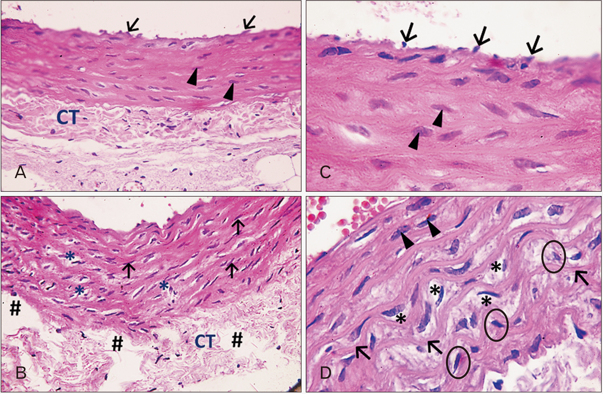
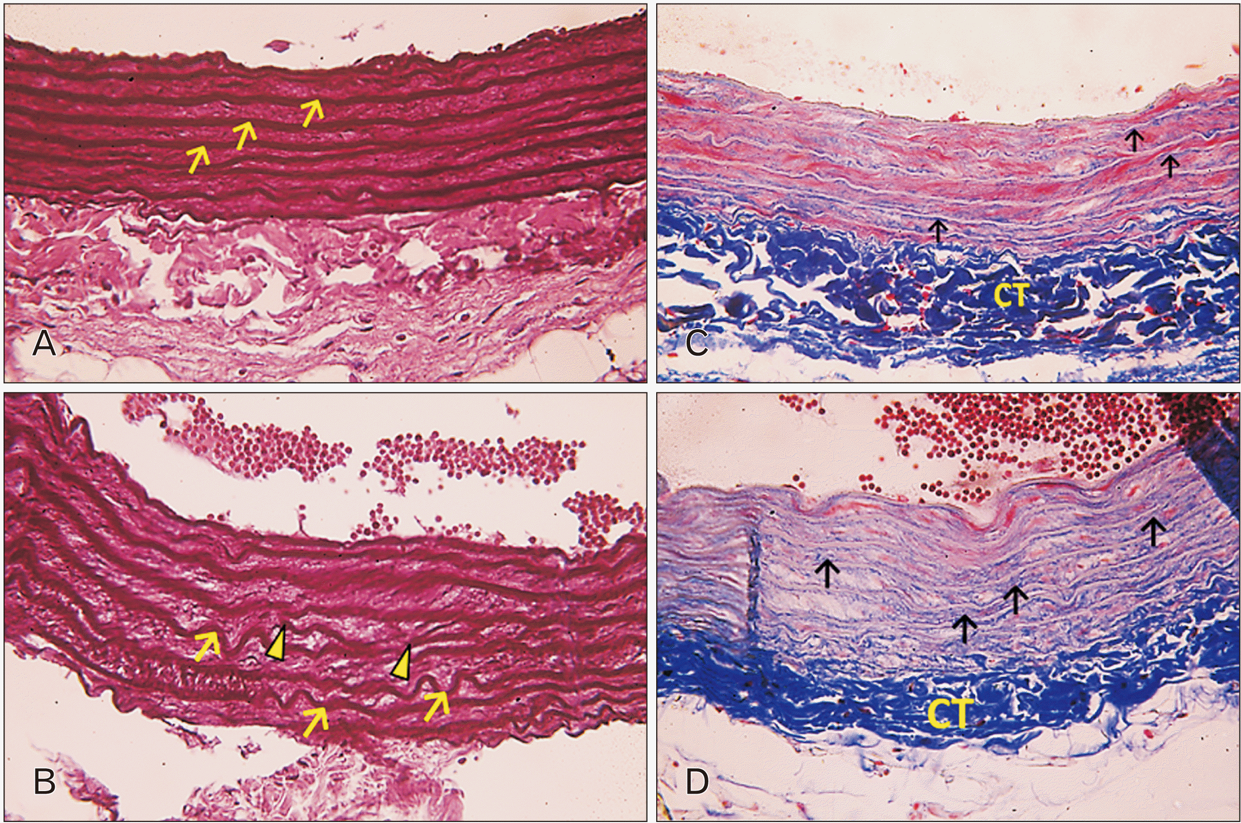
H&E stained sections of the adult control group (group I) revealed the classical structure of the aortic wall formed of three layers, tunica intima, tunica media, and tunica adventitia. Innermost tunica intima was composed of simple squamous (endothelial) cells with flat basophilic nuclei resting on a basement membrane with a sub-endothelial layer of elastic and collagen fibers and limited by a continuous layer of elastic fibers (internal elastic lamina). Intima was very thin and hence difficult to be distinguished from next layer, media. Tunica media constituted the main bulk of the vessel wall. It showed spindle shaped smooth muscle cells (SMCs) with oval nuclei and dark acidophilic cytoplasm. SMCs showed a regular concentric arrangement alternating with the elastic laminae. The tunica adventitia was composed of packed, wavy connective tissue (Fig. 1A, C).
Examination of H&E stained sections of the adult ciprofloxacin treated rats (group II) showed some histological changes. The intima revealed uneven surface with numerous foldings. Tunica media showed many areas of tissue loss (vacuolations) and the nuclei of SMCs took different orientations and some of them were darkly stained and shrunken (pyknotic). The adventitial connective tissue appeared loosely packed with multiple tissue loss (Fig. 1B, D).

Orcein stained sections

In orcein stained sections of the adult control group (group I), the tunica media showed dark brown continuous elastic fibers (laminae). They were thick and parallel with regular concentric orientation, in line with SMCs (Fig. 2A).
In group II, partial loss of the regular arrangement was observed. Widening between the concentric elastic laminae was seen. In addition, some elastic laminae appeared markedly corrugated. Some laminae showed obvious fragmentation and interruption. Acidophilic stained SMCs were well observed between elastic laminae (Fig. 2B).

Masson’s trichrome stained sections

Masson’s trichrome stained sections of the adult control group (group I) showed distribution of wavy collagen fibers in the tunica media making horizontal concentric arrangement alternating with SMCs in between. The tunica adventitia showed its packed connective tissue (Fig. 2C).
In group II, there was an obvious increase in collagen fibers deposition in the tunica media that was encroaching on SMCs. Collagen fibers didn’t show regular arrangement. SMCs in the tunica media appeared severely damaged with faint acidophilic stain (Fig. 2D).

Immunohistochemical staining for alpha smooth muscle actin

Immunohistochemical staining for the adult control group (group I) showed homogenous packed layers of dense alpha actin positive SMCs located between elastic and collagen fibers lamellae (Fig. 3A).
In group II, weak positive immune reaction for alpha smooth muscle actin was observed involving wide areas of the muscle bulk of the tunica media (Fig. 3B).

Scanning electron microscope results

Examination of aortae in adult control group (group I) revealed well adherent three layers of the aortic wall; intima, media, and adventitia. Luminal endothelial surface appeared smooth and continuous showing nuclei of lining endothelium protruding into the vessel lumen with distinct boundaries between adjacent cells. The cells were aligned parallel to the long axis of the vessel. Tunica media was the thickest layer followed by the outer adventitia (Fig. 4A, B).
In adult ciprofloxacin treated rats (group II), sections showed rough lining endothelial surface with numerous irregular folds between the endothelial cells (Fig. 4C).

Senile groups (groups III and IV)

Examination of stained sections of the thoracic aortae of the senile control subgroup (III-b) revealed similar findings as compared to the senile control subgroup (III-a).

Hematoxylin and eosin stained sections
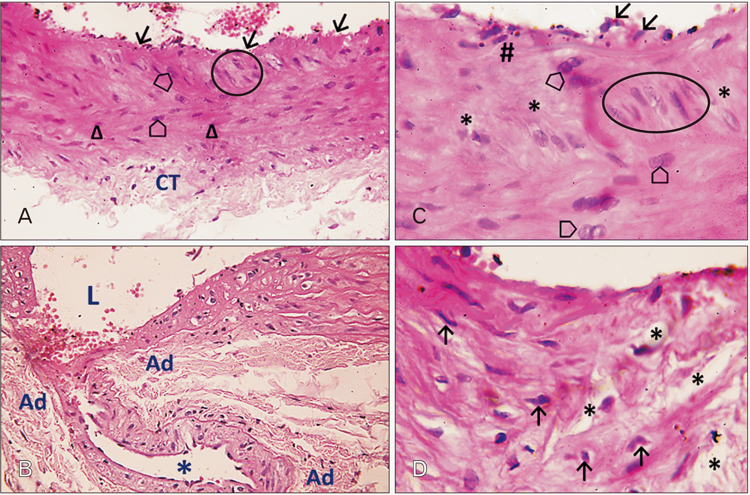
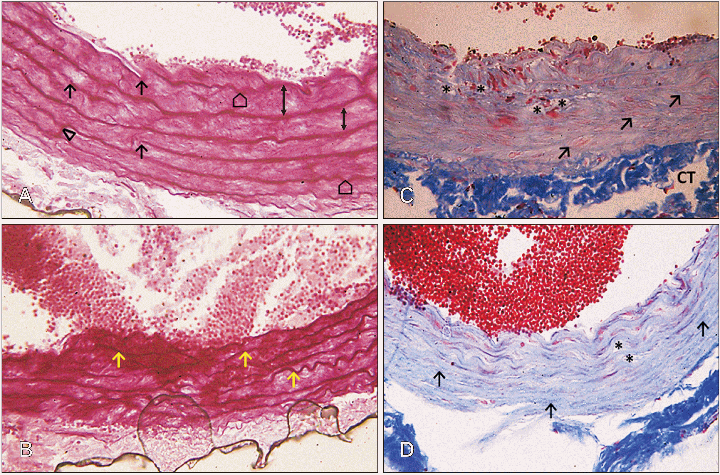
Examination of H&E stained sections of aortae of the senile control group (group III) demonstrated that the intima showed focal desquamation of the lining endothelium, multiple interruptions of the internal elastic lamina, and areas of intimal thickening. Tunica media showed loss of the normal concentric horizontal arrangement of the SMCs. It showed vertical, horizontal and oblique orientations of SMCs. Frequent areas of muscle degeneration were observed. Some smooth muscle fibers appeared proliferating with multiple nuclei. The tunica adventitia was composed of sparse, loose connective tissue (Fig. 5A, C).
In senile ciprofloxacin treated rats (group IV), similar findings were noted as those in (group III). In addition, there was aberrant decrease in the number of the SMCs. Multiple spaces between the smooth muscle fibers were noted (Fig. 5B, D). Moreover, some sections showed severe damage of the aortic wall. Interrupted, discontinuous intima and media was noted. In addition, large pouch made up of an invagination of the inner layers of aortic wall outwards was observed. The pouch was connected to aortic lumen by a narrow area of intimal tear. The tunica media was severely damaged with numerous pyknotic nuclei and necrotic SMCs. The elastic laminae were totally disorganized (Fig. 5B).

Orcein stained sections

In orcein stained sections of the senile control group (group III), the elastic fibers appeared thin, widely spaced, and occasionally fragmented. Some fibers were corrugated and sometimes branched (Fig. 6A).
In group IV, severe disruption of the regular arrangement of the elastic laminae was noticed. Most of the elastic fibers appeared disorganized, wavy, and fragmented (Fig. 6B).

Masson’s trichrome stained sections

Masson’s trichrome stained sections of the senile control group (group III) showed collagen fibers associated with elastic fiber lamellae that revealed obviously disorganized arrangement. Many unstained spaces (vacuoles) were noticed between lamellae. Interrupted, irregular subintimal region was also seen. Weak acidophilic stained SMCs were hardly seen (Fig. 6C).
In group IV, there was aberrant increase in the collagen fibers deposition encroaching on the muscle layer in tunica media. Vacuoles in interlamellar spaces were observed occupying interlamellar spaces with aberrant decrease in the vessel wall thickness (Fig. 6D).

Immunohistochemical staining for alpha smooth muscle actin

Alpha smooth muscle actin immunostaining of the senile control group (group III) revealed moderate to weak immune reactivity through whole extent of SMCs of tunica media (Fig. 7A).
In group IV, there was almost negative immune reactivity. Only some sporadic areas showed weak immune reactivity (Fig. 7B).

Scanning electron microscope results

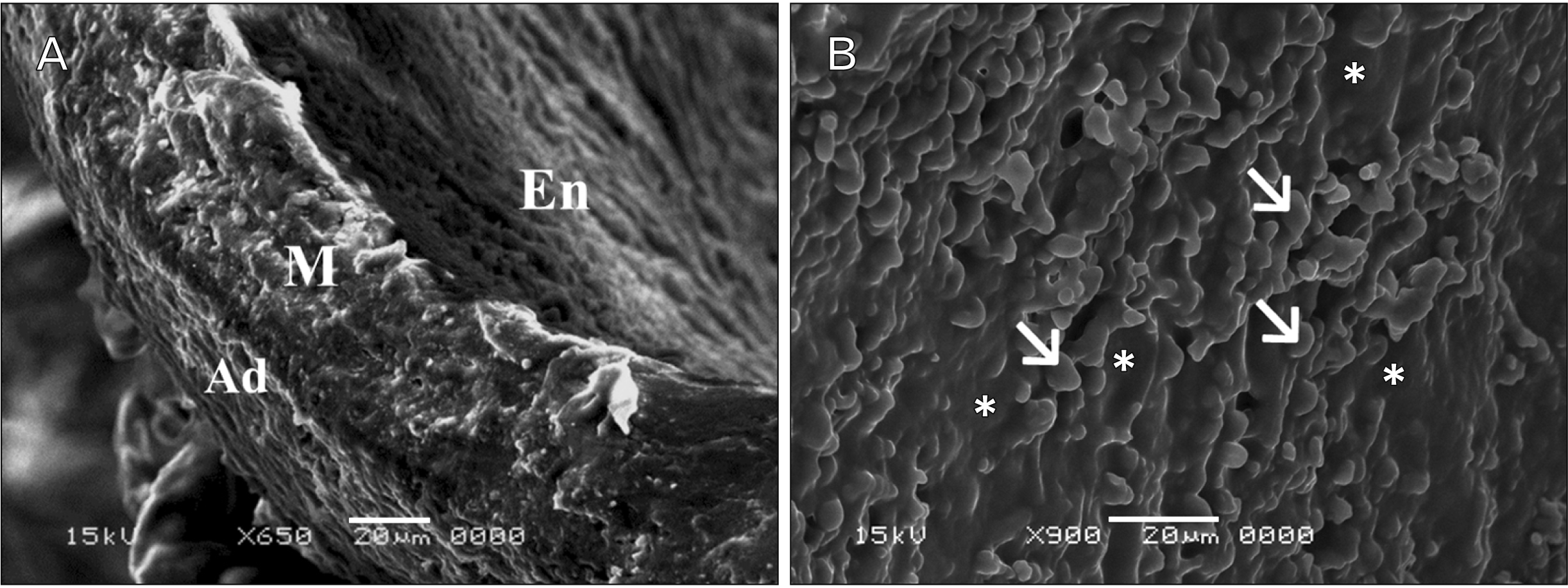
Examination of aortae in senile control group (group III) revealed rough endothelial lining with irregular folds between the endothelial cells with thinning of the endothelium in some areas. The media was the thickest layer, with outer adventitial layer. The three layers were tightly opposed to each other (Fig. 8).
In senile ciprofloxacin treated rats (group IV), the intima revealed many cracks, one of these led to large outpouching that passed through both media and adventitia and projected towards the outer wall of the aorta (Fig. 9A). Moreover, there was rough endothelial surface and many fissures or cracks between endothelial cells. Some sections showed intercellular spaces and desquamations with adherent blood cells and accumulation of fibrin (Fig. 9B, C).

Morphometric results and statistics

A morphometric study was conducted and statistically analyzed. No significant differences were noted in the adult control subgroup (I-b) in comparison with the adult control subgroup (I-a). Also, no significant differences were noted in the senile control subgroup (III-b) in comparison with the senile control subgroup (III-a).

The total thickness of the wall of the thoracic aorta

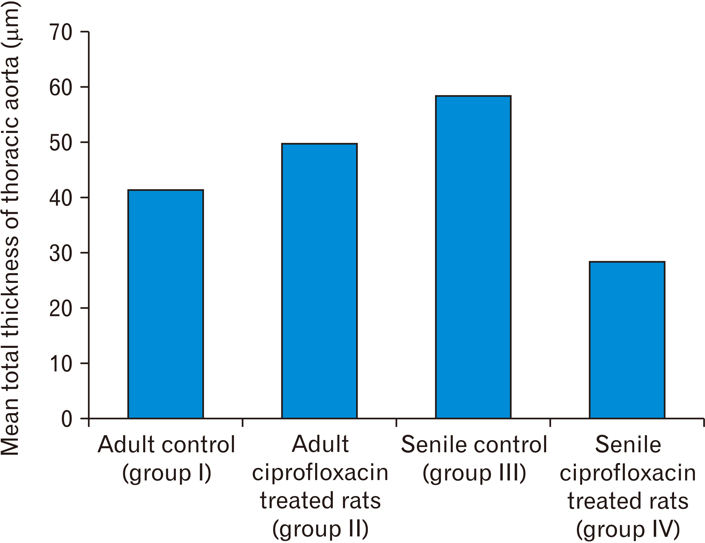
The mean total thickness of the wall of thoracic aorta in group II revealed a highly significant increase in comparison with the adult control groups (Table 1, Fig. 10). Moreover, the mean total thickness in group III recorded the highest value among the groups with highly significant increase in comparison with both groups I and II.
As regard group IV, there was a highly significant decrease as compared with other groups as the mean total thickness in this group recorded the lowest value.

Mean area percentage of alpha smooth muscle actin staining per microscopic field

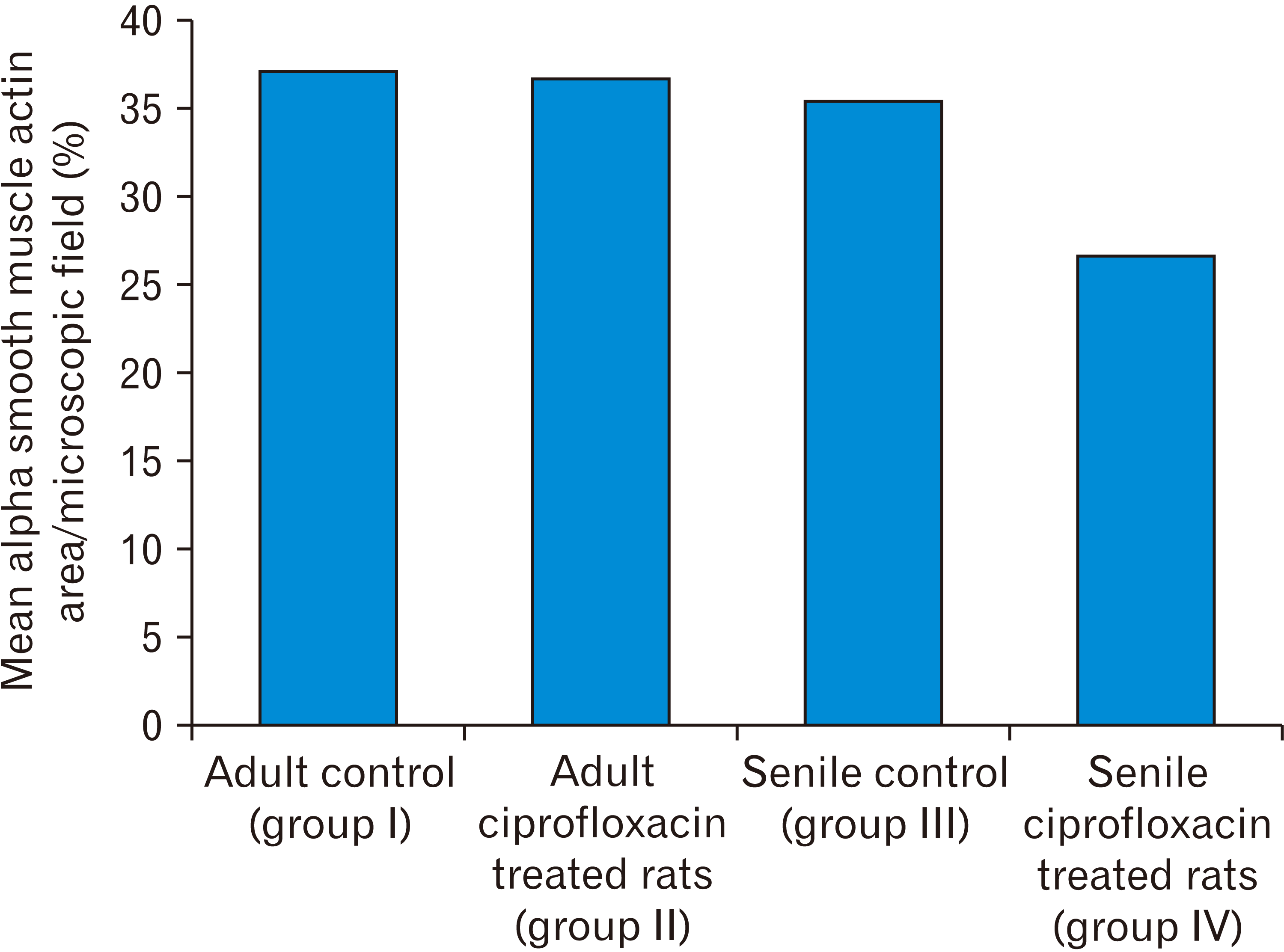
The mean area percentage of alpha smooth muscle actin staining per microscopic field of the thoracic aorta of group II showed non-significant decrease in comparison with adult control groups (Table 2, Fig. 11). However, the mean area percentage in group III showed a significant decrease as compared with group I and non-significant decrease as compared with group II.
Furthermore, there was a highly significant decrease in group IV as compared with other groups.

Discussion

To our knowledge, the present in vivo experimental work investigated, for the first time, the effect of ciprofloxacin (FQs antibiotic) administration on the structure of the wall of the thoracic aorta in adult and senile albino rats. Light microscopy, immunohistochemistry, morphometry, and statistical analysis, and scanning electron microscopy were employed in this study.
The present study focused on thoracic aorta rather than abdominal aorta due to higher incidence of AD and AA in the thoracic aorta. These two aortic diseases (AD and AA) are among the deadliest cardiovascular diseases with a mortality rate reaching 100% if not promptly diagnosed and treated [20].
An annual incidence of AA of 3 to 13.7 and AD of 3 to 20 per 100,000 population was recorded [21]. Moreover, the annual incidence of AA for the elderly was reported to be much higher, reaching about 130 per 100,000 population [22].
In the present study, the wall of the control adult thoracic aorta consisted of three layers, innermost intima, middle media, and outer adventitia.
Tunica intima was made up of flattened endothelial cell lining with flat nuclei that rested on a sub-endothelial layer composed of few elastic and collagen fibers and an outermost boundary formed by a continuous layer of elastic fibers (the internal elastic lamina).
Tunica media constituted the main bulk of the vessel wall. It was composed of regular concentric elastic and collagen fiber lamellae with intervening SMCs with oval nuclei, all immersed in ECM components.
Tunica adventitia consisted of loose connective tissue with plenty of collagen fibers but few elastic fibers.
Scanning electron microscopic examination revealed compact three layers of aortic wall, smooth continuous luminal endothelium, thick tunica media and adventitia. These observations coincide with previous studies [23].
The effect of aging on the structure of the thoracic aorta was seen in the control senile group. In the present work, the wall of control senile thoracic aorta demonstrated evident histopathological changes compared to control adult aorta. The intima showed focal epithelial loss (ulcer) or thickening (neointima) and interrupted internal elastic lamina. Tunica media revealed widening, fragmentation or splitting of the elastic laminae. SMCs showed damage, disrupted arrangement, and were immersed in excessive amount of ECM material and fibrous tissue deposition. Scanning electron microscopic observation confirmed these results. The present observations are in line with previous reports [23].
In the present study, thoracic aortae of all rats treated with ciprofloxacin revealed evident histopathological changes compared to aortae of control groups. Ciprofloxacin treatment induced the following structural changes; in the intima, irregularities of lining endothelium with frequent ulceration (ulcers together with interruption and damage of internal elastic lamina led to cracking of the wall). And reactive thickening of the intima (neointima) was noticed. In the media, there was thinning and damage of the elastic laminae and collagen fibers leading to disruption of normal arrangement of SMCs that also showed degeneration and damage (proved by immune staining with alpha-smooth muscle actin and morphometric analysis), and excessive deposition of matrix material and collagen fibers was seen on the expense of the muscle mass. In severe cases (mostly senile rats), dissection of the aortic wall and development of aneurysm like pouch was observed by light and scanning electron microscopy. The overall degree of damage observed was most intense in senile treated rats rather than adult treated group.
This was justified by Zarkovic et al. [24] who stated that the structure of the aortic wall was altered in an age-related manner with a significant decrease of SMCs and elastic fibers accompanied by increased interlamellar space due to excessive formation of connective tissue between the elastic laminae. Komutrattananont et al. [25] recorded that the mean percentage density of elastic fibers decreased in aortic wall with ageing and abdominal aorta showed the highest correlation with age followed by the thoracic aorta, the aortic arch, and the ascending aorta, respectively. They stated that these changes in the percentage density of elastic fibers can add information to age estimation purposes in human. In addition, Fritze et al. [26] reported that aging was accompanied by fragmentation and thinning of the elastic laminae.
Greenberg [27] explained that, with advancing age cross linking microfibrillar type of elastin increases on the expense of the thick regularly arranged ones.
In the present work, ciprofloxacin treatment led to thinning out, fragmentation, and increased spacing between elastic laminae of aortic tunica media.
Nakashima [28] believed that elastic laminae are crucial for maintaining aortic wall integrity. He added that, in normal human and animals, the elastic fibers of aortic tunica media are composed of concentric elastic laminae with connecting vertically oriented inter-laminar elastic fibers that are, in the meantime, densely adherent and connected with the SMCs of the tunica media providing the strength and integrity to tunica media and the aortic wall as a whole. Nakashima [28] observed by 3-dimensional scanning electron microscope of autopsied patients of AD decrease of inter-laminar elastic fibers of tunica media.
In the current study, ciprofloxacin treatment resulted in, loss of the classical concentric arrangement of the SMCs of tunica media, degeneration, and necrosis with patches of muscle tissue loss. This was also detected by alpha-smooth muscle actin immune staining and was confirmed by the morphometric analysis. The adult treated group (group II) revealed statistically significant decrease of immune staining density compared to adult control group. On the other hand, the senile treated group (group IV) showed highly statistically significant decrease of immune staining density compared to all other groups.
Findings of the present study as regards damage of the elastic laminae and SMCs were previously described by Nakashima [28] as cystic media necrosis (CMN) which the author considered a pre-requisite for the development of AD. Interestingly, CMN was observed in autopsied patients of AD in variable percentages; 10%, 18%, and 19% by Wilson and Hutchins [29], Larson and Edwards [30], and Nakashima [28], respectively.
The pathogenesis of AD because of CMN was explained by Carino et al. [31]. They reported that damage of the structural components of the media (elastic lamellae constituting the framework of aortic wall and SMCs responsible for the muscular contractile property) make the force caused by motion of the aorta or by the blood flow inside aortic lumen led to strain which can easily induce dissection of the aortic media.
The present findings could be explained in view of the mechanism of action of FQs antibiotics. These antibiotics cause degeneration of elastin, collagen, and other structural components of the ECM by stimulating the activity of MMPs [13, 14, 20], and by reducing the de novo production of collagen [15] leading to many unwanted adverse effects because collagen is abundant in the thoracic aortic wall [31].
Ishii and Asuwa [32] reported increased expression of MMP 1, 2, and 9 in aortic specimens of patients with AD.
In the present study, intimal tear, dissection of the aortic tunica media (AD), and outpouching of aortic wall (AA) was observed by light and scanning electron microscopy in some aortic specimens of senile ciprofloxacin treated rats. These observations were justified by Macura et al. [33] who stated that intimal tear penetrates the aortic media allowing blood flow from the entry site into the false lumen and this entry tear occurs at sites of greatest wall tension. Vilacosta and San Román [34] added that, blood at high pressure then splits tunica media creating a false lumen that runs alongside the true lumen.
In the present study, the aortic wall of adult ciprofloxacin treated rats and senile control rats revealed excess deposition of matrix material and collagen fibers in the tunica media as observed by Masson’s trichrome staining. Morphometric analysis confirmed observational findings and revealed highly statistically significant increase in the total thickness of the thoracic aortic wall compared with adult control group. On the contrary, senile treated group showed highly statistically significant decrease in the total thickness of thoracic aortic wall compared with all other groups.
These observations were justified by Sandison et al. [35] who stated that damage of vascular SMCs is associated with transition from contractile property to a migratory, synthetic phenotype that lay down matrix material and increase deposition of collagen fibers. Taking that in mind, in the present study, the wall of aorta showed increase in thickness because of excessive matrix material deposition while in senile ciprofloxacin treated group, collagen fibers deposition reached maximum on the expense of SMCs leading to contraction and decrease wall thickness. Similarly, Atkinson [36] reported progressive increase in the thickness of the tunica media with aging accompanied by decrease cellularity.
Ishii and Asuwa [32] reported in their electron microscopic and immunohistochemical analysis of aortic structure from patients with AD the presence of spirally thickened collagen fibers, damage of basement membrane of SMCs.
In a study using a mouse model of AA and dissection, LeMaire et al. [17] reported other mechanisms contributing to the effect of ciprofloxacin on susceptibility to aortic dissection and rupture. They stated that ciprofloxacin led to decreased Lysyl oxidase expression in aortic media with increased apoptosis and necroptosis in the aortic wall. Moreover, they recorded that ciprofloxacin caused mitochondrial DNA and nuclear DNA damage, leading to mitochondrial dysfunction. Furthermore, they expected that the ciprofloxacin-induced disruption of ECM integrity may be mediated by the activation of the cytosolic DNA sensor stimulator of interferon genes.
More recently, James et al. [37] reported that ciprofloxacin caused reduced cell viability and proteoglycan synthesis in a whole tissue explant model. Their study demonstrated that these cellular changes are not rapidly reversed after cessation of ciprofloxacin treatment.
Findings of the present experimental study confirmed the clinical observation of cases reported to FDA in four published studies [7-9, 20].

Limitations of the study

Although the results of this study were informative about the deleterious effects of ciprofloxacin on the structure of the thoracic aorta in rats, it has certain limitations. This study is the first, to our knowledge, that conducted to assess the effect of FQs on thoracic aorta on histological, immunohistochemical, and morphometric levels. The study used morphological methods only and further experiments are needed to confirm possible mechanisms related to the effect of ciprofloxacin on the thoracic aorta. The study also included the therapeutic dose and duration of the treatment without assessing the reversibility of the resulting structural changes.

Conclusion

It was concluded that oral administration of ciprofloxacin caused deleterious structural changes in the thoracic aortae of senile more than adult male albino rats giving a possible explanation for FQs-induced risk of AD. This could demonstrate valuable for individuals who deal with FQs prescriptions.

Recommendations

‒ Extreme caution should be taken when prescribing FQs (ciprofloxacin) particularly to high-risk cases.
‒ Mandatory call for medical help in case of any cardiac complaint in those under FQs treatment.
‒ Conduct further studies including different types of FQs to compare the resulting risk from each drug.

Acknowledgements

The authors would like to thank Mr. Abd Elmageed for his good preparation of histological specimens.

Notes

Author contributions

Conceptualization: NEEN, HYR. Data acquisition: AFAN, NEEN, HYR. Data analysis or interpretation: AFAN, NEEN, HYR. Drafting of the manuscript: AFAN. Critical revision of the manuscript: AFAN. Approval of the final version of the manuscript: all authors.

Conflict of interest

No potential conflict of interest relevant to this article was reported.

References

1. Etminan M, Sodhi M, Ganjizadeh-Zavareh S, Carleton B, Kezouh A, Brophy JM. 2019; Oral fluoroquinolones and risk of mitral and aortic regurgitation. J Am Coll Cardiol. 74:1444–50. DOI: 10.1016/j.jacc.2019.07.035. PMID: 31514945.
2. Michalak K, Sobolewska-Włodarczyk A, Włodarczyk M, Sobolewska J, Woźniak P, Sobolewski B. 2017; Treatment of the fluoroquinolone-associated disability: the pathobiochemical implications. Oxid Med Cell Longev. 2017:8023935. DOI: 10.1155/2017/8023935. PMID: 29147464. PMCID: PMC5632915.
crossref
3. Singh S, Nautiyal A. 2017; Aortic dissection and aortic aneurysms associated with fluoroquinolones: a systematic review and meta-analysis. Am J Med. 130:1449–57.e9. DOI: 10.1016/j.amjmed.2017.06.029. PMID: 28739200.
crossref
4. Jackson MA, Schutze GE. Committee on Infectious Diseases. 2016; The use of systemic and topical fluoroquinolones. Pediatrics. 138:e20162706. DOI: 10.1542/peds.2016-2706. PMID: 27940800.
crossref
5. Stephenson AL, Wu W, Cortes D, Rochon PA. 2013; Tendon injury and fluoroquinolone use: a systematic review. Drug Saf. 36:709–21. DOI: 10.1007/s40264-013-0089-8. PMID: 23888427.
crossref
6. Raguideau F, Lemaitre M, Dray-Spira R, Zureik M. 2016; Association between oral fluoroquinolone use and retinal detachment. JAMA Ophthalmol. 134:415–21. DOI: 10.1001/jamaophthalmol.2015.6205. PMID: 26967005.
crossref
7. Lee CC, Lee MT, Chen YS, Lee SH, Chen YS, Chen SC, Chang SC. 2015; Risk of aortic dissection and aortic aneurysm in patients taking oral fluoroquinolone. JAMA Intern Med. 175:1839–47. DOI: 10.1001/jamainternmed.2015.5389. PMID: 26436523.
crossref
8. Daneman N, Lu H, Redelmeier DA. 2015; Fluoroquinolones and collagen associated severe adverse events: a longitudinal cohort study. BMJ Open. 5:e010077. DOI: 10.1136/bmjopen-2015-010077. PMID: 26582407. PMCID: PMC4654346.
crossref
9. Pasternak B, Inghammar M, Svanström H. 2018; Fluoroquinolone use and risk of aortic aneurysm and dissection: nationwide cohort study. BMJ. 360:k678. DOI: 10.1136/bmj.k678. PMID: 29519881. PMCID: PMC5842359.
crossref
10. Clouse WD, Hallett JW Jr, Schaff HV, Spittell PC, Rowland CM, Ilstrup DM, Melton LJ 3rd. 2004; Acute aortic dissection: population-based incidence compared with degenerative aortic aneurysm rupture. Mayo Clin Proc. 79:176–80. DOI: 10.4065/79.2.176. PMID: 14959911.
crossref
11. Kent KC. 2014; Clinical practice. Abdominal aortic aneurysms. N Engl J Med. 371:2101–8. DOI: 10.1056/NEJMcp1401430. PMID: 25427112.
12. Goldfinger JZ, Halperin JL, Marin ML, Stewart AS, Eagle KA, Fuster V. 2014; Thoracic aortic aneurysm and dissection. J Am Coll Cardiol. 64:1725–39. DOI: 10.1016/j.jacc.2014.08.025. PMID: 25323262.
crossref
13. Corps AN, Harrall RL, Curry VA, Fenwick SA, Hazleman BL, Riley GP. 2002; Ciprofloxacin enhances the stimulation of matrix metalloproteinase 3 expression by interleukin-1beta in human tendon-derived cells. A potential mechanism of fluoroquinolone-induced tendinopathy. Arthritis Rheum. 46:3034–40. DOI: 10.1002/art.10617. PMID: 12428247.
14. Tsai WC, Hsu CC, Chen CP, Chang HN, Wong AM, Lin MS, Pang JH. 2011; Ciprofloxacin up-regulates tendon cells to express matrix metalloproteinase-2 with degradation of type I collagen. J Orthop Res. 29:67–73. DOI: 10.1002/jor.21196. PMID: 20602464.
crossref
15. Chang HN, Pang JH, Chen CP, Ko PC, Lin MS, Tsai WC, Yang YM. 2012; The effect of aging on migration, proliferation, and collagen expression of tenocytes in response to ciprofloxacin. J Orthop Res. 30:764–8. DOI: 10.1002/jor.21576. PMID: 22021103.
crossref
16. Paget GE, Barnes JM. Laurenc DR, Bacharach AL, editors. 1964. Chapter 6- toxicity tests. Evaluation of Drug Activities. Academic Press;New York: p. 135–66. DOI: 10.1016/B978-1-4832-2845-7.50012-8.
17. LeMaire SA, Zhang L, Luo W, Ren P, Azares AR, Wang Y, Zhang C, Coselli JS, Shen YH. 2018; Effect of ciprofloxacin on susceptibility to aortic dissection and rupture in mice. JAMA Surg. 153:e181804. DOI: 10.1001/jamasurg.2018.1804. PMID: 30046809. PMCID: PMC6233654.
crossref
18. Bancroft JD, Gamble M. 2008. Theory and practice of histological techniques. 6th ed. Churchill Livingstone/Elsevier;Philadelphia: p. 440.
19. Graham L, Orenstein JM. 2007; Processing tissue and cells for transmission electron microscopy in diagnostic pathology and research. Nat Protoc. 2:2439–50. DOI: 10.1038/nprot.2007.304. PMID: 17947985. PMCID: PMC7086545.
crossref
20. Lee CC, Lee MG, Hsieh R, Porta L, Lee WC, Lee SH, Chang SS. 2018; Oral fluoroquinolone and the risk of aortic dissection. J Am Coll Cardiol. 72:1369–78. DOI: 10.1016/j.jacc.2018.06.067. PMID: 30213330.
21. Wang SW, Huang YB, Huang JW, Chiu CC, Lai WT, Chen CY. 2015; Epidemiology, clinical features, and prescribing patterns of aortic aneurysm in Asian population from 2005 to 2011. Medicine (Baltimore). 94:e1716. DOI: 10.1097/MD.0000000000001716. PMID: 26469911. PMCID: PMC4616784.
crossref
22. Howard DP, Banerjee A, Fairhead JF, Handa A, Silver LE, Rothwell PM. 2015; Population-based study of incidence of acute abdominal aortic aneurysms with projected impact of screening strategy. J Am Heart Assoc. 4:e001926. DOI: 10.1161/JAHA.115.001926. PMID: 26289347. PMCID: PMC4599457.
crossref
23. Abu-Dief EE, Abdelrahim EA, Abdelrahim KM. 2016; Histological modifications aging aorta in male albino rat. J Cytol Histol. 7:408. DOI: 10.4172/2157-7099.1000408.
crossref
24. Zarkovic K, Larroque-Cardoso P, Pucelle M, Salvayre R, Waeg G, Nègre-Salvayre A, Zarkovic N. 2015; Elastin aging and lipid oxidation products in human aorta. Redox Biol. 4:109–17. DOI: 10.1016/j.redox.2014.12.008. PMID: 25553420. PMCID: PMC4309857.
crossref
25. Komutrattananont P, Palee P, Prasitwattanaseree S, Mahakkanukrauh P. 2020; The estimation of age from elastic fibers in the tunica media of the aortic wall in a thai population: a preliminary study using aorta image analysis. Anat Cell Biol. 53:284–91. DOI: 10.5115/acb.20.094. PMID: 32727957. PMCID: PMC7527121.
crossref
26. Fritze O, Romero B, Schleicher M, Jacob MP, Oh DY, Starcher B, Schenke-Layland K, Bujan J, Stock UA. 2012; Age-related changes in the elastic tissue of the human aorta. J Vasc Res. 49:77–86. DOI: 10.1159/000331278. PMID: 22105095.
crossref
27. Greenberg SR. 1986; The association of medial collagenous tissue with atheroma formation in the aging human aorta as revealed by a special technique. Histol Histopathol. 1:323–6.
28. Nakashima Y. 2010; Pathogenesis of aortic dissection: elastic fiber abnormalities and aortic medial weakness. Ann Vasc Dis. 3:28–36. DOI: 10.3400/avd.ctiia09001. PMID: 23555385. PMCID: PMC3595817.
crossref
29. Wilson SK, Hutchins GM. 1982; Aortic dissecting aneurysms: causative factors in 204 subjects. Arch Pathol Lab Med. 106:175–80.
30. Larson EW, Edwards WD. 1984; Risk factors for aortic dissection: a necropsy study of 161 cases. Am J Cardiol. 53:849–55. DOI: 10.1016/0002-9149(84)90418-1.
crossref
31. Carino D, Zafar MA, Singh M, Ziganshin BA, Elefteriades JA. 2019; Fluoroquinolones and aortic diseases: is there a connection. Aorta (Stamford). 7:35–41. DOI: 10.1055/s-0039-1693468. PMID: 31529426. PMCID: PMC6748841.
crossref
32. Ishii T, Asuwa N. 2000; Collagen and elastin degradation by matrix metalloproteinases and tissue inhibitors of matrix metalloproteinase in aortic dissection. Hum Pathol. 31:640–6. DOI: 10.1053/hupa.2000.7642. PMID: 10872655.
crossref
33. Macura KJ, Corl FM, Fishman EK, Bluemke DA. 2003; Pathogenesis in acute aortic syndromes: aortic dissection, intramural hematoma, and penetrating atherosclerotic aortic ulcer. AJR Am J Roentgenol. 181:309–16. DOI: 10.2214/ajr.181.2.1810309. PMID: 12876003.
crossref
34. Vilacosta I, San Román JA. 2001; Acute aortic syndrome. Heart. 85:365–8. DOI: 10.1136/heart.85.4.365. PMID: 11250953. PMCID: PMC1729697.
crossref
35. Sandison ME, Dempster J, McCarron JG. 2016; The transition of smooth muscle cells from a contractile to a migratory, phagocytic phenotype: direct demonstration of phenotypic modulation. J Physiol. 594:6189–209. DOI: 10.1113/JP272729. PMID: 27393389. PMCID: PMC5088226.
crossref
36. Atkinson J. 1998; Aging of arterial extracellular matrix elastin: etiology and consequences. Pathol Biol (Paris). 46:555–9. French.
37. James S, Schuijers J, Daffy J, Cook J, Samiric T. 2021; Ciprofloxacin reduces tenocyte viability and proteoglycan synthesis in short-term explant cultures of equine tendon. PeerJ. 9:e12003. DOI: 10.7717/peerj.12003. PMID: 34540363. PMCID: PMC8411937.
crossref

Fig. 1
Photomicrographs of sections in the thoracic aortae of adult rats (H&E). (A) Group I (adult control; ×400): the tunica intima shows simple squamous endothelial cells having flattened nuclei bulging into the lumen (arrows). The tunica media shows smooth muscle cells (SMCs) having single oval nuclei (arrowheads). Notice the packed, wavy connective tissue (CT) in the tunica adventitia. (B) Group II (adult rats treated with ciprofloxacin; ×400): tunica intima reveals uneven surface with many folds. Tunica media shows many vacuoles of lost tissue (asterisks), many nuclei of SMCs are darkly stained (pyknotic; arrows). The adventitia shows loosely packed CT with multiple tissue loss (hashtags). (C) Group I (adult control; ×1,000): the simple squamous cells of the tunica intima appear having flattened nuclei bulging into the lumen (arrows). The tunica media shows the oval nuclei (arrowheads) of SMCs that have regular concentric arrangement alternating with elastic laminae. (D) Group II (adult rats treated with ciprofloxacin; ×1,000): the inner part of the tunica media shows the classical concentric arrangement of the SMCs (arrowheads). The outer part of the tunica media exhibits attenuation of the SMCs (circles) with multiple areas of tissue loss (asterisks ) and folding of the elastic laminae (arrows).
acb-55-1-79-f1.tif
Fig. 2
Photomicrographs of sections in the thoracic aortae of adult rats. (A) Group I (adult control; Orcein, ×400): the laminae of elastic fibers are thick, parallel, and organized (arrows). (B) Group II (adult rats treated with ciprofloxacin; Orcein, ×400): the laminae of elastic fibers are corrugated (arrows) and fragmented (arrowheads). Also note the widening between the elastic laminae with partial loss of their regular arrangement. (C) Group I (adult control; Masson’s Trichrome, ×400): wavy collagen fibers (arrows) are seen with the smooth muscle cells (SMCs) distributed in between. Notice the packed connective tissue (CT) in tunica adventitia. (D) Group II (adult rats treated with ciprofloxacin; Masson’s Trichrome, ×400): note the aberrant increase in the collagen fibers deposition (arrows) that is encroaching on the SMCs in the tunica media. The SMCs are few, interrupted and show very faint acidophilic stain. Tunica adventitia shows very dense deposition of CT.
acb-55-1-79-f2.tif
Fig. 3
Photomicrographs of sections in the thoracic aortae of adult rats (immune staining with anti-alpha smooth muscle actin antibody, ×400). (A) Group I (adult control): notice homogenous packed layers of strong positive alpha actin filaments of smooth muscle fibers (arrows) in tunica media located between elastic laminae. (B) Group II (adult rats treated with ciprofloxacin): weak positive immune reaction for alpha smooth muscle actin is observed (asterisks).
acb-55-1-79-f3.tif
Fig. 4
Scanning electron microscope (SEM) photographs of sections in the thoracic aortae of adult rats. (A) Group I (adult control; ×250; scale bar, 100 μm): the vessel wall is formed of well adherent three layers; intima (I), media (M), and adventitia (Ad). The media is the thickest layer. Endothelial surface (En) appears smooth and homogenous. (B) Group I (adult control; ×450; scale bar, 50 μm): luminal En appears smooth and continuous showing nuclei (arrows) of the lining endothelium protruding into the vessel lumen with distinct boundaries between adjacent cells. The cells are aligned parallel to the long axis of the vessel. (C) Group II (adult rats treated with ciprofloxacin; ×900; scale bar, 20 μm): rough En with numerous irregular folds between the endothelial cells are seen.
acb-55-1-79-f4.tif
Fig. 5
Photomicrographs of sections in the thoracic aortae of senile rats (H&E). (A) Group III (senile control; ×400): a photomicrograph showing tunica intima with areas of desquamation (arrows). Notice the area of intimal thickening with vertically aligned nuclei of smooth muscle cells (SMCs; circle). The tunica media reveals lack of concentric arrangement of SMCs with spindle shaped nuclei (triangles). Binucleated proliferating SMCs can be seen (pentagons). Notice also the sparse, loose connective tissue (CT) in the tunica adventitia. (B) Group IV (senile rats treated with ciprofloxacin; ×400): notice the outpouching of the aortic wall made of an invagination of the inner layers of aortic wall outwards (asterisk). The pouch is connected to aortic lumen by a narrow area of intimal tear. The tunica media is severely damaged with numerous pyknotic nuclei and vacuolated SMCs. The tunica media thins out at the outpouching. Ad, adventitia; L, lumen of the vessel. (C) Group III (senile control; ×1,000): tunica intima reveals un-even endothelial lining with some detached lining cells (arrows). Discontinuous internal elastic lamina is seen (hashtag). An area of intimal thickening with vertically aligned nuclei of SMCs can also be seen (circle). The tunica media shows random arrangement of SMCs nuclei, pale staining degenerating myocytes, and frequent patches of muscle tissue loss (asterisks). Proliferating binucleated SMCs are seen (pentagons). (D) Group IV (senile rats treated with ciprofloxacin; ×1,000): notice the tunica media with severe damage of SMCs with pyknotic nuclei (arrows). Notice the disarrangement of smooth muscle fibers and spacing due to tissue loss (asterisks).
acb-55-1-79-f5.tif
Fig. 6
Photomicrographs of sections in the thoracic aortae of senile rats. (A) Group III (senile control; Orcein, ×400): the elastic fibers appear widely spaced (up down arrows), fragmented (arrows) and corrugated (pentagons). Branching of elastic laminae (triangle) can also be visualized. (B) Group IV (senile rats treated with ciprofloxacin; Orcein, ×400): notice the marked disruption of the regular arrangement of the elastic laminae. Some of them appear severely corrugated or fragmented (arrows). (C) Group III (senile control; Masson’s Trichrome, ×400): many of collagen fibers (arrows) are seen with sparse acidophilic smooth muscle cells in tunica media. Vacuoles (asterisks) could be seen occupying interlamellar spaces. Notice the widely separated connective tissue (CT) in tunica adventitia. (D) Group IV (senile rats treated with ciprofloxacin; Masson’s Trichrome, ×400): notice the aberrant increase in the collagen fibers’ deposition (arrows) encroaching on the muscle layer in tunica media. Vacuoles (asterisks) could be also seen occupying interlamellar spaces. Notice the aberrant decrease in the vessel wall thickness.
acb-55-1-79-f6.tif
Fig. 7
Photomicrographs of sections in the thoracic aortae of senile rats (immune staining with anti-alpha smooth muscle actin antibody, ×400). (A) Group III (senile control): moderate to weak immune reactivity (asterisks) is seen in many patches through the tunica media. (B) Group IV (senile rats treated with ciprofloxacin): almost negative immune reactivity of smooth muscle fibers is seen through the whole extent of the tunica media with only sporadic patches of moderate immune reactivity appearing as brown color (asterisks).
acb-55-1-79-f7.tif
Fig. 8
Scanning electron microscope (SEM) photographs of sections in the thoracic aortae of senile control rats (group III; scale bar: 20 μm). (A) Notice the rough endothelial lining (En) (×650). The media (M) is the thickest layer with outer adventitial layer (Ad). The three layers are tightly opposed to each other. (B) Luminal endothelial surface appears rough with irregular folds between endothelial cells (arrows) (×900). Notice the thinning of endothelium (asterisks) in some areas.
acb-55-1-79-f8.tif
Fig. 9
Scanning electron microscope (SEM) photographs of sections in the thoracic aortae of senile rats treated with ciprofloxacin (group IV). (A) Notice outpouching (asterisk) of the vessel wall extending towards the tunica adventitia (×200; scale bar, 100 μm). The intima reveals many cracks (arrows). L, vessel lumen. (B) Notice the rough endothelial lining (En) with the presence of many fissures or cracks (asterisks) (×1,700; scale bar, 10 μm). Some areas show adherent blood cells (arrows). (C) Notice the accumulation of fibrin (arrows) (×650; scale bar, 20 μm).
acb-55-1-79-f9.tif
Fig. 10
Mean total thickness of the wall of thoracic aorta (×400).
acb-55-1-79-f10.tif
Fig. 11
Mean area percentage of alpha smooth muscle actin staining per microscopic field (×400).
acb-55-1-79-f11.tif
Table 1
Comparison of the total thickness of the wall of thoracic aorta between the experimental groups
Total thickness of the wall of thoracic aorta (μm) Adult control (group I) Adult ciprofloxacin treated rats (group II) Senile control (group III) Senile ciprofloxacin treated rats (group IV)
Mean±standard deviation 44.08±4.7 52.53±3.12
(P=0.0004)a)
61.1±2.46
(P<0.001)a)
(P<0.001)b)
31.15±6.07
(P=0.00012)c)
(P<0.001)d)
(P<0.001)e)

a)Highly significant increase in comparison with group I; b)Highly significant increase in comparison with group II; c)Highly significant decrease in comparison with group I; d)Highly significant decrease in comparison with group II; e)Highly significant decrease in comparison with group III.

Table 2
Comparison of the area percentage of alpha smooth muscle actin staining per microscopic field at magnification (×400) between the experimental groups
Alpha smooth muscle actin staining area per microscopic field (%) Adult control (group I) Adult ciprofloxacin treated rats (group II) Senile control (group III) Senile ciprofloxacin treated rats (group IV)
Mean±standard deviation 38.78±0.9 38.34±1.9 37.09±0.46
(P=0.006)a)
28.31±3.79
(P=0.00014)b)
(P=0.00035)c)
(P<0.00043)d)

a)Significant decrease in comparison with group I; b)Highly significant decrease in comparison with group I; c)Highly significant decrease in comparison with group II; d)Highly significant decrease in comparison with group III.

TOOLS
Similar articles