

 PDF
PDF Citation
 Print
Print


Introduction
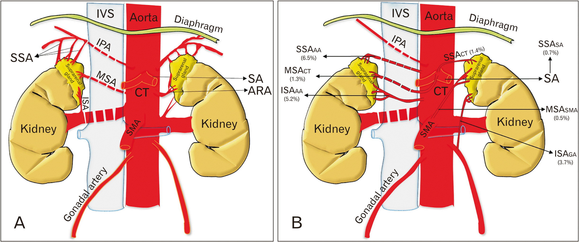 | Fig. 1Schematic diagram of arterial supply of suprarenal gland. (A) Shows normal arterial supply of the suprarenal gland. (B) Shows the most common variant origins of superior, middle, and inferior suprarenal arteries being reported. IVC, inferior vena cava; IPA, inferior phrenic artery; SSA, superior suprarenal artery; MSA, middle suprarenal artery; ISA, inferior suprarenal artery; SMA, superior mesenteric artery; CT, coeliac trunk; SA, splenic artery; ARA, accessory renal artery; SSAAA, superior suprarenal artery originating from the abdominal aorta; MSACT, middle suprarenal artery originating from the coeliac trunk; ISAAA, inferior suprarenal artery originating from the abdominal aorta; SSACT, superior suprarenal artery originating from the coeliac trunk; MSASMA, middle suprarenal artery originating from superior mesenteric artery; ISAGA, inferior suprarenal artery originating from gonadal artery.
|
Materials and Methods
Google Scholar (Google, Inc., Mountain View, CA, USA)
Medline and PubMed (United States National Library of Medicine, Bethesda, MD, USA)
Embase (Ovid Technologies, Inc., New York, NY, USA)
Scopus (Elsevier, Amsterdam, The Netherlands)
Cochrane library
Science direct
Discussion
 | Fig. 2A chronological illustration of hitherto most significant findings in the research history of arterial variations of suprarenal arteries. SSA, superior suprarenal artery; AA, abdominal aorta; RA, renal artery; MSA, middle suprarenal artery; ISA, inferior suprarenal artery.
|
Table 1
| Authors (year) | Population/region | No. and type of specimens | Variations | |||||||
|---|---|---|---|---|---|---|---|---|---|---|
| Superior suprarenal arteries | Middle suprarenal arteries | Inferior suprarenal arteries | ||||||||
| R | L | R | L | R | L | |||||
| Dobbie and Symington (1966) [16] | Scotland | 20 autopsies of human adults, 50 adult patients | S: 100% | M: 100% | I: 100% | |||||
| Lamarque et al. (1973) [14] | France | 255 total aortography, 373 selective arteriography of suprarenal gland | S: 100% | - |
I: 51.5% Iab: 48.5% |
I: 44% Iab: 56% |
||||
| Toni et al. (1988) [15] | Italy | 100 abdominal angiographies |
S: 92% SCT: 5% SAA: 3% |
S: 79% SAA: 16% SIC: 3% SCT: 2% |
M: 91% MCT: 4% MIP: 3% MRA: 2% |
M: 99% MCT: 1% |
I: 96% IPR: 2% IAA: 2% |
I: 95% IAA: 5% |
||
| Bianchi and Ferrari (1991) [4] | Argentina | 50 fetuses | S: 100% |
S: 96% SCT: 4% |
M: 68% MIP: 32% |
M: 68% MIP: 20% MCT: 12% |
I: 60% I+IGA: 12% ISPA: 4% ISPA+IGA: 12% IAA: 4% IGA: 4% IAGA: 4% |
I: 40% IAA: 24% IGA: 8% I+IGA: 8% IAGA: 4% IGA+IAGA: 4% I+ISPA: 4% IAA+ISPA: 4% I SPA+IGA: 4% |
||
| Pityński et al. (1998) [3] | Poland | 40 fetuses | S: 100% |
S: 95% S+SAA: 5% |
M: 32.5% MIP: 27.5% M+MIP: 15% MRA: 17.5% MGA: 7.5% |
M: 47.5% MIP: 17.5% M+MSSA: 2.5% M+MIP: 22.5% MRA: 7.5% MGA: 2.5% |
I: 55% I+IAA: 25% I+ISPA: 5% I+IGA: 7.5% I+IGA+IAA: 2.5% I+IAA+ISPA: 2.5% ISMA: 2.5% |
I: 47.5% I+IAA: 32.5% I+ISPA: 2.5% I+IGA: 12.5% I+IGA+IAA: 10% I+IGA+ISPA: 5% |
||
| Manso and DiDio (2000) [12] | Brazil | 30 pairs of suprarenal glands |
S: 86.7% SCT: 6.7% SAA: 3.3% SAA+SISA: 3.3% |
S: 83.3% SCT: 6.7% SAA: 10% |
M: 53.3% MIP: 26.7% MSSA: 3.3% MISA: 6.7% MSMA: 3.3% MRA: 3.3% MCT: 3.3% |
MAA: 46.7% MIP: 26.7% MSSA: 10.0% MISA: 6.7% MSMA: 3.3% MRA: 3.3% MCT: 3.3% MRA: 3.3% |
I: 70% IAA: 26.7% IAA+ISSA: 3.3% |
I: 50% IAA: 36.7% ISPA: 3.3% IIPA: 3.3% IGA: 6.6% |
||
| Yalçin et al. (2004) [29] | Turkey | 25 years old female cadaver | - | MCT | I | |||||
| Cimen et al. (2007) [30] | Turkey | 45 years old male cadaver | - | MRA | M | I | - | |||
| Deepthinath et al. (2006) [31] | South Indian | 45 years old male cadaver | - | MRA | MCT | I | - | |||
| Dutta (2010) [5] | North Indian | 68 human cadavers | S: 100% |
S: 76% SAA: 18% SSA: 6% |
M: 53% MARA: 18% Absent: 29% |
M: 94% MARA: 6% |
I: 76% IGA: 18% IAA: 6% |
I: 59% IGA: 6% Absent: 35% |
||
| Oztürk et al. (2010) [24] | Turkey | 50 years old male cadaver | Absent | M | IAA | |||||
| Jyothsna et al. (2012) [36] | South Indian | 55 years old male cadaver | - | - | - | IAA | ||||
| Chakravarthi (2014) [21] | South Indian | Middle-aged male cadaver | SAA | SAA | M | M | - | - | ||
| Sushma et al. (2014) [17] | South Indian | 20 cadavers | S: 100% | S: 100% |
M: 80% MRA: 10% MARA: 5% MIP: 5% |
M: 90% MIP: 5% MCT: 5% |
I: 90% IARA: 5% IIP: 5% |
I: 90% IARA: 10% |
||
| Sarkar et al. (2014) [18] | Northeast India | 54 years old male cadaver | S | SCT | M | Absent | I | I | ||
| Ahmed et al. (2015) [20] | South Indian | 25 adult and 50 fetal cadavers |
S: 88% Sab: 12% |
M: 93.34% Mab: 6.66% |
I: 93.34% Iab: 6.66% |
|||||
| Lakshmi and Dhoot (2016) [6] | North Indian | 15 adult human cadavers | S: 100% | S: 100% |
M: 83.33% MARA: 13.33% MRA: 3.33% |
M: 96.66% MCT: 3.33% |
I: 83.33% IARA: 16.66% |
I: 66.66% IARA: 33.33% |
||
| Shanthakumar et al. (2016) [19] | South Indian | 58 years old male cadaver | S | - | M | - | I | IGA | ||
| Olewnik et al. (2018) [22] | Poland | 64 years old male cadaver | SRA | Absent | I | |||||
| Greeff et al. (2019) [27] | South African | 50 fetuses |
S: 98% Absent: 2% |
S: 98% Absent: 2% |
M: 18% MRA: 62% Absent: 20% |
M: 34% MRA: 26% Absent: 40% |
I: 90% IAA: 10% |
I: 92% IAA: 6% IARA: 2% |
||
| Vinitha and Parthasarathy (2020) [37] | South Indian | 48 cadavers |
S: 81.33% SAA: 16.67% SCT: 2% |
M: 98% Absent: 2% |
I: 98% IAA: 2% |
|||||
| Xu et al. (2020) [42] | Chinese | 843 fetuses | - |
M: 89.2% Absent: 10.79% |
- | |||||
| South Indiana) | 147 |
S: 86.8% SAA: 6.2% SCT: 0.8% Sab: 6.2% |
M: 92.8% Mabsent: 0.7% MAB: 3.4% MRA: 1% MARA: 0.3% MCT: 0.7% MIP: 0.7% |
I: 92.9% IAA: 1% IARA: 10.2% IIP: 0.3% Iab: 3.4% IGA: 0.3% |
||||||
| North Indianb) | 83 |
S: 90.4% SAA: 7.2% SSA: 2.4% |
M: 76% MARA: 11% MCT: 0.4% MRA: 0.4% Absent: 12% |
I: 73.5% IGA: 9.7% IAA: 2.4% IARA: 4.7% Iabsent: 9.7% |
||||||
| Caucasiansc) | 919 |
S: 97.5% SAA: 1.3% SCT: 0.8% SIC: 0.1% SAA+SISA: 0.1% S+SAA: 0.1% SRA: 0.1% |
M: 93.5% Mabsent: 0.1% MIP: 3.4% MSSA: 0.2% MISA: 0.2% MSMA: 0.1% MRA: 0.7% MCT: 0.7% M+MSSA: 0.1% M+MIP: 0.8% MGA: 0.2% |
I: 57.7% Iab: 35.7% IIPA: 0.05% IPR: 0.1% IAA: 2.06% I+IGA: 0.9% ISPA: 0.2% IGA: 0.4% ISPA+IGA: 0.4% IAGA: 0.2% IGA+IAGA: 0.1% I+ISPA: 0.3% IAA+ISSA: 0.1% I+IAA: 1.3% I+ISSA: 0.05% I+IGA+IAA: 0.2% I+IAA+ISPA: 0.05% I+IGA+ISPA: 0.1% |
||||||
| Turkishd) | 3 |
S: 66.7% Absent: 33.3% |
M: 66.7% MCT: 33.3% MRA: 33.3% |
I: 66.7% IAA: 33.3% |
||||||
The subscript in the table is denoting the origin of the respective arteries: ab, abnormal origin; AA, abdominal aorta; CT, coeliac trunk; IC, intercostal artery; IP, inferior phrenic artery; RA, renal artery; PR, polar renal artery; GA, gonadal artery; SPA, superior polar artery; AGA, accessory gonadal artery; SMA, superior mesenteric artery; ISA, inferior suprarenal artery; SSA, superior suprarenal artery; IPA, inferior polar artery; SA, splenic artery; ARA, accessory renal artery. a)Average results for the South Indian population include Deepthinath et al. (2006) [31], Jyothsna et al. (2012) [36], Chakravarthi (2014) [21], Sushma et al. (2014) [17], Ahmed et al. (2015) [20], Shanthakumar et al. (2016) [19], Vinitha and Parthasarathy (2020) [37]. b)Average results for North Indian population include Dutta (2010) [5], Lakshmi and Dhoot (2016) [6]. c)Average results for Caucasian population include Dobbie and Symington (1966) [16], Lamarque et al. (1973) [14], Toni et al. (1988) [15], Bianchi and Ferrari (1991) [4], Pityński et al. (1998) [3], Manso and DiDio (2000) [12], Olewnik et al. (2018) [22]. d)Average results for Turkish population includes Yalçin et al. (2004) [29], Cimen et al. (2007) [30], Oztürk et al. (2010) [24]. R, right; L, left; S, normal origin of superior suprarenal artery; M, normal origin of middle suprarenal artery; I, normal origin of inferior suprarenal artery.
![]()
Variations of suprarenal arteries: with regards to the origin
Table 2
| Study type | % of common variations in origin | ||
|---|---|---|---|
| SSA | MSA | ISA | |
| Adult (n=260) | |||
| Fetal (n=983) | |||
| Adult+fetal (n=75) | |||
| Angiographic (n=728) | |||
The subscript in the table is denoting the origin of the respective arteries: ab, abnormal origin; AA, abdominal aorta; CT, coeliac trunk; IC, intercostal artery; IP, inferior phrenic artery; RA, renal artery; PR, polar renal artery; GA, gonadal artery; SPA, superior polar artery; AGA, accessory gonadal artery; SMA, superior mesenteric artery; ISA, inferior suprarenal artery; SSA, superior suprarenal artery; IPA, inferior polar artery; SA, splenic artery; ARA, accessory renal artery.
![]()
Variations of superior suprarenal artery: with regards to the number of arteries
Table 3
| Authors (year) | Population/region | No. and type of specimens | No of arteriesa) | |||||||
|---|---|---|---|---|---|---|---|---|---|---|
| Superior suprarenal arteries | Middle suprarenal arteries | Inferior suprarenal arteries | ||||||||
| R | L | R | L | R | L | |||||
| Miekoś (1979) [43] | Poland | 220 human adrenal glands | 30 (13.63%) | 1 (94%) | 2–3 (6%) | 2–11 (0.9%–5%) | ||||
| Bianchi and Ferrari (1991) [4] | Argentina | 50 fetuses | - | - | - | - | ||||
| Pityński et al. (1998) [3] | Poland | 40 human fetuses | ||||||||
| Manso and DiDio (2000) [12] | Brazil | 30 pairs of suprarenal glands (n=60) | ||||||||
| Bordei et al. (2003) [38] | Romania | 120 cases | - | - | ||||||
| Reddy et al. (2014) [23] | South Indian | 50 fetal specimens and 25 adult specimens | - | - | ||||||
| Ahmed et al. (2015) [20] | South Indian | 25 adult and 50 fetal cadavers | - | - | - | - | ||||
| Greeff et al. (2019) [27] | South African | 50 fetuses | ||||||||
![]()
 XML Download