

 PDF
PDF Citation
 Print
Print


Introduction
Gantzer’s muscle refers to a group of accessory muscles of the flexor compartment of the forearm. It descends beneath with flexor digitorum superficialis (FDS) up to mid-forearm. It takes origin from at the medial epicondyle of humerus (ME) (common flexor origin) or coronoid process of the ulna or fascial sheath of FDS or pronator teres. It inserts onto the deep flexors, i.e., flexor pollicis longus (FPL) and flexor digitorum profundus (FDP) [1]. This muscle was first reported by Albinus in the 18th century and described by Gantzer [2], a German anatomist, in 1813. Based on initial observations, the authors identified two main variants of Ganzter’s muscle, i.e., accessory head of FPL (ahFPL) and accessory head of FDP (ahFDP) [3]. Underneath the FDS, Gantzer’s muscle follows an oblique path from the medial to the lateral aspect of the forearm before joining the FPL [4]. Furthermore, the Gantzer’s muscle can contribute to the FDP muscle through a second tendon. The presence of ahFPL and ahFDP could be explained by the embryological events of the common flexor muscle mass, which splits into two strata: deep and superficial during differentiation [1]. The FPL, FDP, and pronator quadratus muscles are all derived from the deep layer. Gantzer’s muscle is the product of an imperfect cleavage of the deep layer [4]. The human gained the FPL during its evolution. The introduction of FPL into the flexor compartment allowed the thumb to move independently in three different planes [5]. A detailed meta-analysis was conducted by Roy et al. (2015) [6] on the ahFPL variant of Gantzer’s muscle. The authors did not include ahFDP. There was at least a dozen of the manuscript which dealt with ahFDP. So, the pooled prevalence of Gantzer’s muscle in the previous meta-analysis may be considered inaccurate. Finding such deficiency in the previous meta-analysis forced us to conduct the current study to elucidate the pooled estimate of both variants (ahFPL and ahFDP) and unfold the more comprehensive picture. This research aimed to determine the pooled prevalence of these accessory muscles in various populations and their morphometry and relation to the anterior interosseous nerve (AIN). It will be helpful in clinical diagnosis and surgical approaches to the forearm.
Go to : 
Materials and Methods
Search strategy
The authors have conducted a thorough search of the electronic databases PubMed, Google Scholar, Scopus, ScienceDirect, and EMBASE to find papers suitable for inclusion in the meta-analysis. Gantzer’s muscle or accessory head of FPL or ahFPL and accessory head of FDP or ahFDP were among the keywords used in the quest. There were no time or language limitations. We thoroughly reviewed relevant studies or publications to identify potentially qualified articles for the meta-analysis.
Inclusion and exclusion criteria
Studies with extractable data on the occurrence of Gantzer’s muscle in the upper limbs were deemed suitable for inclusion in the meta-analysis. The meta-analysis excluded publications that were case reports, letters to the editor, or conference abstracts, original articles which had insufficient data. During the eligibility appraisal, any disputes among the reviewers are resolved by consensus among all reviewers. The appraisal of quality of each study was conducted with the help of the Anatomy Quality Assessment tool [7].
Data extraction
The authors have collected information on the prevalence of ahFPL or ahFDP, origin, insertion, nerve supply, laterality, relation with nerves of the forearm, and morphological variation from included studies. In addition, we contacted the manuscript authors for more information via email if necessary information was missing.
Statistical analysis
Statistical analysis was performed by R statistical package 4.2.0 (R Foundation for Statistical Computing, Vienna, Austria). The Higgin’s I2 test was used to determine study heterogeneity. A fixed-effects model was used if heterogeneity (Higgin’s I2 statistics) was less than 50%. A random-effects model was used if the heterogeneity (I2 statistics) was greater than 50%. To investigate the causes of heterogeneity, subgroup analysis, sensitivity, and cumulative analysis were used when needed. The unit of analysis was per 100 upper limbs examined.
Go to :
Results
Characteristics of included studies
Fifty-eight studies in the current review have been undertaken to explore the prevalence of Gantzer’s muscle (Fig. 1) [3, 4, 8-55]. These studies examined 5,903 upper limbs for ahFPL variant (Table 1). Only 14 studies have been explored for the prevalence of ahFDP, including the data of 1,627 limbs (Table 1) [3, 8-14, 54]. A total of 5,903 limbs were included in the meta-analysis, which has data from 1868 to 2021. The data of Wagenseil (1936) [54] was bifurcated according to the population because they estimated the prevalence of Gantzer’s muscle in Mongoloid and European populations. These data were collected from June 2020 to February 2021. The study population was predominantly adult cadavers, except for one study, i.e., Kara et al. (2012) [32]. The majority of manuscripts included in the review had wide geographical distribution, and it included data from all subcontinents except Australia.
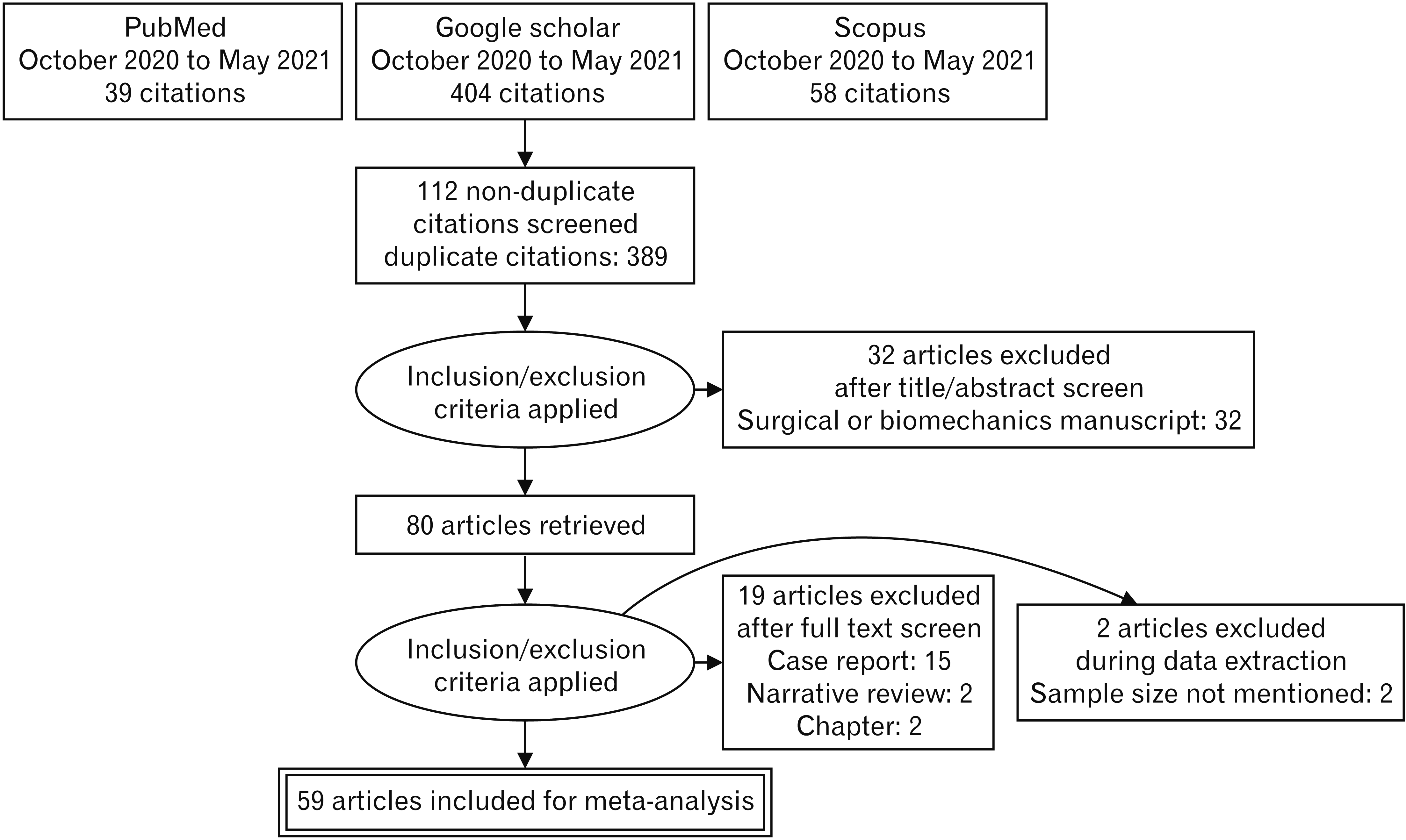 | Fig. 1Preferred Reporting Items for Systematic Reviews and Meta-Analyses flow diagram of search strategy for Gantzer’s muscle.
|
Table 1
Study characteristics of Gantzer’s muscle
| Reference | Year | Prevalence (%) | 95% confidence interval | ahFPL/ahFDP | No. and ethnicity of sample | Risk of bias |
|---|---|---|---|---|---|---|
| Adachi [53]a) | 1910 | 63 | 54–70 | 84 | 134 Asian Mongoloid | Unclear |
| Afroze et al. [18] | 2020 | 24 | 14–38 | 12 | 50 Asian Caucasian | High |
| al-Qattan [15] | 1996 | 52 | 33–70 | 13 | 25 Asian Caucasian | Low |
| Bagoji et al. [16] | 2017 | 29 | 19–42 | 17 | 58 Asian Caucasian | Moderate |
| Bajpe et al. [17] | 2015 | 24 | 14–38 | 12 | 50 Asian Caucasian | High |
| Ballesteros et al. [19] | 2019 | 32 | 24–42 | 34 | 106 South American | Low |
| Bando [53]a) | 1956 | 64 | 59–69 | 217 | 340 Asian Mongoloid | Unclear |
| Bangarayya et al. [20] | 2018 | 40 | 24–58 | 12 | 30 Asian Caucasian | Moderate |
| Bilecenoglu et al. [21] | 2005 | 20 | 9–38 | 6 | 30 Asian Caucasian | Low |
| Burute and Vatsalaswamy [22] | 2017 | 36 | 29–44 | 56 | 156 Asian Caucasian | High |
| Caetano et al. [23] | 2015 | 68 | 57–77 | 54 | 80 South American | Low |
| Chakravarthi et al. [24] | 2014 | 72 | 59–83 | 39 | 54 Asian Caucasian | Moderate |
| Dubois de Monto-Marin et al. [55] | 2021 | 11 | 4–26 | 4 | 36 European Caucasian | Moderate |
| Dellon and Mackinnon [25] | 1987 | 33 | 20–48 | 14 | 43 North American | Low |
| Desai et al. [26] | 2017 | 58 | 46–70 | 35 | 60 Asian Caucasian | High |
| Dolderer et al. [27] | 2011 | 26 | 11–50 | 5 | 19 European Caucasian | Low |
| Dykes and Anson [28] | 1944 | 53 | 45–61 | 80 | 150 North American | Moderate |
| El Domiaty et al. [8] | 2008 | 62 | 47–75 | 26 | 42 African | Low |
| Gunnal et al. [29] | 2013 | 51 | 44–58 | 92 | 180 Asian Caucasian | Moderate |
| Hemmady et al. [30] | 1993 | 67 | 53–78 | 36 | 54 Asian Caucasian | Low |
| Herrold et al. [31] | 2020 | 55 | 49–60 | 148 | 271 South American | High |
| Inoue [53]a) | 1934 | 71 | 61–79 | 71 | 100 Asian Mongoloid | Unclear |
| Jones et al. [3] | 1997 | 45 | 34–56 | 36 | 80 European Caucasian | Low |
| Kara et al. [32] (adult) | 2012 | 38 | 26–52 | 20 | 52 Asian Caucasian | Low |
| Kara et al. [32] (fetal) | 2012 | 32 | 23–43 | 29 | 90 Asian Caucasian | Low |
| Khade et al. [33] | 2020 | 53 | 36–70 | 16 | 30 Asian Caucasian | Moderate |
| Kida [34] | 1988 | 62 | 54–70 | 82 | 132 Asian Mongoloid | Low |
| Kudo and Obata [53]a) | 1957 | 55 | 48–61 | 118 | 216 Asian Mongoloid | Low |
| Kumari et al. [35] | 2017 | 42 | 29–56 | 20 | 48 Asian Caucasian | Moderate |
| Le Double and Berry [36] | 1897 | 33 | 28–39 | 100 | 300 European Caucasian | Moderate |
| Loth [53]a) | 1912 | 89 | 78–95 | 50 | 56 African | Low |
| Mahakkanukrauh et al. [37] | 2004 | 62 | 56–68 | 149 | 240 Asian Mongoloid | Moderate |
| Malhotra et al. [38] | 1982 | 54 | 48–60 | 130 | 240 North American | Moderate |
| Mangini [10] | 1960 | 74 | 63–82 | 56 | 76 North American | Low |
| Matsunaga et al. [39] | 2000 | 35 | 27–43 | 50 | 144 Asian Mongoloid | Low |
| Mohammed [9] | 2018 | 64 | 52–76 | 38 | 59 African | Low |
| Mori [41] | 1964 | 50 | 43–57 | 103 | 205 Asian Mongoloid | Low |
| Mustafa et al. [40] | 2016 | 45 | 25–66 | 9 | 20 Asian Caucasian | Moderate |
| Oh et al. [51] | 2000 | 67 | 55–77 | 48 | 72 Asian Mongoloid | Moderate |
| Oliveira et al. [11] | 2021 | 50 | 34–66 | 17 | 34 South American | Low |
| Pai et al. [12] | 2008 | 46 | 38–55 | 58 | 126 Asian Caucasian | Low |
| Philip and Dakshayani [13] | 2018 | 22 | 13–36 | 11 | 50 Asian Caucasian | Moderate |
| Ravi Prasanna et al. [42] | 2019 | 36 | 24–50 | 18 | 50 Asian Caucasian | High |
| Riveros et al. [43] | 2015 | 10 | 3–27 | 3 | 30 South American | Moderate |
| Sano [53]a) | 1931 | 70 | 38–90 | 7 | 10 Asian Mongoloid | Unclear |
| Sato [44] | 1969 | 25 | 22–29 | 151 | 604 Asian Mongoloid | Moderate |
| Sekizawa [53]a) | 1960 | 54 | 43–64 | 45 | 84 Asian Mongoloid | Unclear |
| Sharma et al. [45] | 2008 | 40 | 28–53 | 24 | 60 Asian Caucasian | Moderate |
| Shayo et al. [46] | 2015 | 42 | 30–54 | 26 | 62 Asian Caucasian | Low |
| Shirali et al. [47] | 1998 | 55 | 42–67 | 33 | 60 North American | Moderate |
| Tamang et al. [48] | 2013 | 25 | 16–37 | 15 | 60 Asian Caucasian | High |
| Tomizawa [53] | 1986 | 54 | 35–73 | 13 | 24 Asian Mongoloid | Moderate |
| Tubbs et al. [49] | 2006 | 20 | 8–43 | 4 | 20 North American | Low |
| Uyaroglu et al. [50] | 2006 | 52 | 39–65 | 27 | 52 Asian Caucasian | Moderate |
| Wagenseil [54] | 1936 | 73 | 65–79 | 103 | 142 Asian Mongoloid | Moderate |
| Wagenseil [54] | 1936 | 55 | 47–62 | 82 | 150 European Caucasian | Moderate |
| Wood [14] | 1868 | 61 | 49–72 | 44 | 72 European Caucasian | Low |
| Yang et al. [4] | 2017 | 48 | 37–59 | 35 | 73 Asian Mongoloid | Moderate |
| Yu et al. [52] | 2018 | 58 | 31–82 | 7 | 12 Asian Mongoloid | Moderate |
| Pooled weighted prevalence | 48 | 44–52 | 2,844 | 5,903 random effect model | ||
| Bando [53]a) | 1956 | 25 | 21–30 | 86b) | 340 Asian Mongoloid | Moderate |
| El Domiaty et al. [8] | 2008 | 14 | 7–28 | 6b) | 42 African | Unclear |
| Inoue [53]a) | 1934 | 29 | 21–39 | 29b) | 100 Asian Mongoloid | Unclear |
| Jones et al. [3] | 1997 | 18 | 11–27 | 14b) | 80 European Caucasian | Low |
| Kudo and Obata [53]a) | 1957 | 20 | 16–26 | 44b) | 216 Asian Mongoloid | Low |
| Mohammed [9] | 2018 | 5 | 2–15 | 3b) | 59 African | Low |
| Mangini [10] | 1960 | 3 | 1–10 | 2b) | 76 North American | Low |
| Oliveira et al. [11] | 2021 | 3 | 0–18 | 1b) | 34 South American | low |
| Pai et al. [12] | 2008 | 14 | 9–22 | 18b) | 126 Asian Caucasians | Low |
| Philip and Dakshayani [13] | 2018 | 22 | 13–36 | 11b) | 50 Asian Caucasians | Moderate |
| Sano [53]a) | 1930 | 23 | 14–36 | 13b) | 56 Asian Mongoloid | Unclear |
| Sekizawa [53]a) | 1960 | 21 | 14–31 | 18b) | 84 Asian Mongoloid | Unclear |
| Wagenseil [54] | 1936 | 26 | 20–34 | 37b) | 142 Asian Mongoloid | Moderate |
| Wagenseil [54] | 1936 | 10 | 6–16 | 15b) | 150 European Caucasian | Moderate |
| Wood [14] | 1868 | 7 | 3–16 | 5b) | 72 European Caucasian | Low |
| Pooled weighted prevalence | 17 | 13–21 | 302b) | 1,627 random effect model |
![]()
Prevalence
The pooled prevalence of Gantzer’s muscle (ahFPL and ahFDP) was found to be 65% (95% confidece interval [CI], 57%–73%) in 5,903 upper limbs. Fifty-eight cadaveric studies (n=5,903 upper limbs) reported the pooled prevalence of only ahFPL to be 48% (95% CI, 44%–52%) (Fig. 2). The sensitivity analysis was conducted to capture the fluctuation in the prevalence after excluding each study. The range of variability of prevalence was 1% (47%–48%). The cumulative analysis was executed to examine the maximum variations in the prevalence estimates by adding each study.
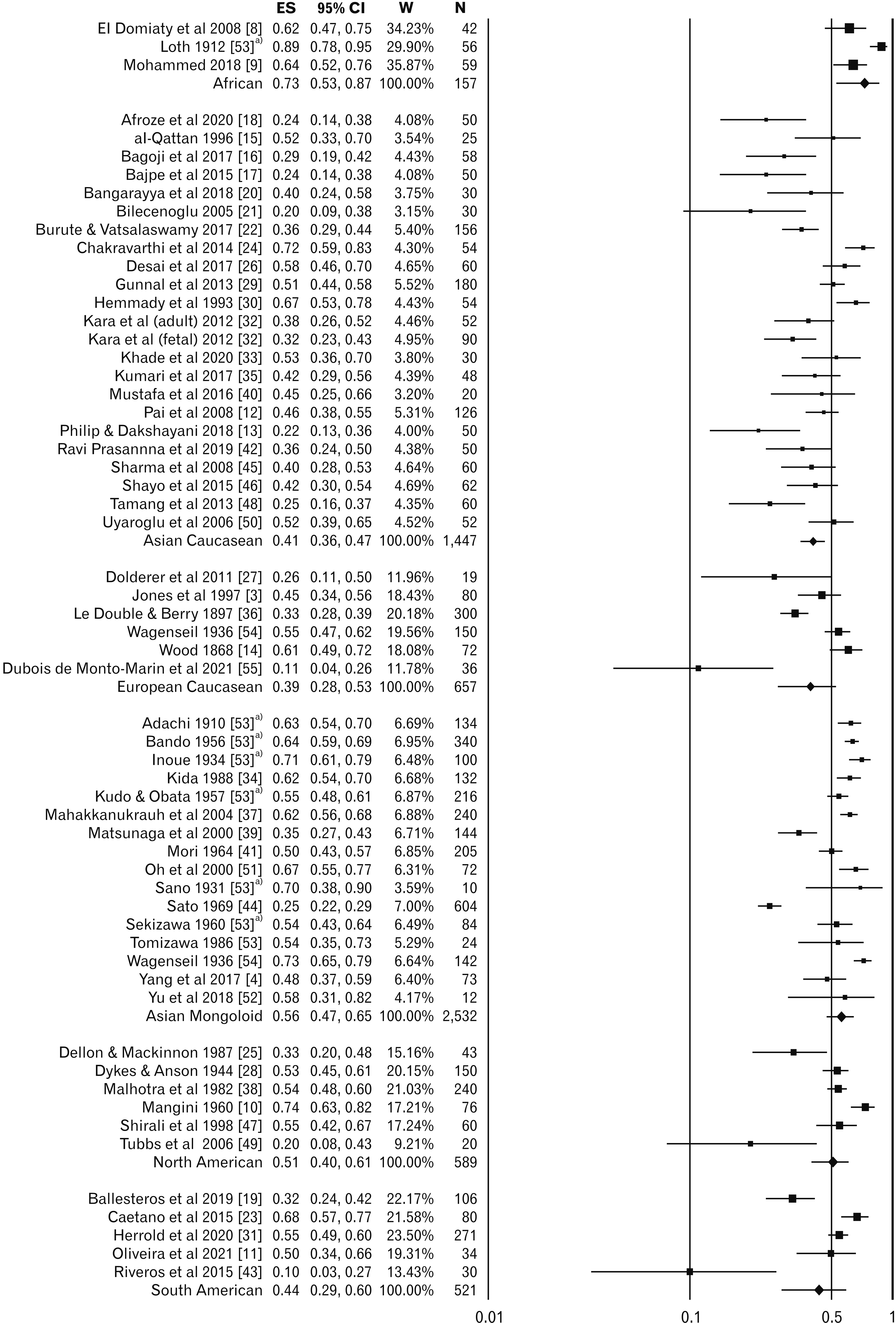 | Fig. 2Pooled weighted prevalence of accessory head of flexor pollicis longus variant. ES, effect size (log-odds ratio); CI, confidence interval; W, weight of study (inverse variance); N, sample size. a)Secondary reference was used because the data collected from secondary reference due to inaccessibility of original manuscript.
|
The pooled estimate of the only ahFDP in 1,627 limbs from 14 studies was 17% (95% CI, 13%–21%) (Fig. 3). The variability of the pooled estimate was 2% in sensitivity analysis and 8% in cumulative analysis. Thus, the heterogeneity of the estimate was 74.3%.
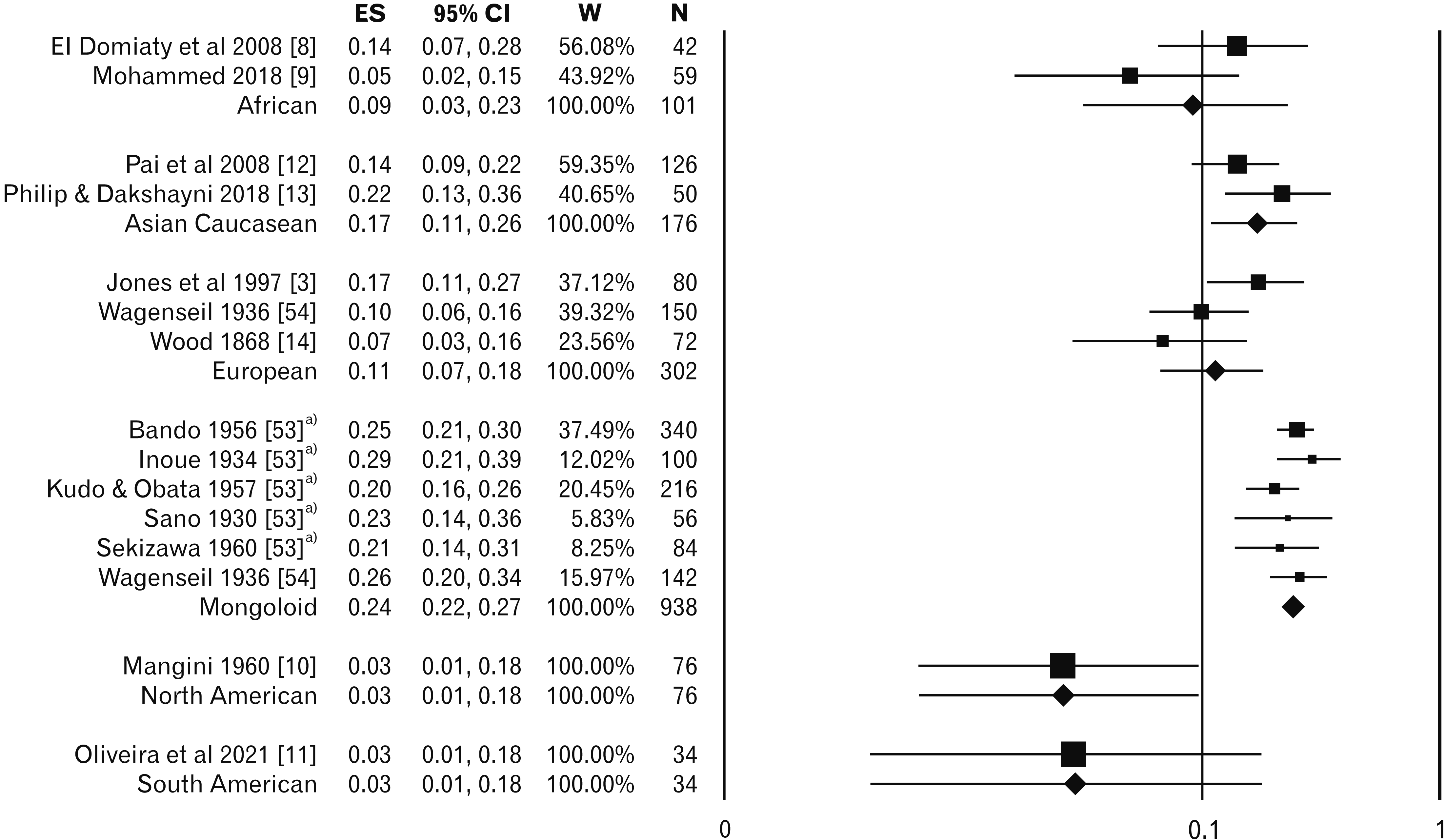 | Fig. 3Pooled weighted prevalence of accessory head of flexor digitorum profundus variant. ES, effect size (log-odds ratio); CI, confidence interval; W, weight of study (inverse variance); N, sample size. a)Secondary reference was used because the data collected from secondary reference due to inaccessibility of original manuscript.
|
Ethnic and geographical distribution
African studies have demonstrated the highest prevalence, 73% (95% CI, 53%–87%) of ahFPL in 157 limbs with nil heterogeneity. The Mongoloid population 56%, (95% CI, 47%–65%) in 2,532 limbs and North American population 51%, (95% CI, 40%–61%) in 589 limbs have similar prevalence. South American studies, including 521 limbs, have a prevalence of 44% (95% CI, 29%–60%). The Caucasian population (Asian 41% in 1,447 limbs and European 39 % in 657 limbs) has a lower prevalence of ahFPL than the ethnicities mentioned earlier. No studies were reported from the Australian population. The heterogeneity among studies of other ethnic groups varied from 76% to 94%.
The prevalence of ahFDP was 24% (95% CI, 22%–27%) in Mongoloid population without any heterogeneity of estimate. The same prevalence in African, Caucasian of Asian and European origin were 9% (95% CI, 3%–23%), 17% (95% CI, 11%–26%) and 11% (95% CI, 7%–18%), respectively. The prevalence in North and South American populations were based on only a single study, and they were 3% (95% CI, 0.7%–10%) and 3% (95% CI, 0.41%–18%), respectively. Most of the estimates have wider confidence intervals due to the low sample size.
Laterality and sex distribution
The laterality of ahFPL was examined in 1,275 limbs (Table 2). The occurrence of ahFPL was more frequent in right side (49%) (95% CI, 46%–53%) than left side (45%) (95% CI, 42%–49%) with rate difference of 5% (95% CI, 0.2%–11%, P=0.043). Almost, similar occurrences of ahFDP were in right and left upper limbs, i.e., 9% (95% CI, 5%–14%) and 10% (95% CI, 6%–15%), respectively. The unilateral occurrence of ahFDP was 8%, and bilateral occurrence was slightly higher, i.e., 10%. The data on sex distribution was inadequate. The prevalence of ahFPL was 38% in males and 13% in females in 402 limbs which would be misleading. The distribution of ahFDP in males and females was 12% and 23%, respectively. So, females have a double prevalence rate of ahFDP.
Table 2
Characteristics of variants of Gantzer’s muscle: laterality, sex, anatomical and morphological distribution
![]()
Anatomical distribution
The origin of ahFPL was evaluated in 1,283 limbs (Table 2). The commonest site of its origin was ME in 37% (95% CI, 35%–40%) followed by coronoid process of ulna (CP) in 24% (95% CI, 22%–26%), and muscle sheath of FDS in 15% (95% CI, 13%–17%). The dual origin from ME and CP has been observed in 8% (95% CI, 7%–10%). Antebrachial fascia also gave origin to ahFPL in 4% (95% CI, 3%–5%). The fascial sheath of FDS was the predominant site of origin for ahFDP, which was 74% (95% CI, 65%–82%). The origin ahFDP from ME, CP, and pronator teres were 15% (95% CI, 9%–23%), 6% (95% CI, 3%–13%) and 4% (95% CI, 2%–10%), respectively in 214 samples.
Gantzer’s muscle was inserted either in the muscle belly or tendon of FPL and FDP. The insertion of ahFPL was examined in 345 limbs (Table 3). The ahFPL was inserted in the muscle belly of FPL in 1/2nd to 2/3rd of the sample, and remaining samples were inserted on the tendinous part of FPL. The extent of ahFPL in the upper 1/3rd of the forearm was observed in 71% of the sample, followed by 23% in the middle 1/3rd and the remaining 6% extended up to the lower 1/3rd of the forearm. The insertion of ahFDP was predominantly on the tendon of the index finger, i.e., 47% (95% CI, 37%–57%), followed by the tendon of middle finger, i.e., 20% (95% CI, 13%–29%) (Table 3).
Table 3
Characteristics of variants of Gantzer’s muscle: insertion of both variants
![]()
The innervation of ahFPL was examined in 1,237 limbs (Table 2). AIN was the predominant supply of ahFPL in 2/3rd of samples (95% CI, 64.1%–69.3%). The median nerve supplied ahFPL in 1/3rd samples (95% CI, 30.6%–35.9%). Ulnar nerve innervated it in 0.1% samples (95% CI, 0%–4%). The innervation of ahFDP was AIN in 55.6% and medial nerve (MN) in 44.4% (Table 1).
Morphological distribution
The morphology of ahFPL was examined in a sample of 655 limbs (Table 2). The fusiform shape was the predominant shape of muscle which was observed in almost 3/4th of samples. The length of ahFPL varied from 6.9 to 12 cm, and width varied from 0.3 to 0.7 cm. The adequate data was unavailable to estimate the morphological distribution of ahFDP. However, the Fusiform shape was predominant in ahFDP.
Risk of bias
Most of the studies did not provide adequate information about sex distribution. The studies may have a high risk of bias (ROB) because the authors did not report adequate anatomical and morphological details [16, 17, 20, 25, 30, 41, 47]. The studies with a higher ROB reported less prevalence of ahFPL, i.e., 37% (95% CI, 27%–48%) than moderate and low ROB studies, i.e., 47% and 52%. The prevalence of ahFDP was similar in both moderate and low risk. None of the studies was categorized into a high ROB for ahFDP.
Publication bias
The funnel plot of the current meta-analysis was symmetrical. Egger’s linear regression test for publication bias was conducted, refuting the possibility of publication bias (P-value=0.858). Trim and fill analysis was undertaken to estimate pooled prevalence. The observed pooled prevalence was similar to the estimated pooled prevalence.
Go to :
Discussion
Summary of findings
In the current meta-analysis, the prevalence of Gantzer’s muscle was 65% in 5,903 upper limbs, which is inconsistent with the results of the prior meta-analysis. The pooled prevalences of ahFPL and ahFDP variants were 48% and 17%, respectively. The pooled prevalence that varied in the cumulative analysis was 4% (48% to 52%). The overall heterogeneity was 89%, which was much lower than earlier meta-analysis. The African, Mongoloid, and North American ethnicities had a higher prevalence than other ethnic groups for ahFPL. It was more frequent on the right side. ME was the commonest site of origin for ahFPL, and the muscle belly of FPL was the most common site of its insertion.
Similarly, the fascial sheath of FDS was the commonest site of origin, and FDP tendon for the index finger was the commonest insertion site for ahFDP. AIN predominantly innervated both variants. The fusiform shape was most frequent in both variants.
The ahFPL is a wide variation in modern humans, and it has clinical significance, especially in AIN and median nerve compression. For example, the Gantzer’s muscle, or ahFPL, which acts as an additional head of the FPL, would enhance thumb flexion, indicating a functional difference from other primates [12]. Similarly, authors speculate that ahFDP might be improving pinching action or flexion of other fingers.
Agreement or disagreement with other studies
The prevalence of Gantzer’s muscle has been shown to be 44.2%, with a 95% confidence interval of 34.7% to 54% in a previous meta-analysis conducted by Roy et al. (2015) [6]. The authors have computed only the prevalence of the ahFPL variant in 2,358 upper limbs. We considered both variants for pooled estimation. The prevalence of ahFPL in the present meta-analysis was 48% (95% CI, 44%–52%) in 5,903 upper limbs. The difference in prevalence between both meta-analyses is attributed to higher sample size. The present meta-analysis examined more than double the sample size of the previous meta-analysis. The authors [3, 12, 37, 53] reported Gantzer’s muscle prevalence, which varied from 60% to 71%. These authors reported both variants. The studies [14, 15, 25, 28, 38, 41, 47] reported lower prevalence (39%–55%) and they only included ahFPL variant. The second variant, i.e., ahFDP, might have been missed due to ignorance. Such ignorance may be dealt with in the classification of these variants. The variants of Gantzer’s muscle may be classified as per its morphology and attachment (Fig. 4). They were classified into three types. The suggested classification is as follows, based on the review of various literatures, which could be helpful in the future to study the relationship with the nearby structure.
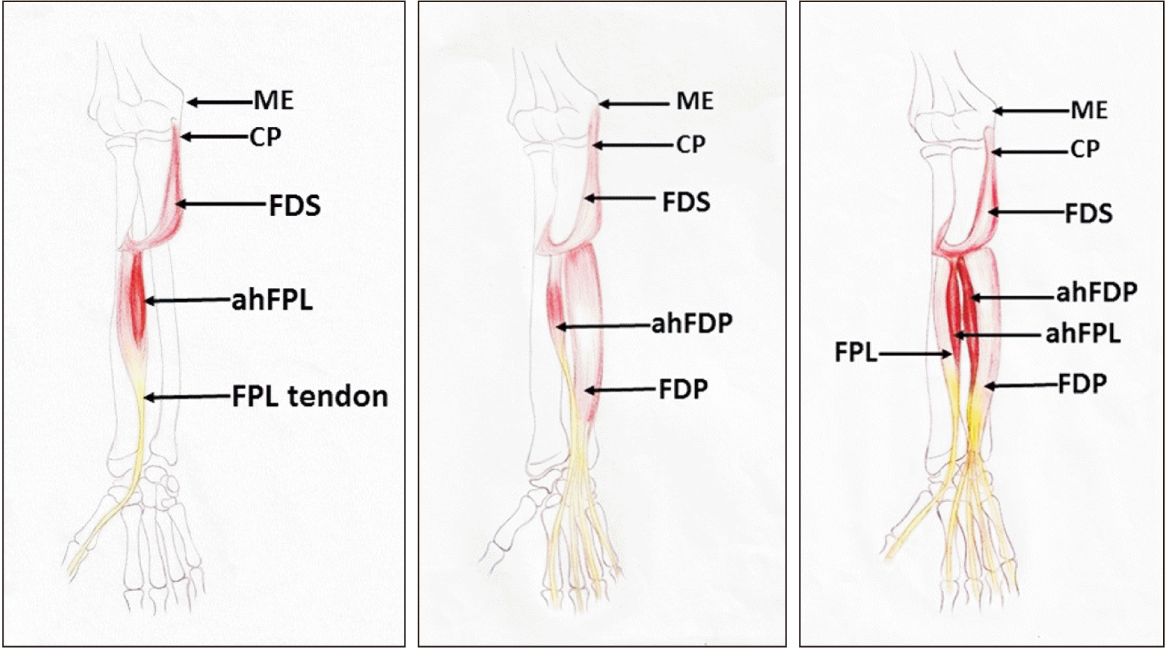
Type I: ahFPL
Type Ia: Insertion into the belly of FPL
Type Ib: Insertion into the tendon of FPL
Type II: ahFDP
Type IIa: Insertion into the first tendon of FDP (index finger)
Type IIb: Insertion into the second tendon of FDP (middle finger)
Type IIc: Insertion into the third tendon of FDP (ring finger)
Type IId: Insertion into the fourth tendon of FDP (little finger)
Combination of any of two or more may be denoted as IIbcd or IIab, etc.
The sub-category of type III will be developed in the future with the availability of adequate data.
Type III is rare, and this subtype was not included for the pooled prevalence of variants of Gantzer’s muscle due to inadequate description and data. The forearm muscle blastema develops from Interzone blastema over cartilage of developing radius and ulna at the 4th week of intrauterine life [6, 8]. The superficial muscle blastema migrates earlier than the blastema of the deeper muscle. FDS, FDP, and FPL are phylogenetically newer muscles that develop from volar hand blastema, and they ascend upwards to reach the definitive origin [12]. The fascial sheath of superficial muscles like FDS or pronator teres a guide for deeper FPL and FDP. The variants of Gantzer’s muscle might be developmental errors [3, 12]. The FPL is the newer muscle (phylogenetically) among the forearm flexors, which could be the reason for the higher prevalence of ahFPL.
Clinical implications
These muscles generally lie deep to MN and are innervated by AIN [56]. The Gantzer’s muscle has long been debated as a cause of neurological compression of AIN or MN. Tabib et al. [57] documented AIN syndrome caused by Gantzer’s muscle. The patient had isolated weakness of the FPL and was unable to pinch between thumb and index finger. The pronated and extended elbow may cause characteristic pain in front of the mid-forearm. Electrodiagnostic investigation revealed moderate slowing of conduction velocity. On surgical exploration, Gantzer’s muscle along with swollen AIN. The surgical removal led to the resolution of pain within a month. Similar reports were also noted in many other literatures [57-60]. Such syndrome was named as Kiloh–Nevin syndrome or AIN syndrome. This disorder also often leads to loss of pinching [12, 56].
Limitation & potential bias
The high heterogeneity of pooled prevalence and inadequate data of sex distribution were the significant limitations. The high heterogeneity was mainly attributed to the variable population of studies. Most old studies lack sex-based data, and retrieving such data from the author's communication was impossible. It is the scope of further research. The strength of the current meta-analysis is that the present study has a double sample size than the previous one.
Go to :
Conclusion
The prevalence of Gantzer’s muscle is 65%. It has two major variants: ahFPL and ahFDP. Both variants have population and sex variations. The origin of both variants is almost similar, but their insertions vary. Accessory head of FPL inserts on belly or tendon of FPL. Still, the other variant (ahFDP) inserts on the tendon of the FDP for the index and middle finger.
Go to :
 XML Download