

 PDF
PDF Citation
 Print
Print


Introduction
Spleen is the largest lymphatic organ in the body and its main function is to produce blood (hematopoiesis), to store blood, to filter the blood and to provide immunological responses [1, 2]. The spleen’s becoming larger and heavier explains splenomegaly as a gold standard definition [3]. Radiological assessment of the changes in spleen size is important in hematological and lymphatic system diseases [4]. Multidetector computed tomography (MDCT), ultrasonography (USG), and magnetic resonance imaging (MRI) techniques are used in the radiological assessment of the spleen [5].
The rapid removal of blood cells with the increase in the physiological functions of the spleen is called hypersplenism [6, 7]. Hypersplenism is generally characterized by the decrease in shaped elements of the blood with the enlargement of the spleen [8]. Studies conducted have reported that the strongest correlation between the change in the size of spleen and hematological findings is with the platelet (PLT) level [9-12]. There are also studies conducted on different disease groups which have associated spleen volume with the level of white blood cell (WBC), hematocrit (HCT) level, hemoglobin (HGB) level, lymphocyte percentage (LYM%), and level of aspartate aminotransferase (AST) and alanine aminotransferase (ALT) [10, 12-16].
Cavalieri principle, which is a stereological method, enables a reliable and efficient prediction of the volumes of objects [17, 18]. In this method, which is also used to calculate the volume of irregularly shaped structures, the structure is divided into parallel slices with equal thickness. By taking sections from radiological imaging methods of MDCT and MRI, Cavalieri principle makes these applicable in radiological images [19].
In our literature review, although there are studies examining the relationship between spleen volume and blood parameters in various diseases and by considering various spleen indices, or comparing the splenomegaly group with the normal group, we have not encountered any studies showing stereological methods in healthy individuals. The aim of the present study is to find out whether there are correlations between spleen volume with basic hemogram and biochemical parameters in healthy individuals. Another aim of the study is to compare the mean spleen volume in terms of sex and age.
Go to : 
Materials and Methods
Study population
The present study was initiated with the October 11th, 2020 dated and 364 protocol numbered approval of our university’s non-invasive ethics committee. The sample of this study was formed from people aged 25 to 55 years who were found to be healthy between January 2019 and January 2020. A total of 74 individuals (34 male and 40 female) who had received MDCT and whose basic hemogram and biochemistry tests had been done were included in the study. Individuals who had undergone trauma or operation in the related area, and those with an oncological or hematological history were excluded from the study.
MDCT protocol
The radiological images used in the study were obtained from supine CT images with a slice thickness of 2.0 mm by using a 16-slice MDCT device (Aquilion 16; Toshiba Medical Systems, Tokyo, Japan). Imaging protocol values were as tube voltage: 120 kV, gantry rotation time: 0.75 seconds and pitch: 1.0 mm.
Basic hemogram and biochemical parameters
In this retrospective study, basic hemogram and biochemistry parameters formed as a result of the laboratory analysis of venous blood taken from the individuals were used. The basic hemogram parameters used were red blood cell (RBC) level, WBC level, LYM%, monocyte percentage (MON%), neutrophil percentage (NEU%), eosinophil percentage (EOS%), HGB level, HCT level, PLT level, mean corpuscular volume (MCV) and mean corpuscular HGB concentration (MCHC). Biochemical parameters used were creatinine, AST, ALT, sodium (Na), potassium (K) and chlorine (Cl).
Estimation of spleen volume from computed tomography images
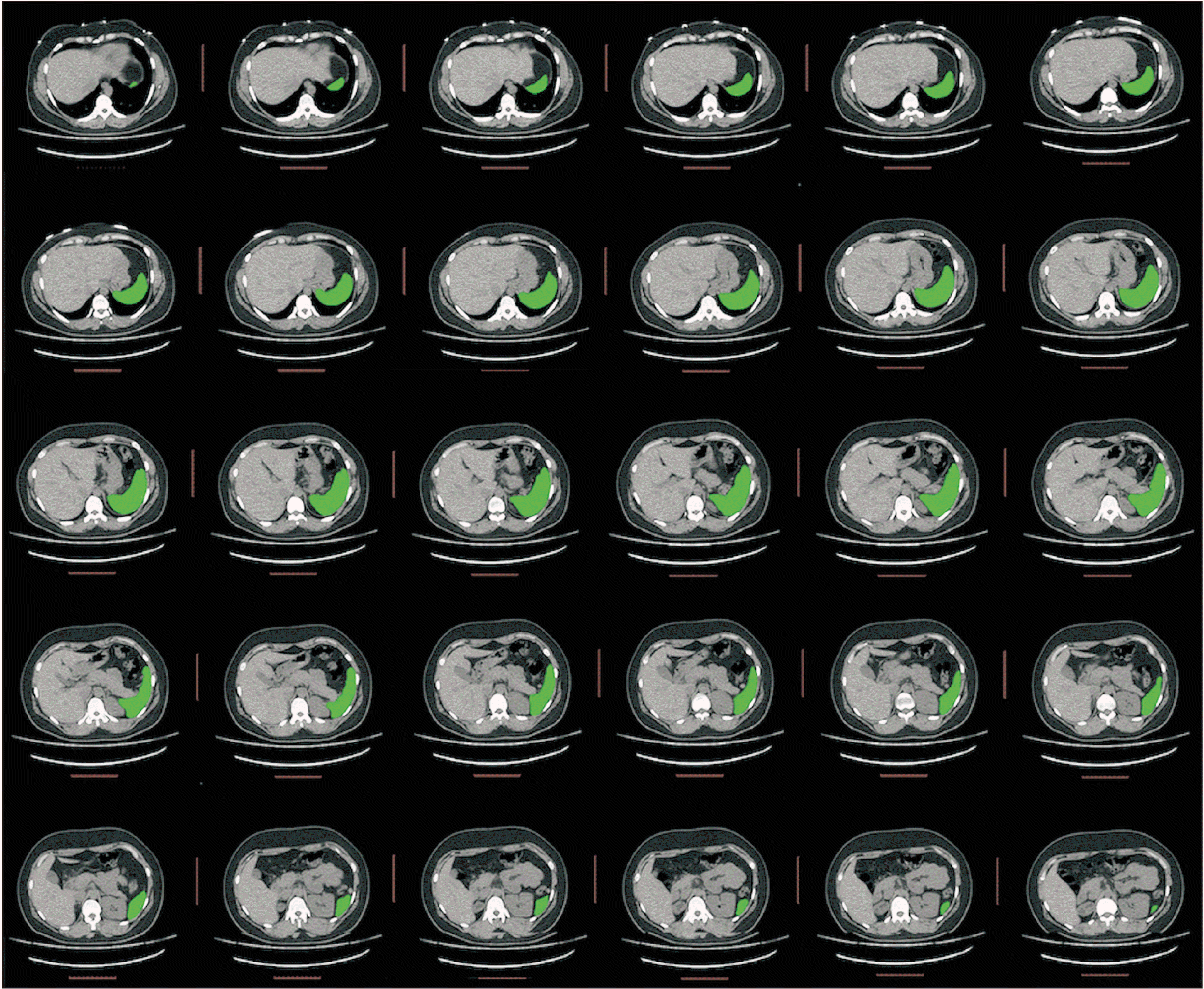
All images of the individuals were transferred to an imagining analysis program ImageJ (version 1.53d; National Institutes of Health, Bethesda, MD, USA) with Digital Imaging and Communications in Medicine (DICOM) format. An abdominal imaging series that had 2 mm space and axial plan were used. Spleen volume measurements were performed from CT images by using the planimetry approach of Cavalieri principle. In all the sections with spleen, surface areas of the spleen were calculated with planimetry approach (Fig. 1). This procedure was carried out by drawing the boundaries of the spleen through polygon selection tool. Projection area of the spleen at each cross section was obtained from the program. The surface area values obtained were recorded on the Excel (Microsoft, Redmond, WA, USA) as electronic table. By using the Excel program, surface area values calculated with ImageJ program were summed and spleen volume was estimated in cm3 by multiplying the slice thickness of 0.2 [17].
Statistical analysis
The data obtained in the study were analyzed by using Minitab v.17 program. In defining the data, numbers were used for categorical variables, mean±standard deviation was used for parametric continuous variables and median, minimum (min) and maximum (max) values were used for nonparametric continuous variables. Normality distribution of continuous variables was evaluated with Anderson Darling test. In the comparison of continuous variables between the two groups, those with normal distribution were evaluated with Student t-test, and those which were not normally distributed were evaluated with Mann–Whitney U-test. The linear relationship between the continuous variables was evaluated with Pearson correlation test. P<0.05 value was considered as statistically significant.
Intra-rater reliability
In this study, spleen volumes were measured 10 times from CT images of 3 different individuals to evaluate in-class reliability and intra-observer agreement, and the measurements were evaluated with the intraclass correlation test.
Coefficient of error
In the application of the Cavalieri principle, coefficient of error (CE) is used to evaluate the reliability of the point density and cross-section spacing of grids. The CE, or relative standard error, represents the precision of the volume value estimated using the Cavalieri principle [17]. The calculated CE value should be less than 10% [17, 20, 21]. In this study, CE was calculated according to the formulas created by Gundersen and Jensen [22].
Go to :
Results
By using the correlation coefficient values observed in accordance with the literature scan, power analysis performed with the G*Power (version 3.1). The minimum number of individuals to be included in the study was calculated as 67 in order to complete our study with 95% confidence level and 80% power.
The CE values of the spleen volumes calculated in this study were between 0.35% and 1.17%, and the average CE value was calculated as 0.76%. This value is in the acceptable range.
As a result of the measurements, the intraclass reliability coefficient of the calculated spleen volumes was calculated as 0.976.
Results regarding demographic characteristics
It was found that the individuals were between 25 and 55 years of age (41.89±8.86 years), mean age of the female participants was 41.55±9.28 years, while mean age of the male participants was 42.29±8.46 years. No statistically significant difference was found between the mean ages of the sex (P=0.90).
Radiological findings
Student t-test was conducted to find out whether spleen volume differed significantly in terms of sex variable. Mean spleen volume was found as 303.4 cm3 in male, while it was found 232.8 cm3 in female. Spleen volume of male was found to be 30.32% larger than those of female (P<0.05).
Basic hemogram findings
MON%, HCT and MCHC values, the basic hemogram values which were not distributed normally, were found to be higher in male when compared with female and this difference was significant (P<0.05). No statistically significant difference was found in EOS% between the sex (P>0.05).
HGB and RBC values, the basic hemogram values which were distributed normally, were found to be higher in male when compared with female and this difference was significant (P<0.05). No statistically significant difference was found in WBC, LYM%, NEU%, PLT and MCV between the sex (P>0.05).
Biochemical findings
ALT values, the biochemical values which were not distributed normally, were found to be higher in male when compared with female and this difference was significant (P<0.05). No significant difference was found in AST parameter in terms of sex (P>0.05).
Creatinine and Na values, the biochemical values which were distributed normally, were found to be higher in male when compared with female and this difference was significant (P<0.05). No significant difference was found in K and Cl parameters in terms of sex (P>0.05).
Correlation analysis findings regarding spleen volume
As a result of the Pearson correlation analysis we conducted to find out the correlation between spleen volume with basic hemogram and biochemistry parameters, a negative, significant and linear correlation was found between spleen volume and LYM% and PLT level. In other words, it was found that spleen volume showed a tendency to get smaller when these parameters increased (r<0; P<0.05). Spleen volume was found to have a positive, significant and linear correlation with HGB, HCT, RBC, and ALT parameters. In other words, it was found that spleen volume showed a tendency to get larger when these parameters increased (r>0; P<0.05). No significant correlation was found between WBC, MON%, NEU%, EOS%, MCV, MCHC, AST, Na, K, Cl and creatinine level and spleen volume (P>0.05). A negative correlation was found between spleen volume and age (r=–0.145; P<0.05); in other words, it was found that spleen volume showed a tendency to get smaller with aging (Table 1, Figs. 2, 3).
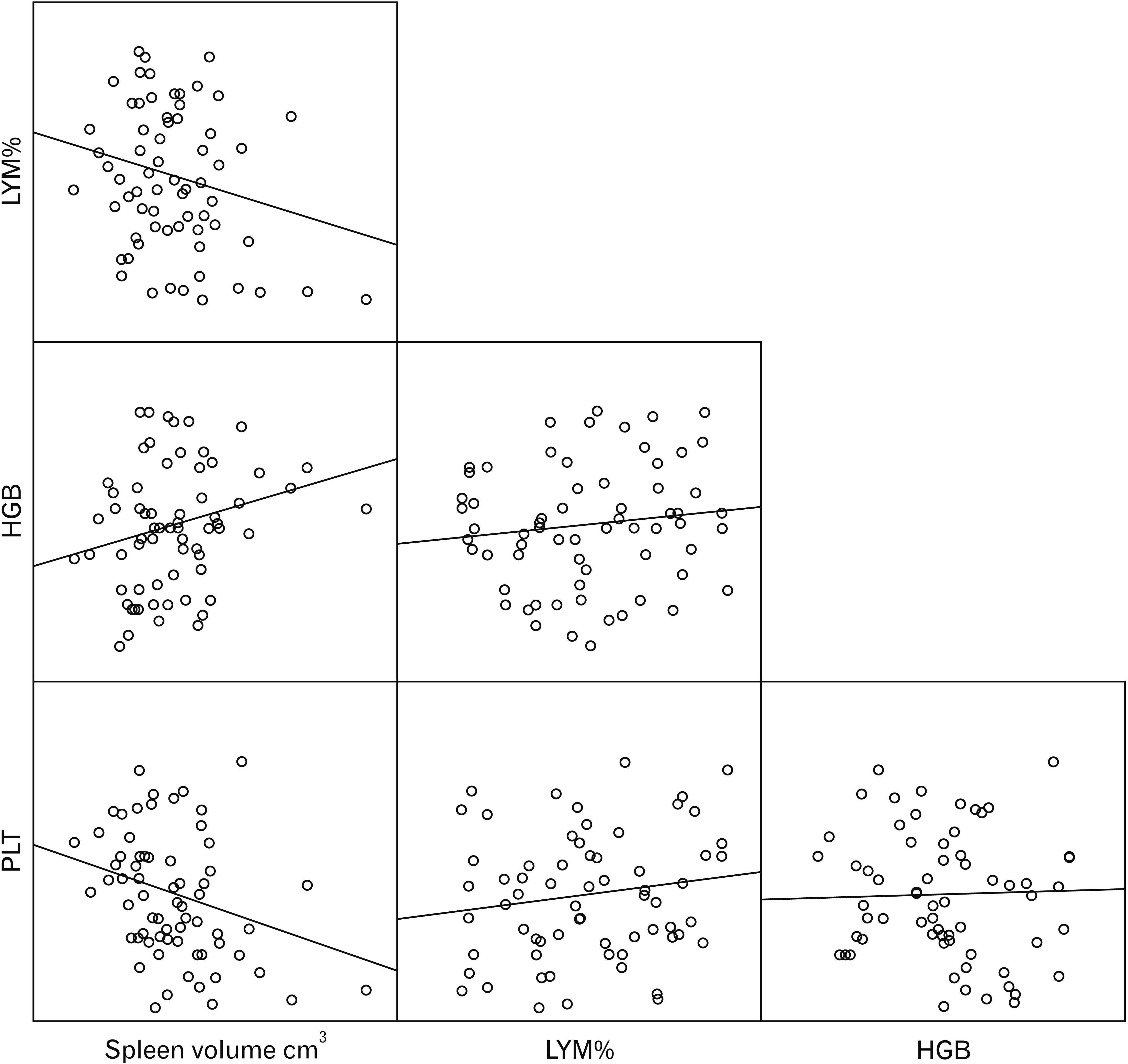 | Fig. 2Visual matrix of correlations between spleen volume, platelet (PLT), hemoglobin (HGB), and lymphocyte percentage (LYM%).
|
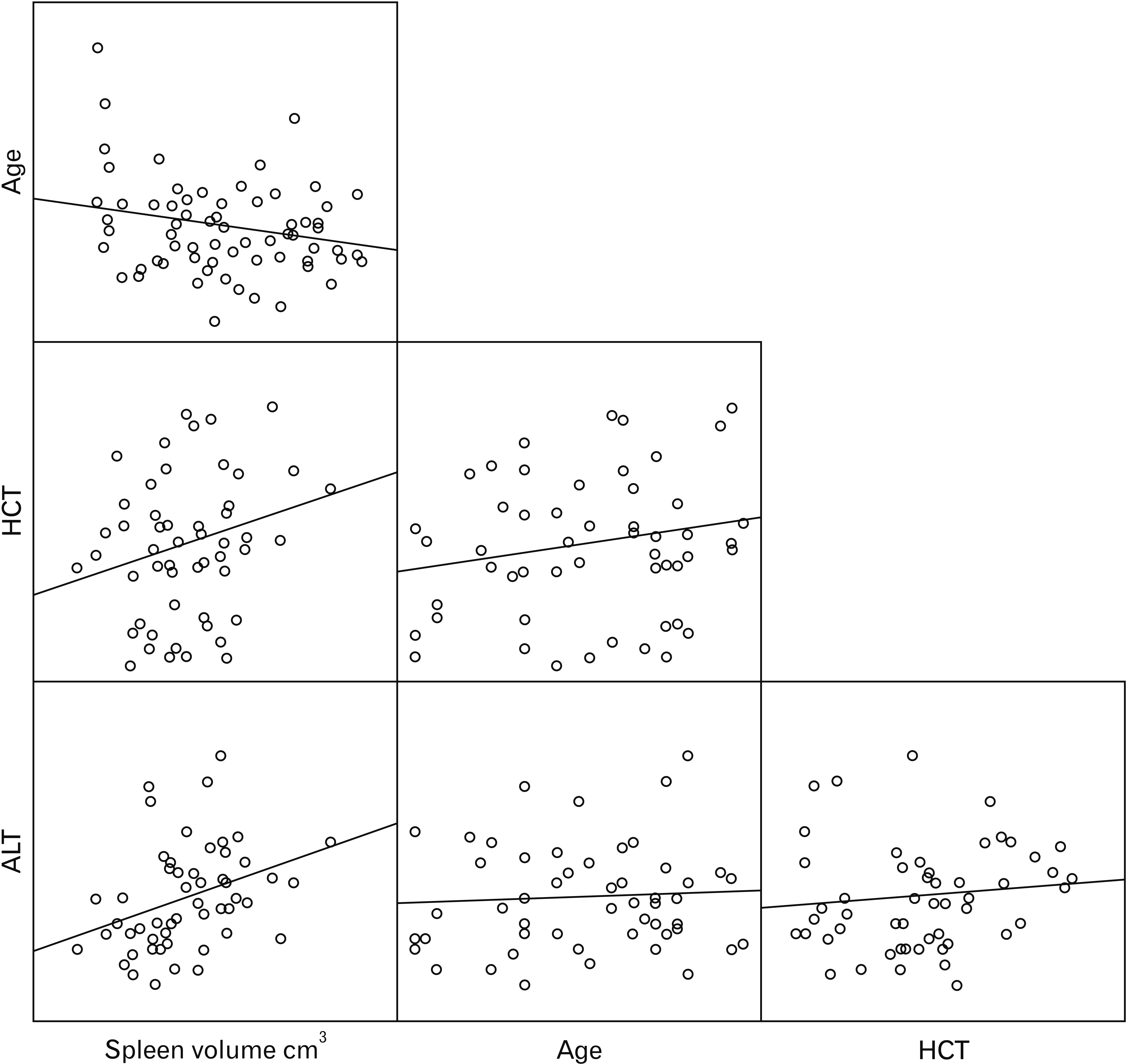 | Fig. 3Visual matrix of correlations between spleen volume, age, alanine aminotransferase (ALT), and hematocrit (HCT) level.
|
Table 1
Correlation findings regarding the spleen volume
| Parameter | Spleen volume | |
|---|---|---|
| r | P-value | |
| Age | –0.145 | 0.041* |
| WBC | –0.111 | 0.349 |
| LYM% | –0.224 | 0.049* |
| MON% | 0.207 | 0.077 |
| NEU% | 0.171 | 0.144 |
| EOS% | –0.014 | 0.906 |
| HGB | 0.228 | 0.050* |
| HCT | 0.237 | 0.042* |
| PLT | –0.271 | 0.020* |
| MCV | –0.111 | 0.344 |
| MCHC | 0.048 | 0.682 |
| RBC | 0.375 | 0.001* |
| Creatinine | –0.009 | 0.940 |
| AST | 0.179 | 0.168 |
| ALT | 0.345 | 0.007* |
| Na | 0.254 | 0.078 |
| K | 0.136 | 0.350 |
| Cl | 0.057 | 0.749 |
r, correlation coefficient; WBC, leukocyte level; LYM%, lymphocyte percentage; MON%, monocyte percentage; NEU%, neutrophil percentage; EOS%, eosinophil percentage; HGB, hemoglobin level; HCT, hematocrit level; PLT, platelet level; MCV, mean corpuscular volume; MCHC, mean corpuscular hemoglobin concentration; RBC, red blood cell; AST, aspartate aminotransferase; ALT, alanine aminotransferase; Na, sodium; K, potassium; Cl, chlorine. *Statistical significance (P<0.05).
![]()
Go to :
Discussion
In this study which aimed to examine the possible correlations between spleen volume with basic hemogram and biochemical parameters, it was found that spleen volume was correlated with sex and male had larger spleen volume when compared with female. It was found that spleen volume showed a tendency to get smaller with age. It was concluded that while spleen volume was positively correlated with HGB, HCT, RBC, and ALT parameters, it was negatively correlated with LYM% and PLT level. No association was found between spleen volume and WBC, MON%, NEU%, EOS%, MCHC, MCV, AST, Na, K, Cl and creatinine level.
According to studies conducted, spleen volume shows a tendency to get smaller with increasing age [23-25]. The negative correlation found between age spleen volume and age in this study supports this correlation (r=–0.145).
Some studies in literature have shown that spleen volume differs in terms of sex and male have larger spleen than female [23, 25, 26]. On the contrary, Harris et al. [24] reported no significant difference between sex in terms of spleen volume. They found that mean ages of sex groups were different and reported that they found correlation between spleen volume and sex when they formulized in terms of age and formed spleen indices. In the present study, according to the results obtained, we found that spleen volume and sex were correlated and male had larger spleen when compared with female (P=0.006). This finding has shown that spleen volume is a criterion showing sexual dimorphism.
In this study, we found a negative significant correlation between spleen volume and PLT level (r=–0.271). There are studies in literature showing this association and their results support this study [10-12, 27]. The increase in PLT pools with the enlargement of spleen physiologically and the fact that PLTs are stored more in the spleen have been explained with the decrease in PLT count in the circulation [28]. On the other hand, in a study conducted on 24 individuals by Singh et al. [15] and in a study conducted on 61 individuals by Vasconcellos et al. [29], no correlation was reported between spleen volume and PLT level. We think that the reason why these researchers could not find a correlation was due to the fact that the study populations consisted of fewer individuals when compared with other studies.
There are different findings in literature on the association between spleen volume and PLT count. The differences in the main reasons underlying splenomegaly cause the results to show differences [5]. There are some studies reporting a negative correlation between spleen volume and PLT count [10, 15, 29, 30]. However, there are also studies which do not report a correlation between spleen volume and PLT count [12, 14]. As a result of the study we conducted, we also found that there was no significant correlation between spleen volume and PLT count (P=0.020).
Wadenvik and Kutti [12] reported that erythrocyte pools would also increase with the increase in spleen size and as a result there would be a decrease in the RBC level in blood. They also stated that HCT level would accompany this decrease. On the other hand, a positive correlation was found between spleen volume and RBC level in the present study (r=0.375). We believe that this difference may be due to secondary polysemy of the RBC level of our study population, which may have been caused by erythrocytosis due to tissue hypoxia caused by environmental factors. Blood viscosity increases as a result of the increase in erythrocyte mass [31]. The spleen enlarges due to increased blood flow [32]. Positively correlated HCT and HGB parameters showed a positive correlation due to the correlation of the spleen with RBC.
In their study they evaluated spleen volume following acute stroke, Chiu et al. [14] found a negative correlation between lymphocyte percentage and spleen volume. As a result of the present study which was conducted in healthy individuals, we found negative correlation. Also, Chiu et al. [14] could not find any correlations between spleen volume and MON%, and our study supports this. We think that these correlation found by Chiu et al. [14] may not be a correlation directly resulting from the post-acute stroke period. The negative correlation between spleen volume and lymphocyte level has been explained with the number of lymphocytes sent by the spleen to the circulation in case of the spleen getting smaller and contracting and this result is similar to the results of experiments conducted on animals [33].
In their study they examined the correlation between splenomegaly and hematological findings in patients with hepatosplenic schistosomiases, Leite et al. [13] found a significant difference in AST and ALT values between the control group and the hepatosplenic group. In the present study, we could find a positive correlation between spleen size and ALT value (r=0.345; P=0.007). Present study, which we conducted in healthy people, also shows that spleen volume is associated with ALT and suggests that spleen size and liver size may also be associated.
The relationship between spleen volume and WBC finds different answers in the literature. Some studies mention the existence of a relationship between them [10, 15, 26, 27]. However, there are also studies stating that there is no relationship [12, 14]. We did not observe a significant relationship between them in this study. The fact that the results are different in studies conducted in different diseases suggests that there may be other factors determining this relationship.
Dellenback et al. [34] performed blood tests before and after splenectomy in hunting dogs and stated that there was a significant decrease in HCT, RBC and MCHC values, and a significant increase in the number of MCV and PLT in the measurements they made after splenectomy. When we interpreted the splenectomy procedure as a decrease in volume, these findings were consistent with our finding that the HCT and RBC values obtained in our study had a positive correlation with the spleen volume, and the PLT value had a negative correlation. In the present study, although the direction of correlations between MCV and MCHC and spleen volume showed the same direction as their study, it was not found significant (P>0.05).
There are a large number of methods used in the prediction of spleen volume [4, 35, 36]. Splenic index and ellipsoid formulas, which use the width, length and height measurements of the spleen in three planes, is used for a quick result. However, calculations by using formulas on irregularly shaped structures cause deviations from the actual volume value. On the other hand, calculations with Cavalieri principle, which is a stereological method, is a method with high efficiency and accuracy that can be applied to calculate the volumes of irregularly shaped structures [19]. We think that using Cavalieri principle in this study increased the accuracy of the study. Other strength of our study is that we have many parameters. On the other hand, the limitations are the fact that it was a retrospective study and the study did not discuss a single disease, such as splenomegaly.
In conclusion, the present study shows the correlation between spleen volume determined with Cavalieri principle and hematological and biochemical parameters. Our findings show that spleen volume is associated with PLT, LYM, RBC, HGM, HCT, and ALT parameters. It is concluded that this situation, which has found different answers in the literature, is affected by environmental factors and pathophysiological mechanisms. As a conclusion, we believe that, monitoring the changes in spleen volume in pathologies of the spleen and the hematological and biochemical parameters that may occur due to these changes may be a reference for clinicians working in the field.
Go to :
 XML Download