See the reply "READER’S FORUM" in Volume 52 on page 247.
Abstract
Objective
The purpose of this study was to compare the differences in dentoskeletal and soft tissue changes following conventional tooth-borne rapid maxillary expansion (RME) between adolescents and adults.
Methods
Dentoskeletal and soft tissue variables of 17 adolescents and 17 adults were analyzed on posteroanterior and lateral cephalograms and frontal photographs at pretreatment (T1) and after conventional RME using tooth-borne expanders (T2). Changes in variables within each group between T1 and T2 were analyzed using Wilcoxon signed-rank test. Mann–Whitney U test was used to determine the differences in the pretreatment age, expansion and post-expansion durations, and dentoskeletal and soft tissue changes after RME between the groups. Spearman’s correlation between pretreatment age and transverse dentoskeletal changes in the adolescent group was calculated.
Results
Despite similar amounts of expansion at the crown level in both groups, the adult group underwent less skeletal expansion with less intermolar root expansion after RME than the adolescent group. The skeletal vertical dimension increased significantly in both groups without significant intergroup difference. The anteroposterior position of the maxilla was maintained in both groups, while a greater backward displacement of the mandible was evident in the adult group than that in the adolescent group after RME. The soft tissue alar width increased in both groups without a significant intergroup difference. In the adolescent group, pretreatment age was not significantly correlated with transverse dentoskeletal changes.
Rapid maxillary expansion (RME) is a method for orthopedic expansion of the maxillary arch by opening the midpalatal suture in patients with transverse maxillary discrepancy.1,2 Unlike the mandible, wherein skeletal expansion is practically impossible without surgical interventions,3 skeletal expansion of the maxilla can be successfully performed using a conventional non-surgical tooth-borne expander,1 such as the commonly used hyrax-type expander.4
Since transverse growth of the maxillary complex is completed before the anteroposterior and vertical growth,5 transverse maxillary discrepancy should be corrected relatively early. Patients older than 15 years of age usually cannot undergo successful skeletal expansion using a conventional tooth-born expander because closure of the midpalatal suture would have begun and the resistance to orthopedic expansion is considerably greater.6,7 If orthopedic expansion with a conventional tooth-borne expander fails, heavy forces are transmitted to the anchor teeth and the surrounding periodontium within a short period. Therefore, conventional RME using a tooth-borne expander in late adolescents and adults may lead to potential complications, including pain, gingival recession, bone loss, and root resorption as well as questionable success of orthopedic expansion.8
Consequently, surgically-assisted rapid maxillary expansion (SARME)9 and bone-borne expanders using skeletal anchorage10,11 were introduced to overcome the potential shortcomings of the conventional tooth-borne expanders. However, these procedures increase the costs and risks of infection because of the need for additional invasive surgical procedures.12
Previous studies have analyzed dentoskeletal and soft tissue changes following midpalatal suture opening by conventional RME in growing patients. In adolescents, conventional RME results in triangular expansion with the nasal bone as a hinge in the frontal plane,2,6,13 an increase in the vertical dimension of the face, backward rotation of the mandible,1,14,15 and an increase in soft tissue alar width.16,17 Clinically successful correction of transverse maxillary deficiency using conventional RME in adults has also been reported.18,19 However, information on the dentoskeletal and soft tissue changes due to conventional RME in adults is limited because most studies have used plaster models for the analyses.8,19 Considering the progressive ossification and interdigitation of the midpalatal suture with aging, conventional RME may result in different dentoskeletal and soft tissue changes with aging. Therefore, the aim of this study was to evaluate the differences in dentoskeletal and soft tissue changes between adolescents and adults following conventional RME using tooth-borne expanders. The null hypothesis was that there would be no significant differences in dentoskeletal and soft tissue changes following RME using conventional tooth-borne expanders between the two groups.
This retrospective two-group observational study was performed according to the STrengthening the Reporting of OBservational studies in Epidemiology (STROBE) guidelines.20 Consecutive patients of pretreatment age less than 15 years and those older than 18 years of age who underwent RME using a hyrax-type expander at the Department of Orthodontics, Seoul National University Dental Hospital between 2009 and 2019 were included in this study. Patients with available complete series of posteroanterior (PA) and lateral cephalograms and frontal photographs at pretreatment (T1) and after expansion (T2) were included. All patients were diagnosed with transverse maxillary discrepancy. The records at T1 were collected for routine pretreatment evaluation, and the records at T2 were collected before commencing the second phase of treatment (at least 2 months after cessation of expansion). Cephalograms and frontal photographs were obtained with the patient in the resting lip and natural head positions. All cephalograms were obtained using Asahi CX-90SP II (Asahi Roentgen, Kyoto, Japan) under the same scanning conditions (76 kVp, 80 mA, 0.32 seconds of exposure, and magnification ratio of 110%). Patients with the following conditions were excluded: (1) craniofacial syndromes; (2) history of trauma; (3) temporomandibular disorders; (4) history of orthodontic treatment; and (5) gingival recession or possible bony dehiscence around the anchor teeth. The study design was approved by the Institutional Review Board of the University (S-D20190027).
Overall, 34 patients were included in this study. Of these, 17 patients younger than 15 (11.2–14.6) years were categorized as the adolescent group; those who were older than 18 (18.2–26.7) years were included in the adult group. The age criterion was based on the fact that somatic growth in Koreans is completed by the age of 17 years.21 When cervical vertebral maturation (CVM) was used to assess skeletal maturation,22 all participants in the adolescent group had CVM stage 4 (circumpubertal) or less and those in the adult group had CVM stage 5 or more (completion of active growth). Power analysis was performed using G*Power 3.1 (Heinrich-Heine, Düsseldorf, Germany).23 Based on a previous study,8 at least 12 patients per group were required to determine differences between the groups with an alpha error of 0.05 and a power of 0.8. Consequently, 17 patients were included in each group.
After fitting bands on the maxillary first premolars and molars of patients, an expander with a jackscrew (Dentaurum GmbH & Co. KG, Ispringen, Germany) was fabricated in the conventional way. Adolescents were instructed to activate the expander by two-quarter turns per day (0.50-mm widening per day), while adults were instructed to activate it by two-quarter turns per day and reduce it to one-quarter turn per day (0.25-mm widening per day) after suture opening. Successful opening of the midpalatal suture was presumed by the appearance of midline diastema6 and PA cephalogram at T2. Transverse maxillary deficiency was overcorrected until the palatal cusp tip of the maxillary molar contacted the buccal cusp tip of the mandibular molar just before achieving a scissor-bite relationship. During the expansion period, the patients were recalled at 1-week intervals to assess the progress. After the completion of expansion, the expander’s jackscrew was fixed, and the expander was retained for more than 2 months.
An investigator blinded to patient information assessed the dentoskeletal and soft tissue variables on cephalograms and frontal photographs. Dentoskeletal variables were assessed using V-Ceph 8.0 (Osstem Implant, Seoul, Korea) on PA and lateral cephalograms. Previously described soft tissue variables16 were analyzed on frontal photographs using the picture archiving and communication system viewer (Infinitt Healthcare, Seoul, Korea). All soft tissue variables are presented as a percentage of the interpupillary distance to nullify the effect of size differences on photographs. The landmarks, reference planes, measured variables, and abbreviations used in this study are presented in Figures 1–4. The dentoskeletal and soft tissue changes following RME within a group were analyzed. Additionally, the differences in the dentoskeletal and soft tissue changes between the groups were compared after the amount of change was calculated by subtracting the values at T1 from those at T2.
To evaluate the intra-examiner reliability, four patients were randomly selected from each group at 4-week intervals. When the same investigator repeated all the measurements, the intraclass correlation coefficients of all measurements exceeded 0.898. When the Dahlberg’s formula was used, the error of the linear measurement was in the range of 0.32–0.47 mm and the angular measurement was 2.40°.
The chi-square test was used to analyze the differences in the sex distribution between the two groups. Most variables were not distributed normally according to the Shapiro–Wilk test; therefore, non-parametric analyses were used. The changes in the dentoskeletal and soft tissue variables within a group between T1 and T2 were analyzed using Wilcoxon signed-rank test. Mann–Whitney U test was used to analyze the differences in the pretreatment age, expansion duration, post-expansion duration, and dentoskeletal and soft tissue changes between the two groups. To evaluate the relationship between transverse dentoskeletal changes and pretreatment age in the adolescent group, Spearman’s correlation was calculated. All analyses were performed using SPSS ver. 25 (IBM Corp., Armonk, NY, USA). The significance level was set at an alpha value of 0.05.
Table 1 demonstrates that there were no significant differences in the sex distribution, expansion duration, and post-expansion duration between the two groups; however, the pretreatment age differed significantly between the two groups. Additionally, there was no significant difference between the groups in terms of pretreatment transverse dentoskeletal variables including the nasal width, maxillary width, intermolar root and crown widths, and intermolar angle; therefore, both groups had similar transverse maxillary deficiency at T1 (data not shown).
Changes in transverse dentoskeletal variables are presented in Table 2. After maxillary expansion, the width of the maxillary arch at the crown of the maxillary first molar significantly increased in both groups (intermolar crown width); however, the amount of expansion did not differ significantly between the two groups. The width of the maxillary arch at the root of the maxillary first molar also significantly increased in both groups (intermolar root width); however, the increase in intermolar root width was significantly greater in the adolescent group than that in the adult group. The intermolar angle increased significantly after RME in comparison with the pretreatment angle in the adult group only. Both groups exhibited a significant increase in maxillary width and nasal width; however, the amount of expansion was greater in the adolescent group than that in the adult group (Table 2).
Table 3 shows the changes in the sagittal skeletal variables due to RME. Both groups demonstrated a significant increase in the vertical dimension after RME (Frankfort-mandibular plane angle [FMA], sella-nasion to mandibular plane angle [SN-MP], and lower anterior facial height [LAFH]) without significant intergroup differences. The anteroposterior maxillary position did not change significantly (sella-nasion-A point [SNA], and A point to nasion perpendicular [A to N perp]) in either group; however, the mandible shifted posteriorly after treatment in the adult group only (sella-nasion-B point [SNB], A point-nasion-B point [ANB], and pogonion to nasion perpendicular [Pog to N perp]), which resulted in significant differences in the mandibular position between the two groups (SNB, ANB, and Pog to N perp) (Table 3).
The changes in soft tissue variables following RME are presented in Table 4. Both groups demonstrated a significant increase in the alar width after RME; however, there was no significant difference in the alar width change between the two groups. The vertical soft tissue variables including the nose length, upper lip length, and lip chin length did not change significantly in either group after RME; these changes were not significantly different between the groups (Table 4).
The analysis of the relationship between transverse dentoskeletal changes and pretreatment age in the adolescent group revealed no statistically significant variables (Table 5).
Previous studies have reported successful expansion of the maxilla in adults; however, dentoskeletal changes following RME are not fully understood because the expansion was measured in plaster models.8,19 Additionally, few studies have investigated soft tissue changes following conventional RME in adults. Considering that the maturation of the midpalatal sutures is influenced significantly by aging, the dentoskeletal and soft tissue changes may be significantly different between growing and non-growing patients. In this study, age-related differences in dentoskeletal and soft tissue changes following conventional RME using a tooth-borne expander were analyzed on PA and lateral cephalograms and frontal photographs.
This study showed that there were no significant differences in the durations of expansion and post-expansion between the two groups (Table 1). Moreover, conventional RME increased the width of the maxilla in the region of the maxillary first molar crown (intermolar crown width) significantly, by approximately 6 mm, in both groups; while no significant difference was shown between the two groups (Table 2). Considering the similar sex distribution between the groups, these results indicate that both groups had similar clinical conditions except the pretreatment age (Table 1).
The effects of conventional RME on skeletal expansion in adults remain controversial. A previous study that analyzed plaster models8 presumed that the expansion of the adult maxillary arch was mainly caused by the displacement of the alveolar process, while another study that included the results of bone scintigraphy24 reported that the midpalatal suture opened following RME. In the present study, significant increases in the maxillary, nasal, and intermolar root widths were observed after conventional RME in the adult group (Table 2), which suggests that conventional RME may expand not only the alveolar process but also the maxillary basal bone in adults.
In both groups, the amount of expansion decreased from the maxillary first molar crown region to the nasal cavity (Table 2), thus resulting in a triangular expansion in the frontal plane (Figure 5). However, there were significant differences in the expansion patterns above the crown level between the two groups. The intermolar root width, maxillary width, and nasal width increased to a lesser extent in the adult group than they did in the adolescent group (Table 2 and Figure 5). Additionally, there was a significant difference in the intermolar angle changes between the two groups. The amount of buccal tipping of the molar was significantly increased only in the adult group following conventional RME (intermolar angle, Table 2). These results indicate that skeletal expansion was greater in the adolescent group than in the adult group despite similar amounts of expansion at the crown level. The smaller amount of skeletal expansion in the adult group in comparison with that in the adolescent group may be due to the increased resistance to expansion due to ossification of the midpalatal and circummaxillary sutures with aging.6,7
In this study, the expansion schedule was different after suture opening between the groups. Before suture opening, both adolescent and adult patients were instructed to activate the expander by two-quarter turns per day (0.50-mm widening per day). After suture opening, the adolescents were instructed to continue the same schedule, while adults were instructed to activate one-quarter turn per day (0.25-mm widening per day). As the expansion duration between the two groups was not significantly different (Table 1), more expansion was expected in the adolescent group than that the adult group. However, there was no significant difference in the intermolar crown width increase between the two groups (Table 2), probably due to no significant difference in the amount of expansion after suture opening between two groups. The expander was used as a retainer prior to commencing the second phase of treatment, and there was no significant difference in the post-expansion duration between the two groups; therefore, there may be no significant difference in the magnitude of relapse between the two groups.
After RME, the vertical dimension increased significantly (FMA, SN-MP, and LAFH) in both groups, without a significant intergroup difference (Table 3). In adolescent patients, a significant increase in vertical dimension after maxillary expansion has been reported to be associated with premature occlusal contacts.1,15 The maxillary buccal segments expand after RME; therefore, premature contacts may occur between the maxillary and mandibular posterior teeth, which results in an increase in LAFH and vertical dimension in both adolescent and adult groups. In contrast, another study that compared the effects of conventional RME between adolescents and adults8 reported that there was no change in the mandibular plane angle in both groups after maxillary expansion; however, LAFH had increased only in adolescents. The previous study8 differs from our study in the design of the expander and the expansion protocol. Additionally, since the records after the completion of fixed orthodontic treatment following maxillary expansion were evaluated in the previous study,8 the effects of fixed orthodontic treatment may have been included as well.
The anteroposterior changes in the mandibular position after RME differed significantly between the two groups. The anteroposterior position of the mandible did not change significantly in the adolescent group, whereas in the adult group, significant backward displacement was evident (SNB, Pog to N perp, and ANB, Table 3). Although there was no significant difference between the two groups, the values of all vertical skeletal variables (FMA, SN-MP, and LAFH) were slightly higher in the adult group than those in the adolescent group (Table 3). The significantly greater increase in the intermolar angle in the adult group (Table 2) may have resulted in more severe premature contacts between the maxillary and mandibular molars, greater increase in the mandibular plane angle, and more backward displacement of the mandible in the adult group. In contrast, more parallel dental expansion observed in the adolescent group than that in the adult group (intermolar angle, Table 2) may have had less influence on the anteroposterior position of the mandible.
The results of the present study demonstrated that the anteroposterior position of the maxilla (SNA and A to N perp) was not significantly influenced by conventional RME in both the adolescent and adult groups (Table 3). This is consistent with the results of a previous study, which demonstrated the absence of any effect of maxillary expansion on the anteroposterior position of the maxilla.25 However, it was suggested that in growing patients, RME may result in forward displacement of the maxilla by affecting the circummaxillary suture.26 Although studies1,27 have reported significant anterior movement of the maxilla after RME, the change was too small (less than 1 mm) to be clinically significant. Therefore, conventional RME may have a greater effect on the anteroposterior position of the mandible than that on the maxilla.
Unlike the dentoskeletal changes, there were no significant differences in soft tissue changes between the two groups (Table 4). The alar width increased significantly following RME in both groups; however, there was no significant difference between them (Table 4). The normal interpupillary distance is approximately 60 mm28; therefore, the increase in alar width by approximately 1–1.5% (Table 4) is less than 1 mm of an actual increase, which may not be clinically relevant.
Soft tissue nose length and other vertical soft tissue variables (upper lip length and lip chin length) did not demonstrate statistically significant changes after RME in either group despite the increase in the skeletal vertical dimension (Table 4). The skeletal vertical changes after RME may be too small to result in soft tissue changes.
The pretreatment age of adolescent patients included in this study ranged from 11.2 years to 14.6 years. Considering that ossification of the midpalatal suture occurs even in adolescents aged 11 years,29 a significant difference in the amounts of skeletal expansion was expected within the adolescent group. However, there was no statistically significant correlation between any transverse dentoskeletal changes and pretreatment age (Table 5), thus indicating that age < 15 years did not affect transverse dentoskeletal changes caused by RME in the adolescent group. This may justify our categorization of patients aged 11–14 years into one group.
Although there are concerns about the periodontal side effects, such as gingival recession or bony dehiscence of conventional RME in adults,8 no periodontal side effects were observed in this study. This is probably because periodontally compromised patients were not included in either group.
Our study demonstrated that the amount of skeletal expansion was greater in the adolescent group than that in the adult group. Skeletal expansion (increase in maxillary width) of approximately 2.7 mm and 1.3 mm was observed in the adolescent and adult groups, respectively, with dental expansion of approximately 6.0 mm for both groups. To aid maximal skeletal expansion in adults, procedures such as SARME9 or expansion using skeletal anchorage,10,11 have been introduced. Particularly, a recent meta-analysis demonstrated that mini-implant-assisted RME can result in an average skeletal expansion of 2.3 mm with dental expansion of approximately 6.6 mm in adults.30 This indicates that similar amounts of skeletal expansion can be expected in both the adult and adolescent groups with skeletal anchorage. However, expansion using skeletal anchorage or SARME may be difficult to perform in patients who do not want surgery or are concerned about additional costs. While it is necessary to consider overexpansion or compensation of posterior teeth inclination due to more dental effects than that in adolescents, the results of our study suggest that conventional RME may be a viable suboptimal option for adult patients who cannot undergo invasive expansion procedures.
In this study, patients were only observed for a short period (average post-expansion period of 3 months). Additional dentoskeletal and soft tissue changes may occur during a retention period of more than 1 year. Additionally, only two-dimensional images of cephalograms and frontal photographs were used in this study; therefore, three-dimensional volume changes could not be measured. Since a cephalogram projects a three-dimensional structure onto a two-dimensional plane, the image may become unclear due to overlapping of structures and distortion. The frontal photograph can also result in unwanted distortion depending on the shooting conditions. Therefore, a long-term study with three-dimensional modalities, such as computed tomography and stereophotogrammetry is warranted to clarify the differences in dentoskeletal and soft tissue changes following conventional RME according to age.
ACKNOWLEDGEMENTS
This study was supported by a grant of Seoul National University Dental Hospital (05-2020-0022).
References
1. Chung CH, Font B. 2004; Skeletal and dental changes in the sagittal, vertical, and transverse dimensions after rapid palatal expansion. Am J Orthod Dentofacial Orthop. 126:569–75. DOI: 10.1016/j.ajodo.2003.10.035. PMID: 15520689.

2. Liu S, Xu T, Zou W. 2015; Effects of rapid maxillary expansion on the midpalatal suture: a systematic review. Eur J Orthod. 37:651–5. DOI: 10.1093/ejo/cju100. PMID: 25700989.

3. Del Santo M Jr, Guerrero CA, Buschang PH, English JD, Samchukov ML, Bell WH. 2000; Long-term skeletal and dental effects of mandibular symphyseal distraction osteogenesis. Am J Orthod Dentofacial Orthop. 118:485–93. DOI: 10.1067/mod.2000.109887. PMID: 11094362.

4. Schuster G, Borel-Scherf I, Schopf PM. 2005; Frequency of and complications in the use of RPE appliances-- results of a survey in the Federal State of Hesse, Germany. J Orofac Orthop. 66:148–61. DOI: 10.1007/s00056-005-0431-6. PMID: 15827702.

5. Nanda R, Snodell SF, Bollu P. 2012; Transverse growth of maxilla and mandible. Semin Orthod. 18:100–17. DOI: 10.1053/j.sodo.2011.10.007.

6. Bishara SE, Staley RN. 1987; Maxillary expansion: clinical implications. Am J Orthod Dentofacial Orthop. 91:3–14. DOI: 10.1016/0889-5406(87)90202-2. PMID: 3541577.

7. Melsen B. 1975; Palatal growth studied on human autopsy material. A histologic microradiographic study. Am J Orthod. 68:42–54. DOI: 10.1016/0002-9416(75)90158-X. PMID: 1056143.
8. Handelman CS, Wang L, BeGole EA, Haas AJ. 2000; Nonsurgical rapid maxillary expansion in adults: report on 47 cases using the Haas expander. Angle Orthod. 70:129–44. DOI: 10.1043/0003-3219(2000)070<0129:NRMEIA>2.0.CO;2. PMID: 10833001.
9. Koudstaal MJ, Poort LJ, van der Wal KG, Wolvius EB, Prahl-Andersen B, Schulten AJ. 2005; Surgically assisted rapid maxillary expansion (SARME): a review of the literature. Int J Oral Maxillofac Surg. 34:709–14. DOI: 10.1016/j.ijom.2005.04.025. PMID: 15961279.

10. Ramieri GA, Spada MC, Austa M, Bianchi SD, Berrone S. 2005; Transverse maxillary distraction with a bone-anchored appliance: dento-periodontal effects and clinical and radiological results. Int J Oral Maxillofac Surg. 34:357–63. DOI: 10.1016/j.ijom.2004.10.011. PMID: 16053842.

11. Carlson C, Sung J, McComb RW, Machado AW, Moon W. 2016; Microimplant-assisted rapid palatal expansion appliance to orthopedically correct transverse maxillary deficiency in an adult. Am J Orthod Dentofacial Orthop. 149:716–28. DOI: 10.1016/j.ajodo.2015.04.043. PMID: 27131254.

12. Gunyuz Toklu M, Germec-Cakan D, Tozlu M. 2015; Periodontal, dentoalveolar, and skeletal effects of tooth-borne and tooth-bone-borne expansion appliances. Am J Orthod Dentofacial Orthop. 148:97–109. DOI: 10.1016/j.ajodo.2015.02.022. PMID: 26124033.

13. Kanomi R, Deguchi T, Kakuno E, Takano-Yamamoto T, Roberts WE. 2013; CBCT of skeletal changes following rapid maxillary expansion to increase arch-length with a development-dependent bonded or banded appliance. Angle Orthod. 83:851–7. DOI: 10.2319/082012-669.1. PMID: 23488528. PMCID: PMC8744536.

14. Sandikçioğlu M, Hazar S. 1997; Skeletal and dental changes after maxillary expansion in the mixed dentition. Am J Orthod Dentofacial Orthop. 111:321–7. DOI: 10.1016/S0889-5406(97)70191-4. PMID: 9082855.

15. Lione R, Franchi L, Cozza P. 2013; Does rapid maxillary expansion induce adverse effects in growing subjects? Angle Orthod. 83:172–82. DOI: 10.2319/041012-300.1. PMID: 22827478. PMCID: PMC8805530.

16. Berger JL, Pangrazio-Kulbersh V, Thomas BW, Kaczynski R. 1999; Photographic analysis of facial changes associated with maxillary expansion. Am J Orthod Dentofacial Orthop. 116:563–71. DOI: 10.1016/S0889-5406(99)70190-3. PMID: 10547518.

17. Pangrazio-Kulbersh V, Wine P, Haughey M, Pajtas B, Kaczynski R. 2012; Cone beam computed tomography evaluation of changes in the naso-maxillary complex associated with two types of maxillary expanders. Angle Orthod. 82:448–57. DOI: 10.2319/072211-464.1. PMID: 22032536. PMCID: PMC8865835.

18. Handelman CS. 1997; Nonsurgical rapid maxillary alveolar expansion in adults: a clinical evaluation. Angle Orthod. 67:291–305. discussion 306–8. DOI: 10.1043/0003-3219(1997)067<0291:NRMAEI>2.3.CO;2. PMID: 9267578.
19. Northway WM, Meade JB Jr. 1997; Surgically assisted rapid maxillary expansion: a comparison of technique, response, and stability. Angle Orthod. 67:309–20. DOI: 10.1043/0003-3219(1997)067<0309:SARMEA>2.3.CO;2. PMID: 9267580.
20. von Elm E, Altman DG, Egger M, Pocock SJ, Gøtzsche PC, Vandenbroucke JP. STROBE Initiative. 2014; The strengthening the reporting of observational studies in epidemiology (STROBE) statement: guidelines for reporting observational studies. Int J Surg. 12:1495–9. DOI: 10.1016/j.ijsu.2014.07.013. PMID: 25046131.

21. Kim JH, Yun S, Hwang SS, Shim JO, Chae HW, Lee YJ, et al. 2018; ; Committee for the Development of Growth Standards for Korean Children and Adolescents; Committee for School Health and Public Health Statistics, the Korean Pediatric Society; Division of Health and Nutrition Survey, Korea Centers for Disease Control and Prevention. The 2017 Korean National Growth Charts for children and adolescents: development, improvement, and prospects. Korean J Pediatr. 61:135–49. DOI: 10.3345/kjp.2018.61.5.135. PMID: 29853938. PMCID: PMC5976563.

22. Baccetti T, Franchi L, McNamara JA Jr. 2005; The cervical vertebral maturation (CVM) method for the assessment of optimal treatment timing in dentofacial orthopedics. Semin Orthod. 11:119–29. DOI: 10.1053/j.sodo.2005.04.005.

23. Faul F, Erdfelder E, Lang AG, Buchner A. 2007; G*Power 3: a flexible statistical power analysis program for the social, behavioral, and biomedical sciences. Behav Res Methods. 39:175–91. DOI: 10.3758/BF03193146. PMID: 17695343.

24. Baydas B, Yavuz I, Uslu H, Dagsuyu IM, Ceylan I. 2006; Nonsurgical rapid maxillary expansion effects on craniofacial structures in young adult females. A bone scintigraphy study. Angle Orthod. 76:759–67. DOI: 10.1043/0003-3219(2006)076[0759:NRMEEO]2.0.CO;2. PMID: 17029507.
25. Lagravere MO, Major PW, Flores-Mir C. 2005; Long-term skeletal changes with rapid maxillary expansion: a systematic review. Angle Orthod. 75:1046–52. DOI: 10.1043/0003-3219(2005)75[1046:LSCWRM]2.0.CO;2. PMID: 16448254.
26. Chang JY, McNamara JA Jr, Herberger TA. 1997; A longitudinal study of skeletal side effects induced by rapid maxillary expansion. Am J Orthod Dentofacial Orthop. 112:330–7. DOI: 10.1016/S0889-5406(97)70264-6. PMID: 9294364.

27. Habeeb M, Boucher N, Chung CH. 2013; Effects of rapid palatal expansion on the sagittal and vertical dimensions of the maxilla: a study on cephalograms derived from cone-beam computed tomography. Am J Orthod Dentofacial Orthop. 144:398–403. DOI: 10.1016/j.ajodo.2013.04.012. PMID: 23992812.

28. Kim YC, Kwon JG, Kim SC, Huh CH, Kim HJ, Oh TS, et al. 2018; Comparison of periorbital anthropometry between beauty pageant contestants and ordinary young women with Korean ethnicity: a three-dimensional photogrammetric analysis. Aesthetic Plast Surg. 42:479–90. DOI: 10.1007/s00266-017-1040-7. PMID: 29273931.

29. Tonello DL, Ladewig VM, Guedes FP, Ferreira Conti ACC, Almeida-Pedrin RR, Capelozza-Filho L. 2017; Midpalatal suture maturation in 11- to 15-year-olds: a cone-beam computed tomographic study. Am J Orthod Dentofacial Orthop. 152:42–8. DOI: 10.1016/j.ajodo.2016.11.028. PMID: 28651767.

30. Kapetanović A, Theodorou CI, Bergé SJ, Schols JGJH, Xi T. 2021; Efficacy of miniscrew-assisted rapid palatal expansion (MARPE) in late adolescents and adults: a systematic review and meta-analysis. Eur J Orthod. 43:313–23. DOI: 10.1093/ejo/cjab005. PMID: 33882127. PMCID: PMC8186837.

Figure 1
Transverse dentoskeletal variables assessed on posteroanterior cephalogram. 1, nasal width: the longest distance between the left and right lateral bony walls of the nasal cavity; 2, maxillary width: the distance between the left and right jugal points (intersection of the maxillary tuberosity and outline of the zygomatic buttress); 3, intermolar root width: the distance between the left and right buccal root tips of the maxillary first molars; 4, intermolar crown width: the distance between the most lateral points on the buccal surfaces of the maxillary first molar crowns; and 5, intermolar angle: the angle between the lines connecting the most lateral point of the crown to the buccal root tip of both maxillary first molars.
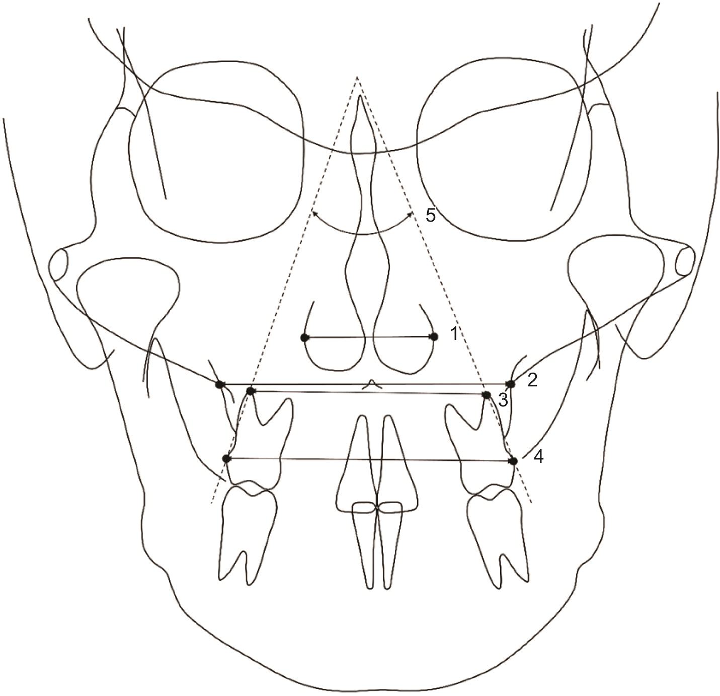
Figure 2
Sagittal landmarks and the vertical reference plane assessed on lateral cephalogram. 1, nasion; 2, sella; 3, orbitale; 4, porion; 5, anterior nasal spine; 6, A point; 7, B point; 8, pogonion; 9, menton; 10, gonion; and 11, nasion perpendicular plane: a line perpendicular to the Frankfort horizontal plane and passing through the nasion.
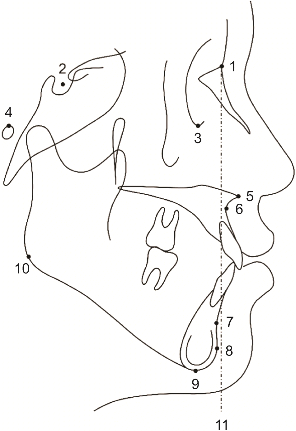
Figure 3
Sagittal skeletal variables assessed on lateral cephalogram. 1, Frankfort-mandibular plane angle (FMA); 2, sella-nasion to mandibular plane angle (SN-MP); 3, lower anterior facial height (LAFH, distance between the anterior nasal spine and the menton parallel to the nasion perpendicular); 4, sella-nasion-A point (SNA); 5, sella-nasion-B point (SNB); 6, A point-nasion-B point (ANB); 7, A point to nasion perpendicular (A to N perp); and 8, pogonion to nasion perpendicular (Pog to N perp). A to N perp, Pog to N perp, and LAFH are linear measurements, while the remaining variables are angular measurements.
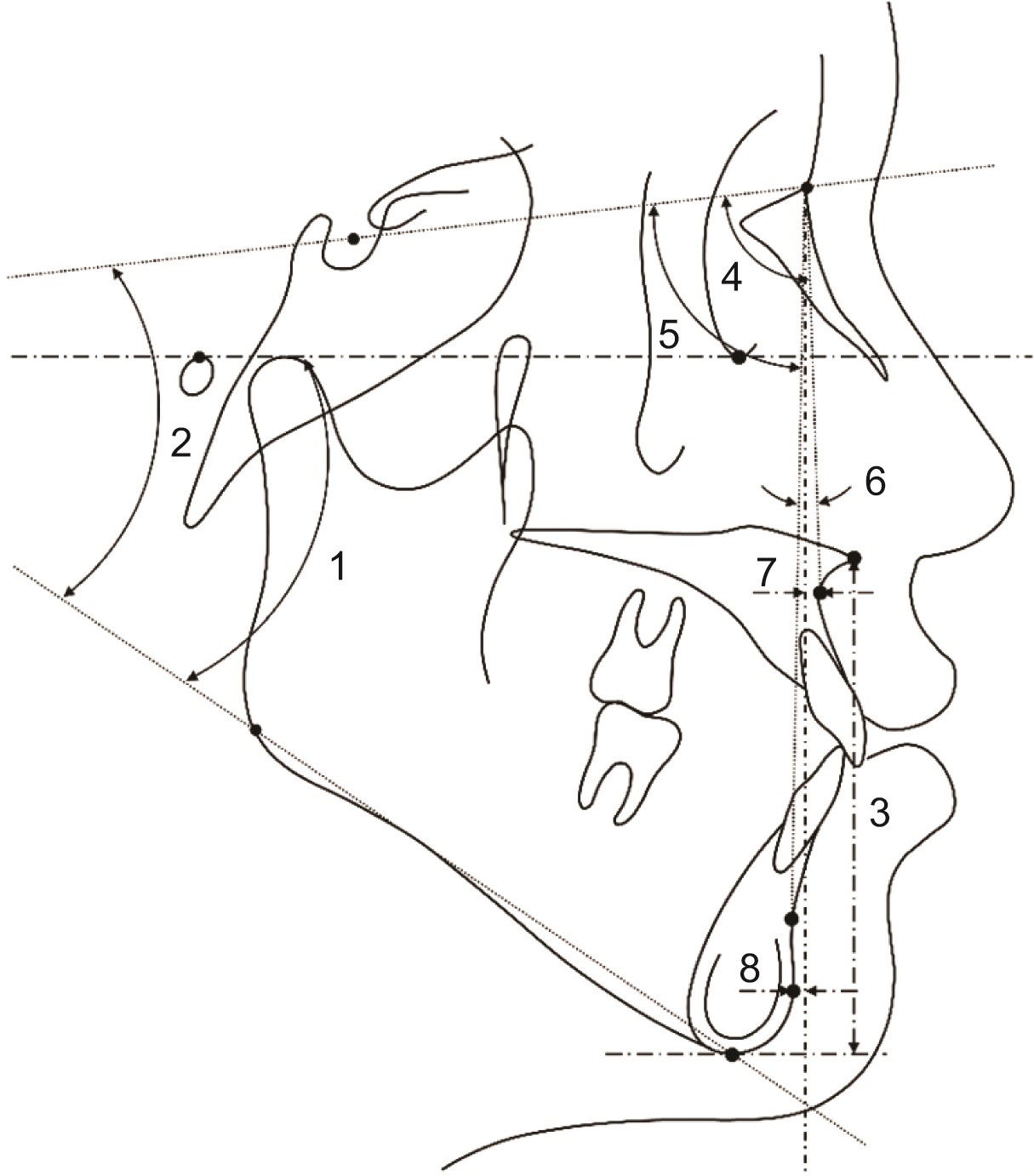
Figure 4
Soft tissue variables assessed on frontal photograph. 1, interpupillary distance: the distance between the left and right pupils; 2, alar width: the distance between the left and right alars; 3, nose length: the distance between the midpoint of the pupils and subnasale; 4, upper lip length: the distance between the subnasale and stomion; and 5, lip chin length: the distance between stomion and menton. Vertical measurements including the nose length, upper lip length, and lip chin length were measured as the distances parallel to the vertical bisector of the pupils.
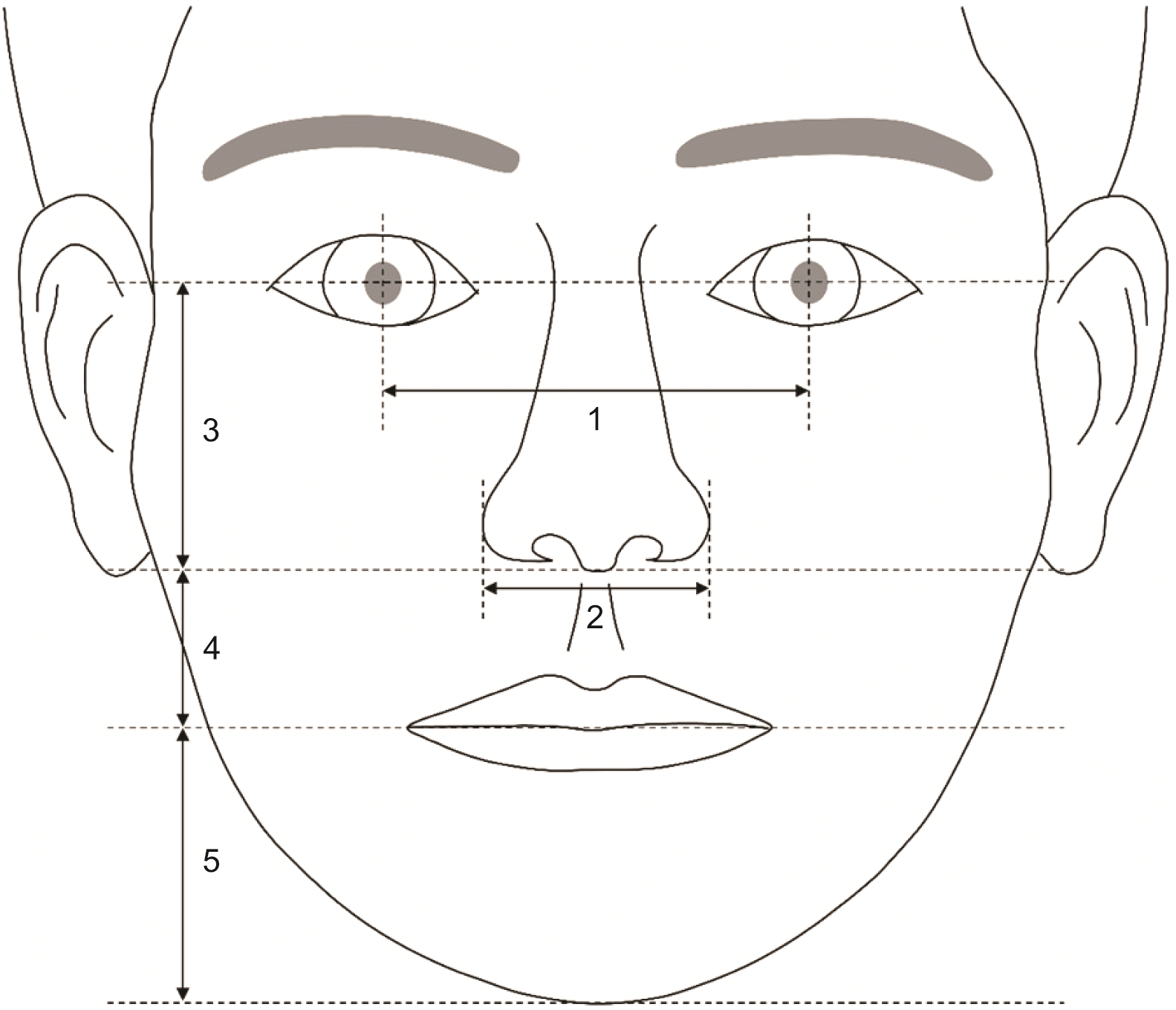
Figure 5
Differences in the transverse dentoskeletal variable changes between the groups after rapid maxillary expansion.
*Statistically significant difference (p < 0.05).
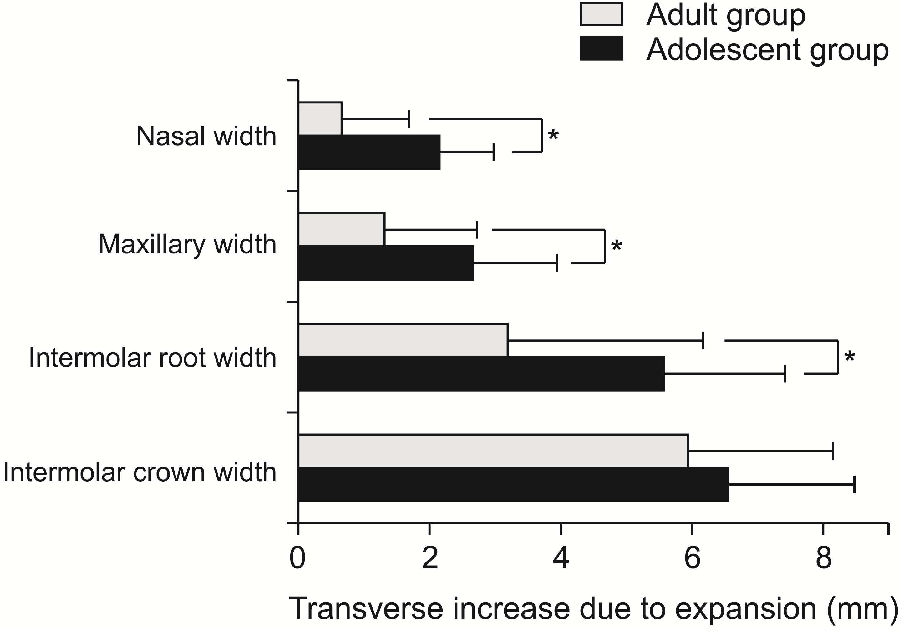
Table 1
Demographic data of patients
| Demographic | Adolescent group | Adult group | p-value |
|---|---|---|---|
| Subjects (% of total) | 17 (50.0) | 17 (50.0) | |
| Sex (% of group)† | 0.724 | ||
| Male | 6 (35.3) | 7 (41.2) | |
| Female | 11 (64.7) | 10 (58.8) | |
| Pretreatment age (yr)‡ | 12.48 ± 1.18 | 20.99 ± 2.49 | < 0.001*** |
| Expansion duration (days)‡ | 23.24 ± 9.10 | 24.12 ± 8.02 | 0.586 |
| Post-expansion duration (mo)‡ | 3.31 ± 0.62 | 2.97 ± 0.55 | 0.182 |
Table 2
Changes in the transverse dentoskeletal variables after rapid maxillary expansion (RME) and differences in the changes between the groups
| Transverse dentoskeletal variable | Adolescent group | Adult group | Intergroup p-value‡ | |||||||
|---|---|---|---|---|---|---|---|---|---|---|
| T1 | T2 | Change | Intragroup p-value† | T1 | T2 | Change | Intragroup p-value† | |||
| Nasal width (mm) | 32.74 ± 3.45 | 34.90 ± 3.89 | 2.15 ± 0.83 | < 0.001*** | 33.88 ± 3.20 | 34.55 ± 3.00 | 0.67 ± 1.02 | 0.028* | < 0.001*** | |
| Maxillary width (mm) | 68.47 ± 3.82 | 71.14 ± 4.17 | 2.67 ± 1.27 | < 0.001*** | 65.82 ± 3.59 | 67.14 ± 3.83 | 1.32 ± 1.41 | 0.001** | 0.002** | |
| Intermolar root width (mm) | 51.27 ± 3.48 | 56.86 ± 4.04 | 5.58 ± 1.84 | < 0.001*** | 49.79 ± 2.83 | 52.99 ± 3.01 | 3.20 ± 2.98 | 0.001** | 0.006** | |
| Intermolar crown width (mm) | 59.18 ± 3.61 | 65.73 ± 4.43 | 6.55 ± 1.92 | 0.002** | 58.83 ± 3.03 | 64.79 ± 3.22 | 5.96 ± 2.20 | < 0.001*** | 0.318 | |
| Intermolar angle (°) | 35.61 ± 7.70 | 38.86 ± 8.00 | 3.25 ± 7.41 | 0.102 | 37.18 ± 8.88 | 47.49 ± 10.18 | 10.31 ± 12.06 | 0.006** | 0.040* | |
Table 3
Changes in the sagittal skeletal variables following rapid maxillary expansion (RME) and differences in the changes between the groups
| Sagittal skeletal variable | Adolescent group | Adult group | Intergroup p-value‡ | |||||||
|---|---|---|---|---|---|---|---|---|---|---|
| T1 | T2 | Change | Intragroup p-value† | T1 | T2 | Change | Intragroup p-value† | |||
| Vertical skeletal variable | ||||||||||
| FMA (°) | 28.37 ± 4.31 | 29.16 ± 4.10 | 0.79 ± 0.69 | 0.001** | 29.96 ± 4.39 | 31.33 ± 4.66 | 1.36 ± 0.96 | 0.001** | 0.076 | |
| SN-MP (°) | 38.24 ± 4.39 | 38.97 ± 4.33 | 0.73 ± 0.79 | 0.004** | 40.18 ± 5.52 | 41.33 ± 5.68 | 1.15 ± 0.97 | 0.001** | 0.228 | |
| LAFH (mm) | 71.88 ± 5.63 | 72.90 ± 5.79 | 1.03 ± 1.32 | 0.002** | 77.37 ± 7.77 | 79.01 ± 7.50 | 1.64 ± 1.26 | 0.001** | 0.102 | |
| Anteroposterior skeletal variable | ||||||||||
| SNA (°) | 80.59 ± 3.20 | 80.82 ± 3.50 | 0.23 ± 0.86 | 0.368 | 78.01 ± 3.38 | 78.76 ± 2.98 | 0.75 ± 1.50 | 0.084 | 0.209 | |
| SNB (°) | 76.77 ± 3.91 | 76.82 ± 3.93 | 0.06 ± 0.75 | 0.356 | 77.25 ± 3.64 | 76.51 ± 3.84 | −0.75 ± 1.01 | 0.016* | 0.024* | |
| ANB (°) | 3.82 ± 2.63 | 4.00 ± 2.47 | 0.17 ± 0.88 | 0.523 | 0.76 ± 1.97 | 2.26 ± 2.34 | 1.50 ± 1.50 | 0.001** | 0.004** | |
| A to N perp (mm) | 0.50 ± 3.60 | 0.71 ± 3.87 | 0.20 ± 1.17 | 0.356 | −2.11 ± 3.18 | −1.51 ± 2.82 | 0.60 ± 1.51 | 0.098 | 0.783 | |
| Pog to N perp (mm) | −6.64 ± 7.68 | −6.70 ± 8.05 | −0.06 ± 1.27 | 0.925 | −5.56 ± 7.07 | −8.05 ± 7.79 | −2.48 ± 1.69 | 0.001** | < 0.001*** | |
T1, pretreatment; T2, after expansion (at least 2 months after cessation of expansion); Change, change in each variable following RME; FMA, Frankfort-mandibular plane angle; SN-MP, sella-nasion to mandibular plane angle; LAFH, lower anterior facial height; SNA, sella-nasion-A point; SNB, sella-nasion-B point; ANB, A point-nasion-B point; A to N perp, A point to nasion perpendicular; Pog to N perp, pogonion to nasion perpendicular.
Table 4
Changes in the soft tissue variables following rapid maxillary expansion (RME) and differences in the changes between the groups
| Soft tissue variable | Adolescent group | Adult group | Intergroup p-value‡ | |||||||
|---|---|---|---|---|---|---|---|---|---|---|
| T1 | T2 | Change | Intragroup p-value† | T1 | T2 | Change | Intragroup p-value† | |||
| Alar width (%) | 60.07 ± 3.91 | 61.71 ± 4.29 | 1.64 ± 1.45 | 0.001** | 58.84 ± 4.22 | 59.82 ± 4.00 | 0.98 ± 1.78 | 0.022* | 0.344 | |
| Nose length (%) | 78.16 ± 4.37 | 78.32 ± 4.26 | 0.16 ± 2.95 | 0.981 | 79.87 ± 5.30 | 79.68 ± 5.87 | −0.19 ± 2.25 | 0.831 | 0.986 | |
| Upper lip length (%) | 38.07 ± 4.54 | 37.89 ± 4.64 | −0.18 ± 1.79 | 0.868 | 36.47 ± 3.42 | 37.41 ± 3.19 | 0.94 ± 1.96 | 0.055 | 0.153 | |
| Lip chin length (%) | 73.61 ± 8.12 | 73.79 ± 5.76 | 0.18 ± 4.63 | 0.435 | 74.70 ± 4.97 | 75.82 ± 5.23 | 1.12 ± 3.18 | 0.163 | 0.221 | |
Table 5
Relationship between changes in transverse dentoskeletal variables and pretreatment age in the adolescent group


 PDF
PDF Citation
 Print
Print


 XML Download