Abstract
Objective
To evaluate if a remote digital monitoring system added at the end of orthodontic treatment could positively influence the retention phase by reducing the occurrence of misfit of removable appliances, number of emergency appointments (EA), and orthodontic relapse.
Methods
Twenty-seven patients who completed active orthodontic treatment were divided into the study and control groups. In addition to the standard chairside follow-up appointments at month 1 (T1), month 3 (T2), month 6 (T3), the study group patients were monitored using Dental Monitoring® with monthly intra-oral scans. Occurrence of misfit of removable retainers, number of EAs, and intercanine width change were recorded for both groups. Differences in EAs and retainer fit were assessed using the chi-square test. Intra-group and inter-group differences in the intercanine width were assessed with Friedman test and Mann–Whitney U test, respectively (α = 0.05).
Results
The study group showed a significantly lower occurrence of misfit of removable retainers (p = 0.027) compared to the control group. No significant inter- and intra-group difference was found in the EAs and intercanine width change at each time-point.
Conclusions
Integrating remote monitoring systems, such as Dental Monitoring®, to the retention phase of the orthodontic treatment may lower the occurrence of misfit of removable retainers. However, a small sample size and a short observation period limit the strength of this evidence. These preliminary results tentatively suggest that remote monitoring technologies may be beneficial, especially during the COVID-19 pandemic, when the regularity of in-office visits might be disrupted.
Successful orthodontic therapy depends not only on good diagnosis and correct treatment planning but also on appropriate retention as a continuation of any active orthodontic phase.1 Retention involves bonding of fixed retainers (mainly for the lower dentition) or the use of removable appliances (mainly for the upper dentition), which relies on patient compliance, which tends to fade over time.2 Irregular use of removable retainers can result in orthodontic relapse.3
Orthodontic relapse can be limited by regularly monitoring the adherence of patients to using their removable appliance. For this purpose, several remote systems have been developed in the last decade, including the embedding of a microsensor into the acrylic of the appliance to record temperature, such as the Smart Retainer (Scientific Compliance, Atlanta, GA, USA)4 and TheraMon Sensor (Handelsagentur Gschladt, Hargelsberg, Austria).5 Other studies on mobile applications providing active reminders6-8 suggest that regular control of patients by the orthodontist can positively influence patient compliance.9
Recently, Dental Monitoring® (DM, Dental Monitoring, Paris, France) was introduced for remote monitoring of patients using dedicated cheek retractors and their smartphone.10 The users can capture pictures of their dentition with and without removable retainers, thus aiding early detection of any potential misfit of the appliance. DM includes another function for three-dimensional (3D) measurements, called 3D Monitoring Light®, which allows 3D calculation of dental movements (e.g., linear and angular measurements)10-12 and two-dimensional (2D) clinical analysis (e.g., monitoring of the fit of the removable retainers), with scans performed once per month.13 Systems of remote monitoring are particularly important in times like the current COVID-19 pandemic, when regular chairside follow-up appointments might be disrupted.14,15
To the best of our knowledge, this is the first study to evaluate the fit of post-orthodontic removable retainers using a monitoring system with a smartphone and a cheek retractor. The aim of this feasibility study was to investigate whether a system of remote monitoring – such as DM – could positively influence the stability of the orthodontic treatment during the retention phase by reducing the occurrence of misfit of the appliances, need for emergency appointments (EA), as well as the incidence of orthodontic relapse.
A sample size of 30 was calculated based on a previous study conducted on DM.12 Thirty consecutive patients were recruited for this prospective feasibility study at the end of their orthodontic treatment, which was performed using aligners or fixed buccal multi-bracket appliance by the same orthodontist (L.S.) between June 2018 and June 2019. The inclusion criteria were access to a smartphone and internet, and recent completion of orthodontic treatment for Class I or mild Class II malocclusion with mild to moderate crowding. The exclusion criteria were a lack of compliance, defined as not attending the appointments for three consecutive months; severe medical history.
Thirty patients were randomly assigned to one of the two groups using a simple randomization method. As three of them declined to participate to the study, a total of twenty-seven participants were included. Informed consent was obtained from the patients and their parents. The ethical committee of the University of Brescia approved the study (DENMON01452).
In both groups, retention was provided through two removable clear full-coverage hard acrylic appliances (1.5 mm of thickness), individually fabricated based on alginate impressions of the upper and lower arches. The removable appliances were given to the patients one week after the end of the active orthodontic treatment, along with written and verbal instructions on retainer management (cleaning and storage). Patients were asked to wear the removable retainers 7 days per week during daytime and night-time in the first month and only at night-time the second month onward. Patients were reminded to bring their removable retainers at each in-office appointment to check the fit of the appliance.
Additionally, a lingual fixed retainer extending between the upper left and right canines was bonded in 9 patients (75%) of the study group and in 13 patients (87%) of the control group, as they presented with a midline diastema before the treatment, residual tongue thrust habit at the end of the active orthodontic therapy, and/or they were cases of retreatment due to relapse.
Both groups underwent a six-month follow-up during the retention phase, with chairside appointments at month 1 (T1), month 3 (T2), and month 6 (T3).
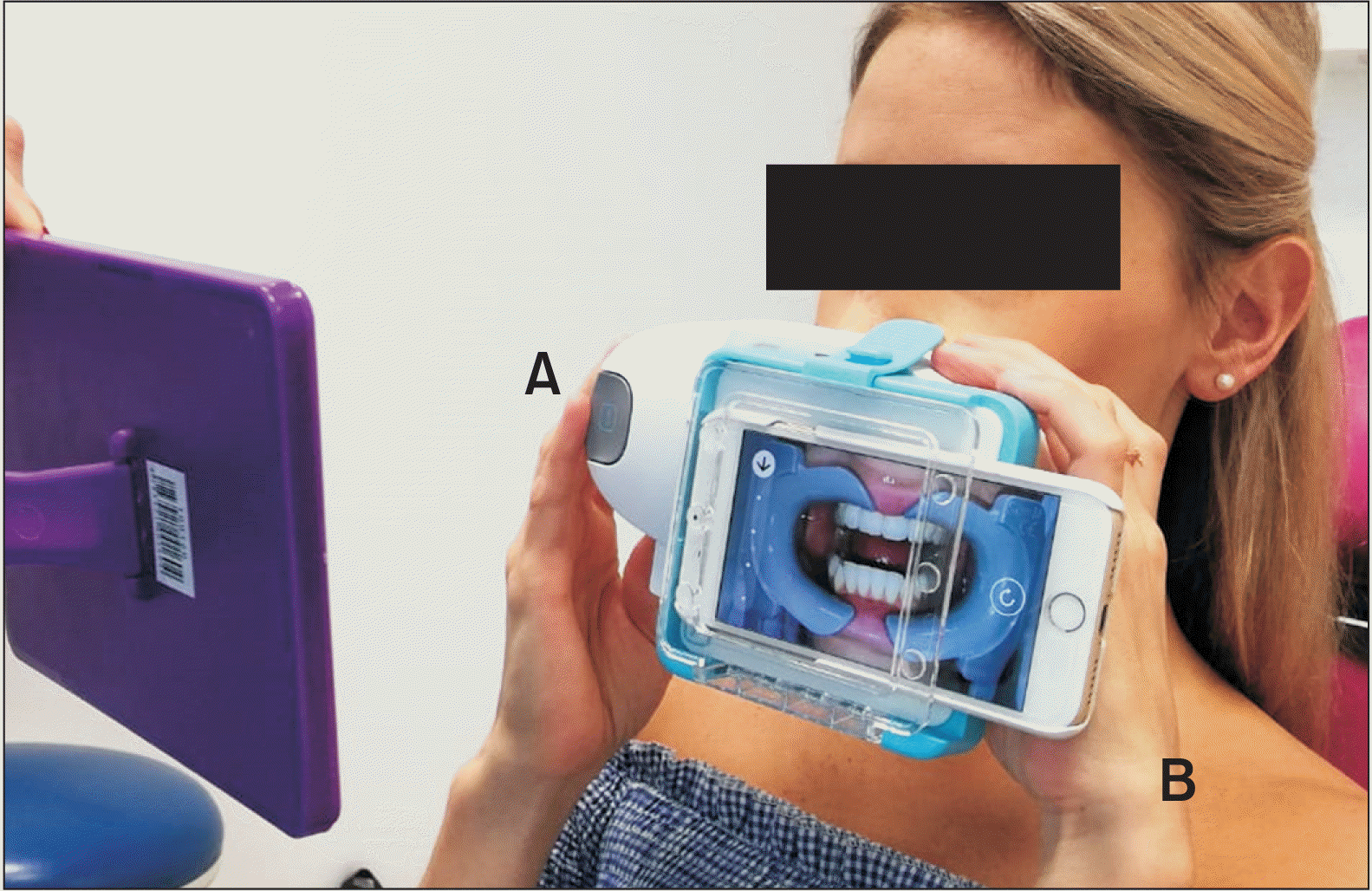
During their first appointment at the beginning of the retention phase (T0), the study group patients were also delivered a ScanBox© (DM, Dental Monitoring) and a dedicated cheek retractor by DM as described elsewhere16 (Figure 1), and they were asked to download the DM app on their smartphone to perform monthly intraoral scans. The first scan was taken together with the orthodontist to ensure proper use of the device. The monthly 2D scans of their mouth (frontal, lateral and occlusal views) were performed with and without the removable retainers in place and were automatically uploaded on the DM app.
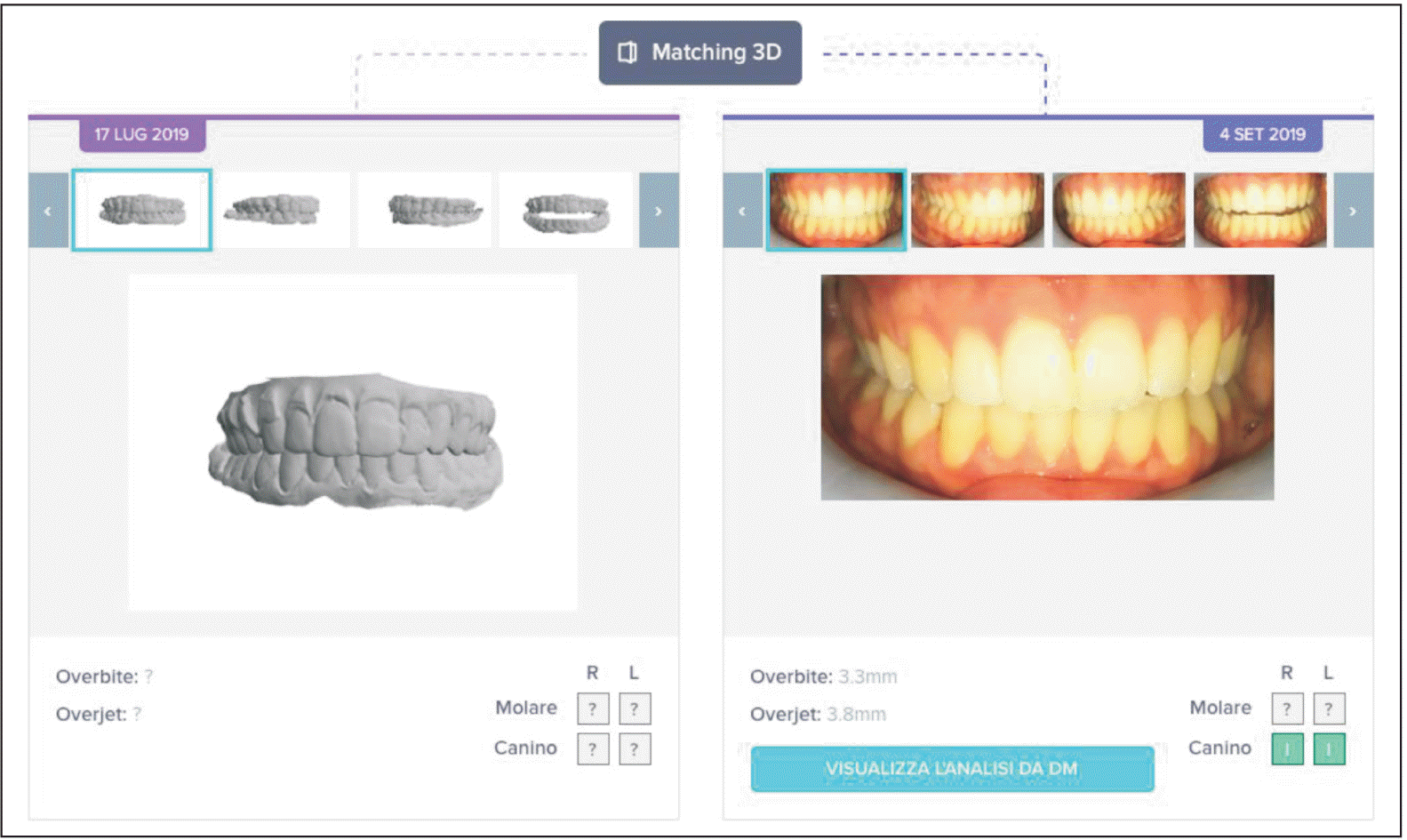
During the same appointment, limited to the study group, poly-vinyl siloxane impressions of maxillary and mandibular arches were taken (Bisico®, Bielefelder Dentalsilicone, Bielefeld, Germany) and poured with type IV extra hard white stone. Dental casts were scanned with a 3D intraoral scanner (TRIOS®, ESM/3ShapeTMR-700, ESM Digital Solutions, Dublin, Ireland) to obtain a stereolithography (.stl) file, which was uploaded to the DM platform as an initial 3D reference model. Each monthly 2D scan uploaded by the study group patients was superimposed on the baseline 3D model and analysed through a software (3D Matching®, Dental Monitoring SAS, Paris, France) (Figure 2). Each superimposition allowed detection of dental movements, including mesio-distal translation (mm), bucco-lingual translation (mm), intrusion-extrusion (mm), mesio-distal angulation (°), bucco-lingual inclination (°), and rotation (°). The software has an error of one tenth of a millimetre for linear movements and of less than 0.5° for angular movements, and the data were compared at different time-points to measure the entity of the relapse.10-12 However, from a clinically relevant standpoint, relapse was defined as any dental movement above a cut-off of 2° and 2 mm to identify its incidence in the data analysis,6 as indicated by Grunheid et al.,17 according to the American Board of Orthodontics model grading system.
As the control group did not undergo 3D monitoring for dental relapse, the lower intercanine width was calculated for both groups at each chairside appointment (T0, T1, T2, T3) by measuring the distance between the cusp tip of the right and left lower canines in millimetres with a manual calliper.18 Each measurement was performed three times by the same examiner, and the mean value was used for data analysis. The intercanine width change was calculated for each time point (ΔT0-T1, ΔT1-T2, and ΔT2-T3) and for the total observation period (ΔT0-T3). Moreover, pre-treatment intercanine width was measured as described above to assess the changes that occurred during the orthodontic treatment.
Lastly, the total number of EAs was recorded for both groups.
Results are reported as the mean values with standard deviation and 95% confidence interval (CI).
The Shapiro–Wilk test showed that the data distribution was not normal; therefore, nonparametric tests were used. The chi-square test and Mann–Whitney U test were used to compare the two groups in relation to the sex proportion, age, type of appliance, number of patients receiving a fixed retainer, and intercanine width at baseline (T0) and at 6 months (T3).
The fit of the removable appliances was visually assessed between the retainer and the teeth. A value of 0 was assigned in presence of more than 1 mm of space between the removable appliance and at least 2 teeth (e.g., not fitting), and a value of 1 was assigned if a smaller space was detected (e.g., proper fit). Inter-group differences in the proportion of not fitting removable appliances and EAs were assessed using the chi-square test. Intra-group differences in the intercanine width were compared among time points with Friedman test for repeated measures. Inter-group differences of intercanine width at each time-point were compared using the Mann–Whitney U test. Spearman’s rho coefficient was used to assess the correlation between change in the pre- and post-treatment intercanine width during the orthodontic treatment and change in the intercanine width during the observation period.
Statistical analysis was performed using SPSS® 27 (IBM Corp., Armonk, NY, USA) at significance level α = 0.05.
Overall, 27 participants were included (10 to 64 years old, average age 22.22 ± 10.28 years, 20 female and 7 men) (Table 1). Twelve patients (4 male and 8 female, mean age 22.6 ± 7.1 years) were assigned to the study group (7 treated with a fixed appliance and 5 with aligners). Fifteen patients (3 male and 12 female, mean age 21.9 ± 12.5 years) were assigned to the control group (7 treated with a fixed appliance and 8 with aligners). The fixed appliance comprised buccal multi-bracket self-ligating Empower© brackets with MBT prescription and a 0.022-inch slot (American Orthodontics, Sheboygan, WI, USA). The aligners used were those by Invisalign® (Align Technology©, San Jose, CA, USA). None of the patients withdrew from the study.
The removable retainers fit appropriately in all study group participants and in 66% of the control group participants (X2 (1) = 4.844; p = 0.027; CI, 3.57 to 58.89). The proportion of EAs was 8% in the study group compared to 13% in the control group (X2 (1) = 0.167; p = 0.682; CI, –23.57 to 30.37).
The change in the intercanine width within the groups was not statistically significant (Table 2). The change in the intercanine width between the two groups at each time-point was not statistically significant (Table 2). No correlation was found between the change in the intercanine width during orthodontic treatment and its change during the observation period (rs = –0.132, p = 0.682 in the study group; rs = 0.250, p = 0.369 in the control group).
3D Matching® detected the occurrence of dental movements in all 12 patients of the study group during the 6-month observation period (Figure 3). The majority and the most severe movements occurred at T1 and were related to changes in the occuredat T1, which were related to changes in the bucco-lingual inclination (Table 3).
To the best of our knowledge, this is the first study that applies DM along with the dedicated ScanBox© to monitor the fit of post-orthodontic removable retainers and its impact on dental relapse at the end of orthodontic treatment.
Remote monitoring systems are part of Artificial Intelligence Driven Remote Monitoring.19,20 In the past few years, the market has assisted an increased availability of orthodontic apps to enhance long-distance monitoring and compliance,21 and a recent study confirmed the positive attitude of patients towards this new digital technology.22 In orthodontics, it can be applied for monitoring of several treatment aspects,23 including patients’ oral hygiene during the therapy,16,24,25 treatment with clear aligners,26,27 rapid palatal expansion,15,28,29 adherence to the use of removable retainers,8 active working time of self-ligating straight-wire appliances,30 and clinical follow-up of corticotomy-accelerated orthodontic therapies.31
In the present work, the integration of DM was found to positively influence the fit of removable retainers at the end of orthodontic treatment, with a significant difference compared to the control group. The retention phase starts at the end of the active orthodontic therapy.1 Each patient is expected to wear the removable retainer at night and to attend regular in-office check-up appointments, especially during the first 6 months when most periodontal remodelling occurs.3 Addition of DM to the standard care, by monitoring of the patients using pictures periodically taken at home, suggested positive reinforcement for patient adherence. However, despite the mere additional monthly monitoring by DM, it is possible that part of the positive effect on the engagement of the participants was derived from the acknowledgement of being regularly monitored and being part of a study, known as Hawthorne effect.32 This positive reinforcement may have partly contributed to the significant difference in the fit of the removable retainers between the two groups, as also demonstrated in other studies in which different telemonitoring systems were applied.4,6,8 Yet, this interpretation is in contrast with a previous study, which did not reveal a significant increase in patient compliance for retainer-use with the integration of a tailored mobile application (“My Retainers” app developed by Al-Moghrabi et al.33) However, the study assessed the median wear time of the retainers over a period of 3 months). Patient compliance is known to cease over a longer timeframe and thus, the lack of difference between the two groups may be explained by the short follow-up of 3 months.34 The present study followed the patients for 6 months; however, this was still a relatively short observation period, and it may have been insufficient to detect significant dental movements. Indeed, the present findings did not reveal any difference in the change of the intercanine width at different time-points between the two groups, despite the significant difference in the fit of the removable retainers.35 Therefore, future studies should aim at extending the observation to one year or more.
Limited to the study group, the 3D Matching® allowed superimposition between the initial 3D model at the time of the removal of the appliance and the following monthly 2D scans taken by the patients, sending a warning message to theirselves in case of variations between consecutive superimposed scans. The use of a 3D Matching® technology permits detection of movements of each dental element that are often clinically imperceptible. In this regard, the picture of a study group patient, where DM detected changes in the position of the upper left central incisor, is significantly explicative (Figure 3). Although no difference can be visually perceived between the two scans, dental movements were measured as a distal rotation by 2.7° and a bucco-lingual inclination by 2.5°. Yet, a criticism against the excessive sensitivity of DM may arise. To counteract this downside, a clinically relevant cut-off of 2 mm and 2° was chosen based on the American Board of Orthodontics model grading system.17 Dental movements greater than 2 mm and 2° were mostly detected on the posterior teeth and mainly occurred at T1, with an average of 7.5 teeth per patient in the study group. This might be reasonably explained as the result of natural settling of the occlusion immediately after the orthodontic appliances are removed.36,37
As for the occurrence of EAs, the results of this study showed that the control group patients requested a higher number of EAs, although the difference between the two groups was not statistically significant.
In the present feasibility study, the use of a technology like DM was not found to be user-friendly as many pictures taken by the patients were rejected and several attempts were needed to obtain acceptable images. This problem was not reported in studies in which this technology was applied on a weekly basis.16 This may suggest that the monthly frequency of scanning leads the patient to forget the method of capturing accurate pictures compared to more frequent monitoring.
Approximately half of the study group patients often failed to adhere to the scheduled time for scanning until a reminder was sent by DM. Additionally, in the actual clinical scenario, few patients would be willing to pay an additional fee to control their retention phase with a sophisticated software, as confirmed by a previous study.22
Despite these practical difficulties, systems of remote monitoring may appear especially helpful in times like the current COVID-19 pandemic, in which regular in-office appointments may be disrupted due to unpredictable lockdowns and the non-urgent nature of some orthodontic procedures.38
The study is not exempt from limitations. First, the small sample size may preclude drawing of strong conclusions. Moreover, the present study has focused on the first 6 months after the removal of orthodontic appliances as it is a critical period for dental stability,39 during which the majority of the periodontal fibers reorganize according to the new dental position, despite the supra-crestal fibers taking a longer time.40 However, dental movement caused by relapse was almost negligible during the 6-month observation period; therefore, the duration of further studies should be extended to at least one year.
An uneven number of patients, who were undergoing retreatment following a relapse, received a fixed lingual retainer, and different types of orthodontic appliances (fixed or removable aligners) were utilized during the active therapy. Future studies should apply stratified randomization methods to equally distribute the participants according to the type of orthodontic treatment received and the need for additional retention systems. Further, cases of retreatment for previous relapse should be excluded. Lastly, the age range of the patients was very broad, although no significant difference was found between mean age of the two groups. It would be interesting for upcoming studies to narrow the participant age to adolescents, who are the main demographic target of orthodontic treatment.
The preliminary results of this feasibility study suggest that adding a remote digital technology to the retention phase at the end of orthodontic treatment may lower the occurrence of misfit of removable retainers over time, compared to the standard care.
Despite the significant difference in proper fitting of the removable retainers, such a result did not influence the occurrence of dental relapse between the two groups, probably due to the short follow-up period (6 months). Therefore, conclusions regarding the effectiveness of remote monitoring technology in reducing treatment relapse should be drawn with caution.
Remote monitoring systems can be useful in times like the current pandemic to minimize in-person visits.
References
1. Bondemark L, Holm AK, Hansen K, Axelsson S, Mohlin B, Brattstrom V, et al. 2007; Long-term stability of orthodontic treatment and patient satisfaction. A systematic review. Angle Orthod. 77:181–91. DOI: 10.2319/011006-16R.1. PMID: 17029533.
2. Littlewood SJ, Millett DT, Doubleday B, Bearn DR, Worthington HV. 2016; Retention procedures for stabilising tooth position after treatment with orthodontic braces. Cochrane Database Syst Rev. 2016:CD002283. DOI: 10.1002/14651858.CD002283.pub4. PMID: 26824885. PMCID: PMC7138206.
3. Miyazaki H, Motegi E, Yatabe K, Isshiki Y. 1998; Occlusal stability after extraction orthodontic therapy in adult and adolescent patients. Am J Orthod Dentofacial Orthop. 114:530–7. DOI: 10.1016/S0889-5406(98)70173-8. PMID: 9810049.
4. Ackerman MB, McRae MS, Longley WH. 2009; Microsensor technology to help monitor removable appliance wear. Am J Orthod Dentofacial Orthop. 135:549–51. DOI: 10.1016/j.ajodo.2008.06.021. PMID: 19361743.
5. Tsomos G, Ludwig B, Grossen J, Pazera P, Gkantidis N. 2014; Objective assessment of patient compliance with removable orthodontic appliances: a cross-sectional cohort study. Angle Orthod. 84:56–61. DOI: 10.2319/042313-315.1. PMID: 23834273. PMCID: PMC8683054.
6. Zotti F, Zotti R, Albanese M, Nocini PF, Paganelli C. 2019; Implementing post-orthodontic compliance among adolescents wearing removable retainers through Whatsapp: a pilot study. Patient Prefer Adherence. 13:609–15. DOI: 10.2147/PPA.S200822. PMID: 31118585. PMCID: PMC6498955.
7. El-Huni A, Colonio Salazar FB, Sharma PK, Fleming PS. 2019; Understanding factors influencing compliance with removable functional appliances: a qualitative study. Am J Orthod Dentofacial Orthop. 155:173–81. DOI: 10.1016/j.ajodo.2018.06.011. PMID: 30712688.
8. Al-Moghrabi D, Pandis N, McLaughlin K, Johal A, Donos N, Fleming PS. 2020; Evaluation of the effectiveness of a tailored mobile application in increasing the duration of wear of thermoplastic retainers: a randomized controlled trial. Eur J Orthod. 42:571–9. DOI: 10.1093/ejo/cjz088. PMID: 31799628.
9. Ackerman MB, Thornton B. 2011; Posttreatment compliance with removable maxillary retention in a teenage population: a short-term randomized clinical trial. Orthodontics (Chic. ). 12:22–7.
10. Roisin LC, Brézulier D, Sorel O. 2016; Remotely-controlled orthodontics: fundamentals and description of the Dental Monitoring system. J Dentofacial Anom Orthod. 19:408. DOI: 10.1051/odfen/2016021.
11. Morris RS, Hoye LN, Elnagar MH, Atsawasuwan P, Galang-Boquiren MT, Caplin J, et al. 2019; Accuracy of Dental Monitoring 3D digital dental models using photograph and video mode. Am J Orthod Dentofacial Orthop. 156:420–8. DOI: 10.1016/j.ajodo.2019.02.014. PMID: 31474272.
12. Moylan HB, Carrico CK, Lindauer SJ, Tüfekçi E. 2019; Accuracy of a smartphone-based orthodontic treatment-monitoring application: a pilot study. Angle Orthod. 89:727–33. DOI: 10.2319/100218-710.1. PMID: 30888840. PMCID: PMC8111833.
13. Caruso S, Caruso S, Pellegrino M, Skafi R, Nota A, Tecco S. 2021; A knowledge-based algorithm for automatic monitoring of orthodontic treatment: the Dental Monitoring system. Two cases. Sensors (Basel). 21:1856. DOI: 10.3390/s21051856. PMID: 33799930. PMCID: PMC7961518. PMID: f11772245cbc4c3292f8fa49c7a10fe3.
14. Giudice A, Barone S, Muraca D, Averta F, Diodati F, Antonelli A, et al. 2020; Can teledentistry improve the monitoring of patients during the COVID-19 dissemination? A descriptive pilot study. Int J Environ Res Public Health. 17:3399. DOI: 10.3390/ijerph17103399. PMID: 32414126. PMCID: PMC7277372. PMID: 73a0f1e6394747ef943ee8418fa4831f.
15. Bianco A, Dalessandri D, Oliva B, Tonni I, Isola G, Visconti L, et al. 2021; COVID-19 and orthodontics: an approach for monitoring patients at home. Open Dent J. 15 Suppl 1:87–96. DOI: 10.2174/1874210602115010087.
16. Sangalli L, Savoldi F, Dalessandri D, Bonetti S, Gu M, Signoroni A, et al. 2021; Effects of remote digital monitoring on oral hygiene of orthodontic patients: a prospective study. BMC Oral Health. 21:435. DOI: 10.1186/s12903-021-01793-9. PMID: 34493255. PMCID: PMC8422366. PMID: 1570871d06134a6a8c7642c9c75321ed.
17. Grünheid T, Loh C, Larson BE. 2017; How accurate is Invisalign in nonextraction cases? Are predicted tooth positions achieved? Angle Orthod. 87:809–15. DOI: 10.2319/022717-147.1. PMID: 28686090. PMCID: PMC8317555.
18. Adamek A, Minch L, Kawala B. 2015; Intercanine width- review of the literature. Dent Med Probl. 52:336–40.
19. Vaid NR. 2021; Artificial Intelligence (AI) driven orthodontic care: a quest toward utopia? Semin Orthod. 27:57–61. DOI: 10.1053/j.sodo.2021.05.001.
20. Hansa I, Katyal V, Semaan SJ, Coyne R, Vaid NR. 2021; Artificial Intelligence Driven Remote Monitoring of orthodontic patients: clinical applicability and rationale. Semin Orthod. 27:138–56. DOI: 10.1053/j.sodo.2021.05.010.
21. Hansa I, Semaan SJ, Vaid NR, Ferguson DJ. 2018; Remote monitoring and "Tele-orthodontics": concept, scope and applications. Semin Orthod. 24:470–81. DOI: 10.1053/j.sodo.2018.10.011.
22. Dalessandri D, Sangalli L, Tonni I, Laffranchi L, Bonetti S, Visconti L, et al. 2021; Attitude towards telemonitoring in orthodontists and orthodontic patients. Dent J (Basel). 9:47. DOI: 10.3390/dj9050047. PMID: 33921925. PMCID: PMC8143570. PMID: e7809cc011d7402a8dbeb378216c3822.
23. Vaid NR, Hansa I, Bichu Y. 2020; Smartphone applications used in orthodontics: a scoping review of scholarly literature. J World Fed Orthod. 9(3S):S67–73. DOI: 10.1016/j.ejwf.2020.08.007. PMID: 33023735.
24. Zotti F, Dalessandri D, Salgarello S, Piancino M, Bonetti S, Visconti L, et al. 2016; Usefulness of an app in improving oral hygiene compliance in adolescent orthodontic patients. Angle Orthod. 86:101–7. DOI: 10.2319/010915-19.1. PMID: 25799001. PMCID: PMC8603968.
25. Savoldi F, Bonetti S, Dalessandri D, Mandelli G, Paganelli C. 2015; Incisal apical root resorption evaluation after low-friction orthodontic treatment using two-dimensional radiographic imaging and trigonometric correction. J Clin Diagn Res. 9:ZC70–4. DOI: 10.7860/JCDR/2015/14140.6841. PMID: 26676099. PMCID: PMC4668528. PMID: 8afb1ab45384426eb7c8628ed13d84ae.
26. Hansa I, Semaan SJ, Vaid NR. 2020; Clinical outcomes and patient perspectives of Dental Monitoring® GoLive® with Invisalign®-a retrospective cohort study. Prog Orthod. 21:16. DOI: 10.1186/s40510-020-00316-6. PMID: 32537723. PMCID: PMC7293972. PMID: 9e46367799694b269618e772223c1c5b.
27. Hansa I, Katyal V, Ferguson DJ, Vaid N. 2021; Outcomes of clear aligner treatment with and without Dental Monitoring: a retrospective cohort study. Am J Orthod Dentofacial Orthop. 159:453–9. DOI: 10.1016/j.ajodo.2020.02.010. PMID: 33573897.
28. Kuriakose P, Greenlee GM, Heaton LJ, Khosravi R, Tressel W, Bollen AM. 2019; The assessment of rapid palatal expansion using a remote monitoring software. J World Fed Orthod. 8:165–70. DOI: 10.1016/j.ejwf.2019.11.001.
29. Savoldi F, Tsoi JKH, Paganelli C, Matinlinna JP. 2017; Evaluation of rapid maxillary expansion through acoustic emission technique and relative soft tissue attenuation. J Mech Behav Biomed Mater. 65:513–21. DOI: 10.1016/j.jmbbm.2016.09.016. PMID: 27669497.
30. Impellizzeri A, Horodinsky M, Barbato E, Polimeni A, Salah P, Galluccio G. 2020; Dental Monitoring Application: it is a valid innovation in the Orthodontics Practice? Clin Ter. 171:e260–7. DOI: 10.7417/CT.2020.2224. PMID: 32323716.
31. Hannequin R, Ouadi E, Racy E, Moreau N. 2020; Clinical follow-up of corticotomy-accelerated Invisalign orthodontic treatment with Dental Monitoring. Am J Orthod Dentofacial Orthop. 158:878–88. DOI: 10.1016/j.ajodo.2019.06.025. PMID: 33129633.
32. Purssell E, Drey N, Chudleigh J, Creedon S, Gould DJ. 2020; The Hawthorne effect on adherence to hand hygiene in patient care. J Hosp Infect. 106:311–7. DOI: 10.1016/j.jhin.2020.07.028. PMID: 32763330.
33. Al-Moghrabi D, Colonio-Salazar FB, Johal A, Fleming PS. 2020; Development of 'My Retainers' mobile application: triangulation of two qualitative methods. J Dent. 94:103281. DOI: 10.1016/j.jdent.2020.103281. PMID: 31987979.
34. Shah N. 2017; Compliance with removable orthodontic appliances. Evid Based Dent. 18:105–6. DOI: 10.1038/sj.ebd.6401268. PMID: 29269817.
35. Steinnes J, Johnsen G, Kerosuo H. 2017; Stability of orthodontic treatment outcome in relation to retention status: an 8-year follow-up. Am J Orthod Dentofacial Orthop. 151:1027–33. DOI: 10.1016/j.ajodo.2016.10.032. PMID: 28554448.
36. Başçiftçi FA, Uysal T, Sari Z, Inan O. 2007; Occlusal contacts with different retention procedures in 1-year follow-up period. Am J Orthod Dentofacial Orthop. 131:357–62. DOI: 10.1016/j.ajodo.2005.05.052. PMID: 17346591.
37. Olive RJ, Basford KE. 2003; A longitudinal index study of orthodontic stability and relapse. Aust Orthod J. 19:47–55. PMID: 14703329.
38. Suri S, Vandersluis YR, Kochhar AS, Bhasin R, Abdallah MN. 2020; Clinical orthodontic management during the COVID-19 pandemic. Angle Orthod. 90:473–84. DOI: 10.2319/033120-236.1. PMID: 32396601. PMCID: PMC8028467.
39. Torkan S, Firth F, Fleming PS, Kravitz ND, Farella M, Huang GJ. 2021; Retention: taking a more active role. Br Dent J. 230:731–8. DOI: 10.1038/s41415-021-2952-9. PMID: 34117428.
40. Edwards JG. 1988; A long-term prospective evaluation of the circumferential supracrestal fiberotomy in alleviating orthodontic relapse. Am J Orthod Dentofacial Orthop. 93:380–7. DOI: 10.1016/0889-5406(88)90096-0. PMID: 3163217.
Table 1
Table 2
Variable | Study group | Control group | p-value* |
---|---|---|---|
ΔT0-T1 (mm) | 0.0 ± 0.0 | −0.1 ± 0.3 | 0.541 |
ΔT1-T2 (mm) | 0.0 ± 0.0 | −0.1 ± 0.3 | 0.548 |
ΔT2-T3 (mm) | 0.0 ± 0.2 | −0.3 ± 0.7 | 0.548 |
ΔT0-T3 (mm) | 0.1 ± 0.1 | −0.4 ± 0.7 | 0.250 |
p-value† | 1.000 | 0.923 |