

 PDF
PDF Citation
 Print
Print


INTRODUCTION
Primary liver cancer is the 5th most common cancer in the world, with hepatocellular carcinoma (HCC) being the most common (accounting for more than 80%) histologic type of primary liver cancer [1]. Most HCCs are associated with chronic liver disease, which is mainly caused by hepatitis B virus infection, hepatitis C virus infection, alcohol abuse, and non-alcoholic fatty liver disease [1,2]. Recently, the incidence of HCC has been increasing in Europe and America [3,4]. Treatment of patients with HCC remains challenging despite enormous efforts. Less than 25% of patients with HCC receive potentially curative therapy such as liver transplantation (LT), partial hepatectomy (PH), and local ablation [5,6]. The prognosis of these patients is not promising, with an overall five-year survival rate of less than 20% [6]. Management of HCC accompanied by cirrhosis is more challenging because a diminished functional hepatic reservoir limits not only standard curative treatment, but also experimental therapeutic trials.
In cases of HCC accompanied by cirrhosis, LT is the primary treatment as it can treat both the tumor and the underlying liver disease simultaneously [7]. However, LT is not always feasible. In addition, it has a few limitations [8,9]. PH can be an alternative option in some cases, for example, when the functional reservoir of the liver is sufficient for PH to be carried out [10]. Recent studies suggest that the prognosis after PH in patients with HCC accompanied by cirrhosis is comparable to that after LT [10-12]. On the other hand, there are negative views of PH as a curative treatment for HCC accompanied by cirrhosis due to a relatively higher recurrence rate after PH than that after LT. However, this argument has been only based on clinical studies carried out in the immediate postoperative period [6,7,11]. Clinicians may underestimate the value of PH as a treatment option for patients with HCC accompanied by cirrhosis because there is little information on changes in prognosis over time. Thus, a more meticulous statistical analysis of PH needs to be conducted. The cure model analysis is a stepping-stone for solving this issue.
A cure model analysis is a useful statistical method for analyzing post-treatment survival data of long-term survivors with specific diseases, for example, long-term survivors with cancer. The cure model is a valuable tool that can analyze cure fraction in patients under various settings. The use of the cure model could explain the long-term effect of a specific treatment for the disease. It may provide the prospect of cure [13,14].
In this study, we attempted to examine the possibility of PH curing patients with HCC accompanied by cirrhosis. We also compared PH cure fraction for patients with HCC with that for patients without cirrhosis to estimate the statistical chance of cure after PH for patients with HCC using a non-mixture cure model. Outcomes were also compared between non-cirrhotic and cirrhotic patients [15].
MATERIALS AND METHODS
Literature search
A comprehensive search of Medline, Embase, and KoreaMed databases was performed for articles published between January 1995 and July 2020 reporting recurrence-free survival (RFS) after PH in patients with HCC and comparing RFS according to the presence or absence of cirrhosis. To include as many relevant articles as possible, we selected “carcinoma, hepatocellular” and “liver cirrhosis” or “fibrosis” and “hepatectomy” as Medical Subject Headings (MeSH) terms. Other keywords such as “hepatocellular carcinoma,” “cirrhosis,” “resect,” and “resection” were also used. We examined titles and abstracts of studies in search results to select relevant ones. For additional screening, two researchers (BB and KK) independently inspected all candidate articles employing inclusion and exclusion criteria. We exclusively abstracted hazard ratios (HRs) from observational studies and then combined them in a meta-analysis. This meta-analysis was conducted following the Preferred Reporting Items for Systematic Reviews and Meta-Analyses (PRISMA).
Eligibility criteria
We selected articles that showed RFS after PH in patients with or without cirrhosis. Studies that provided HRs between cirrhosis and non-cirrhosis groups were included. If HRs were not provided, articles with Kaplan–Meier (KM) survival curve that showed RFS were selected. If two or more studies included identical cohort data, the one with more accumulated data was used for our study. Papers published in languages other than English were excluded.
Reconstruction of Kaplan–Meier data and obtaining cumulative hazard ratios
Data extracted from each original article included the name of the first author, the year of the publication, study design, patient characteristics including the absence or presence of cirrhosis, and outcomes. If possible, HR and 95% confidential interval (CI) were recorded from the original article. If such information was not available, KM survival data were secondarily restored for each group using printed survival curves to calculate HR. Coordinates of time and survival probability were obtained from printed survival curves applying a DigitizeIt software (www.digitizeit.de). The number of patients at risk with regular time intervals and the total number of events (tumor recurrences) were also recorded from the text if available. Survival data for further analysis were secondarily restored employing a unique algorithm proposed in a previous study [16]. Restored KM data of the two groups were combined to calculate secondarily the corresponding study’s HR. Both original and secondarily calculated HRs were used for meta-analysis.
Non-mixture cure model for analyzing long-term survivors
Cure models have been used with the basic premise that a certain portion of patients will never face the event of interest such as disease-specific mortality. They might be particularly appealing to oncologists who believe that a substantial fraction of cancer patients will survive without relapse. This concept can be defined as a cure fraction. What should be noted here is that the estimation of cure is performed at a population level. Practically, when the survival time in a cure model tends to be infinite, it is interpreted as a cure, which can be estimated using a statistical software. In this study, we applied a non-mixture cure model to identify the proportion of patients who could be considered as cured. The non-mixture cure model is a parametric cure model that estimates an asymptote for the survival function at the cure proportion. It was chosen due to its applicability in tumor recurrence modeling [15].
Quality assessment and risk of bias
The quality of included studies was assessed using the modified Newcastle-Ottawa Scale (NOS) that included selection, comparability, and outcome.
Statistical analyses
All statistical analyses were performed using R version 3.6.3 (The R Foundation for Statistical Computing, Vienna, Austria) [17]. “Survminer” and “survival” packages in R were applied for survival analysis, HR calculation, and plotting of survival graph. Meta-analyses were conducted employing the “meta” package. Endpoints in this meta-analysis were evaluated with HRs and 95% CIs using random-effects model. The significance of the combined HR was estimated applying the Z test (p < 0.05). Heterogeneity among enrolled studies was explored using I2 statistics, which was derived from the Q statistic. It was considered significant if the I2 statistic was greater than 50% and when the Q statistic had p < 0.05 [18]. Another package called “flexsurvcure” in R was also used for the non-mixture cure model analysis.
RESULTS
Characteristics and demographics of included studies
Searching results are shown in Supplementary Table 1. A total of 2,542 studies were checked for eligibility and 2,057 irrelevant ones were excluded based on their titles. Abstracts of the remaining 485 studies were reviewed again and 113 studies were selected for full-text review. Of these studies, 18 were qualified for this study [19-36]. Fig. 1 shows a flow diagram summarizing the study selection process. There was no randomized control trial. Selected studies were all retrospective observational studies. Baseline characteristics of patients enrolled in each selected study are summarized in Table 1. Retrospective studies from 2004 to 2018 were included. The 18 studies qualified for the present study included 11 studies from Asian countries, five from European countries, one from Australia, and one from the United States. A total of 5,734 patients were enrolled in data analysis: 3,111 patients in the cirrhosis group and 2,623 patients in the non-cirrhosis group. Most studies had more male patients than female patients. Those 18 studies included for analysis had NOS scores ranging from 4 to 9, indicating a high quality (14 studies with NOS scores of 7–9 and four studies with NOS scores of 4–6) (Table 2).
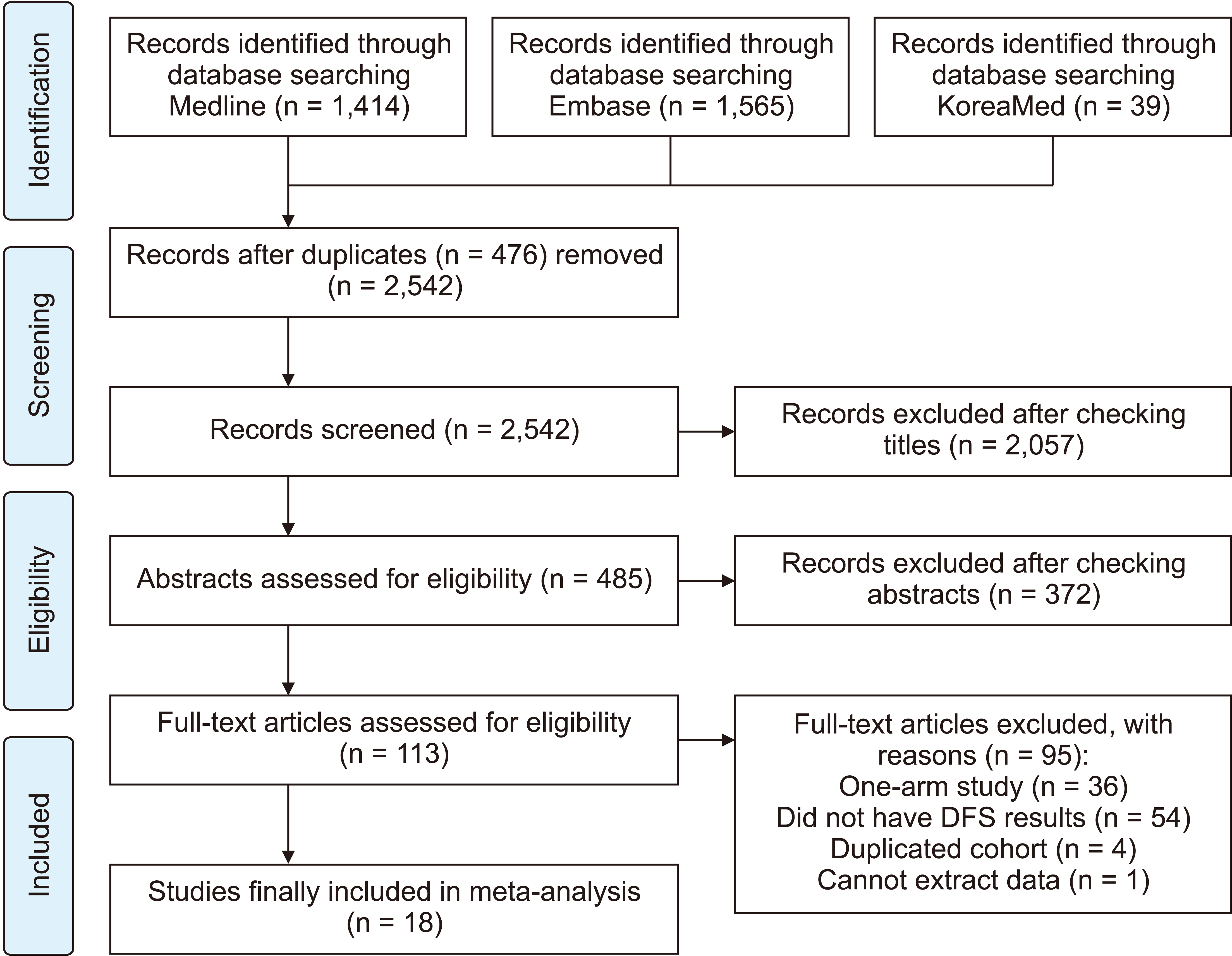
Table 1
Characteristics and baseline demographics of patients in each study included for this research
| Study (year) | Study design (country) | Study period | Cirrhosis (No/Yes) | Diagnosis of cirrhosis | Resection type (major/minor) | Male/female (no. of patients) | Age (yr) | CP class (A/B or C) | Etiology (HBV/other) | Stage (AJCC I/II/III/IV) | Vascular invasion (No/Yes) | Alpha-fetoprotein, mean (ng/mL) |
|---|---|---|---|---|---|---|---|---|---|---|---|---|
| Chang et al. (2004) [19] | Cohort (Taiwan) | 1991–1998 | 75/112 | Histologically confirmed |
16/59 11/101 |
87%/13% | 60 | NA | NA |
60/71/92 58/94/70 |
42/181 58/164 |
13,181 2,716 |
| Yamashita et al. (2007) [20] | Cohort (Japan) | 1985–2004 | 141/180 | Histologically confirmed | NA | 225/66 | 60.7 | 302/19 | 66/225 | NA | NA | 415.8 |
| Chua et al. (2010) [21] | Cohort (Austrailia) | 1991–2009 | 63/34 | Histologically confirmed | 44/53 | 75/22 | 49/48 (< 62/≥ 62) | 82/15 | 33/64 | NA | 93/4 | 67/30 (< 400/≥ 400) |
| Gassmann et al. (2010) [22] | Cohort (Germany) | 1997–2007 | 21/24 | Histologically confirmed | NA |
17/4 19/5 |
65.2 69.6 |
NA 19/5 |
2/19 5/19 |
3/11/7 2/18/4 (T1/T2/T3) |
6/15 6/18 |
NA |
| Fan et al. (2011) [23] | Cohort (China) | 1995–2008 | 97/311 | Histologically confirmed | 124/284 | 322/86 | 314/94 (≤ 65/> 65) | 395/13 | 367/41 | NA | 292/116 | NA |
| Ho et al. (2012) [24] | Cohort (Taiwan) | 2001–2007 | 408/338 | Histologically confirmed | NA | 581/165 | 52.3 | NA | 494/252 | NA | 561/185 | NA |
| Mizuguchi et al. (2013) [25] | Cohort (Japan) | 2006–2010 | 79/70 | Histologically confirmed | NA | NA |
67.8 66.0 |
NA |
34/45 32/38 |
9/48/13/9 16/25/26/3 |
NA | NA |
| Yang et al. (2014) [26] | Cohort (China) | 2002–2012 | 30/128 | Histologically confirmed | NA | 141/173 | 49.2 ± 11.5 | 132/26 | 143/15 | NA | 74/84 | 99/59 (≥ 20/< 20) |
| Yap et al. (2014) [27] | Cohort (Taiwan) | 1992–2002 | 131/93 | Histologically confirmed | 141/83 | 179/45 | 84/140 (< 50/≥ 50) | 192/22 | 151/73 | 224/0/0/0 | NA | 120/58/46 (≤ 20/20–400/≥ 400) |
| Chapman et al. (2015) [28] | Cohort (United States) | 1990–2011 | 151/97 | Histologically confirmed | NA | 170/78 | 66 (17–86) | NA | 57/191 | NA | NA | NA |
| Kluger et al. (2015) [29] | Cohort (France) | 1989–2010 | 166/147 | Histologically confirmed | 175/138 | 250/63 | 59 ± 14 | NA | 77/236 | NA | 155/158 | 226/87 (≤ 200/> 200) |
| Huang et al. (2016) [30] | Cohort (China) | 2001–2009 | 44/96 | Histologically confirmed | NA |
38/6 85/11 |
45 (28–77) 49 (14–79) |
241/15 |
28/16 90/6 |
NA |
41/3 90/6 |
39.2 53 |
| Lee et al. (2017) [31] | Cohort (Korea) | 1996–2012 | 262/387 | Histologically confirmed |
136/126 149/238 |
208/54 303/84 |
54.4 ± 10.8 54.9 ± 9.1 |
NA |
162/100 257/130 |
NA |
141/121 208/179 |
2,185.5 1,560.6 |
| Shehta et al. (2016) [32] | Cohort (Korea) | 2004–2013 | 91/141 | Histologically confirmed |
20/70 17/124 |
65/26 100/41 |
57 (26–79) 57 (31–87) |
87/3 125/16 |
58/33 110/31 |
53/33/4/1 12/48/36/9 (T1/T2/T3/T4) |
59/32 93/48 |
9.1 15.7 |
| Lee et al. (2017) [33] | Cohort (Korea) | 2001–2013 | 686/314 | Histologically confirmed | 529/473 | 807/195 | 760/242 (< 65/≥ 65) | 964/38 | 747/255 | 500/374/123/5 | 571/431 | 660/70/269 (< 200/200–400/≥ 400) |
| Cipriani et al. (2018) [34] | Cohort (Italy, UK, Norway, France, Belgium) | 1998–2014 | 70/333 | Histologically confirmed |
19/51 31/302 |
40/30 124/209 |
70 (7–87) 66 (23–88) |
70/0 308/25 |
NA | NA | NA | NA |
| Famularo et al. (2018) [35] | Cohort (Italy) | 2001–2015 | 63/291 | Histologically confirmed | NA | 280/74 | 66.1 | 330/24 | 74/280 | NA | 234/120 | NA |
| Golse et al. (2018) [36] | Cohort (France) | 2012–2016 | 45/15 | Histologically confirmed |
18/27 7/8 |
37/8 10/5 |
66.6 ± 12 64.1 ± 10 |
NA |
6/39 1/14 |
NA |
24/21 7/8 |
1,107 1,477 |
![]()
Table 2
Quality assessment using the Newcastle-Ottawa Scale
| Study (year) | Selection | Comparability | Outcome | Quality score | ||||||||
|---|---|---|---|---|---|---|---|---|---|---|---|---|
|
|
|
|
||||||||||
| 1 | 2 | 3 | 4 | 1 | 1 | 2 | 3 | |||||
| Chang et al. (2004) [19] | ★ | ★ | ★ | ★ | ★ | ★ | ★ | ★ | 8 | |||
| Yamashita et al. (2007) [20] | - | ★ | ★ | ★ | ★★ | ★ | ★ | ★ | 8 | |||
| Chua et al. (2010) [21] | - | ★ | ★ | ★ | ★★ | ★ | - | ★ | 7 | |||
| Gassmann et al. (2010) [22] | ★ | - | ★ | ★ | ★ | ★ | - | ★ | 6 | |||
| Fan et al. (2011) [23] | - | ★ | ★ | ★ | ★★ | ★ | ★ | ★ | 8 | |||
| Ho et al. (2012) [24] | - | ★ | ★ | ★ | ★ | ★ | ★ | ★ | 7 | |||
| Mizuguchi et al. (2013) [25] | ★ | - | ★ | ★ | - | ★ | - | - | 4 | |||
| Yang et al. (2014) [26] | - | ★ | ★ | ★ | ★★ | ★ | ★ | ★ | 8 | |||
| Yap et al. (2014) [27] | - | ★ | ★ | ★ | ★★ | ★ | ★ | ★ | 8 | |||
| Chapman et al. (2015) [28] | - | - | ★ | ★ | ★ | ★ | ★ | ★ | 6 | |||
| Kluger et al. (2015) [29] | - | ★ | ★ | ★ | ★ | ★ | - | - | 5 | |||
| Huang et al. (2016) [30] | - | ★ | ★ | ★ | ★★ | ★ | ★ | ★ | 8 | |||
| Lee et al. (2017) [31] | - | ★ | ★ | ★ | ★ | ★ | ★ | ★ | 7 | |||
| Shehta et al. (2016) [32] | ★ | ★ | ★ | ★ | ★★ | ★ | ★ | ★ | 9 | |||
| Lee et al. (2017) [33] | ★ | ★ | ★ | ★ | ★★ | ★ | ★ | ★ | 9 | |||
| Cipriani et al. (2018) [34] | - | - | ★ | ★ | ★★ | ★ | ★ | ★ | 7 | |||
| Famularo et al. (2018) [35] | - | ★ | ★ | ★ | ★★ | ★ | - | ★ | 7 | |||
| Golse et al. (2018) [36] | - | ★ | ★ | ★ | ★★ | ★ | - | ★ | 7 | |||
![]()
Recurrence-free survival after partial hepatectomy for hepatocellular carcinoma according to the presence or absence of cirrhosis in each study
Among 18 enrolled studies, the median RFS ranged from 17.8 to 106.8 months for the non-cirrhosis group and 7.1 to 43 months for the cirrhosis group (Table 3). For the non-cirrhosis group, 1-, 3-, and 5-year RFS rates ranged from 70.3% to 93%, 49.3% to 84%, and 20% to 71%, respectively. For the cirrhosis group, 1-, 3-, and 5-year RFS rates ranged from 39% to 85.5%, 36% to 64.5%, and 4% to 50.8%, respectively.
Table 3
Median recurrence-free survival (months) and 1-, 3-, 5-year recurrence-free survival (%)
| Study (year) | Median recurrence free survival (mon) | Recurrence free survival (%) | ||
|---|---|---|---|---|
|
|
||||
| 1-year | 3-year | 5-year | ||
| Chang et al. (2004) [19] | ||||
| Cirrhosis | 34.1 | - | - | 26.9 |
| Non-cirrhosis | 38.2 | - | - | 36.8 |
| Yamashita et al. (2007) [20] | ||||
| Cirrhosis | - | - | - | - |
| Non-cirrhosis | - | - | - | - |
| Chua et al. (2010) [21] | ||||
| Cirrhosis | 8 | - | - | - |
| Non-cirrhosis | 24 | - | - | - |
| Gassmann et al. (2010) [22] | ||||
| Cirrhosis | 7.1 | 39 | - | 4 |
| Non-cirrhosis | 20.7 | 76 | - | 20 |
| Fan et al. (2011) [23] | ||||
| Cirrhosis | 42.8 | - | - | - |
| Non-cirrhosis | 106.8 | - | - | - |
| Ho et al. (2012) [24] | ||||
| Cirrhosis | - | - | - | 35 |
| Non-cirrhosis | - | - | - | 51.8 |
| Mizuguchi et al. (2013) [25] | ||||
| Cirrhosis | - | - | - | - |
| Non-cirrhosis | - | - | - | - |
| Yang et al. (2014) [26] | ||||
| Cirrhosis | - | - | - | - |
| Non-cirrhosis | - | - | - | - |
| Yap et al. (2014) [27] | ||||
| Cirrhosis | - | 81 | 46.7 | 34.4 |
| Non-cirrhosis | - | 83.4 | 64.6 | 54.9 |
| Chapman et al. (2015) [28] | ||||
| Cirrhosis | - | - | - | 18.4 |
| Non-cirrhosis | - | - | - | 30.1 |
| Kluger et al. (2015) [29] | - | |||
| Cirrhosis | 20.2 | - | - | - |
| Non-cirrhosis | - | - | - | - |
| Huang et al. (2016) [30] | ||||
| Cirrhosis | - | 71 | 36 | 25 |
| Non-cirrhosis | - | 93 | 84 | 71 |
| Lee et al. (2017) [31] | ||||
| Cirrhosis | - | 80.4 | 55.6 | - |
| Non-cirrhosis | - | 85.1 | 67.3 | - |
| Shehta et al. (2016) [32] | ||||
| Cirrhosis | 29.3 | - | - | - |
| Non-cirrhosis | 29.1 | - | - | - |
| Lee et al. (2017) [33] | ||||
| Cirrhosis | - | 74.5 | 56.8 | 50.8 |
| Non-cirrhosis | - | 70.3 | 49.3 | 40.6 |
| Cipriani et al. (2018) [34] | ||||
| Cirrhosis | 43 | 85.5 | 64.5 | 37.6 |
| Non-cirrhosis | 55 | 81.4 | 55.1 | 45.3 |
| Famularo et al. (2018) [35] | ||||
| Cirrhosis | - | - | - | - |
| Non-cirrhosis | - | - | - | - |
| Golse et al. (2018) [36] | ||||
| Cirrhosis | 13.7 | - | - | - |
| Non-cirrhosis | 17.8 | - | - | - |
![]()
Cumulative hazard ratios for the two groups from a meta-analysis
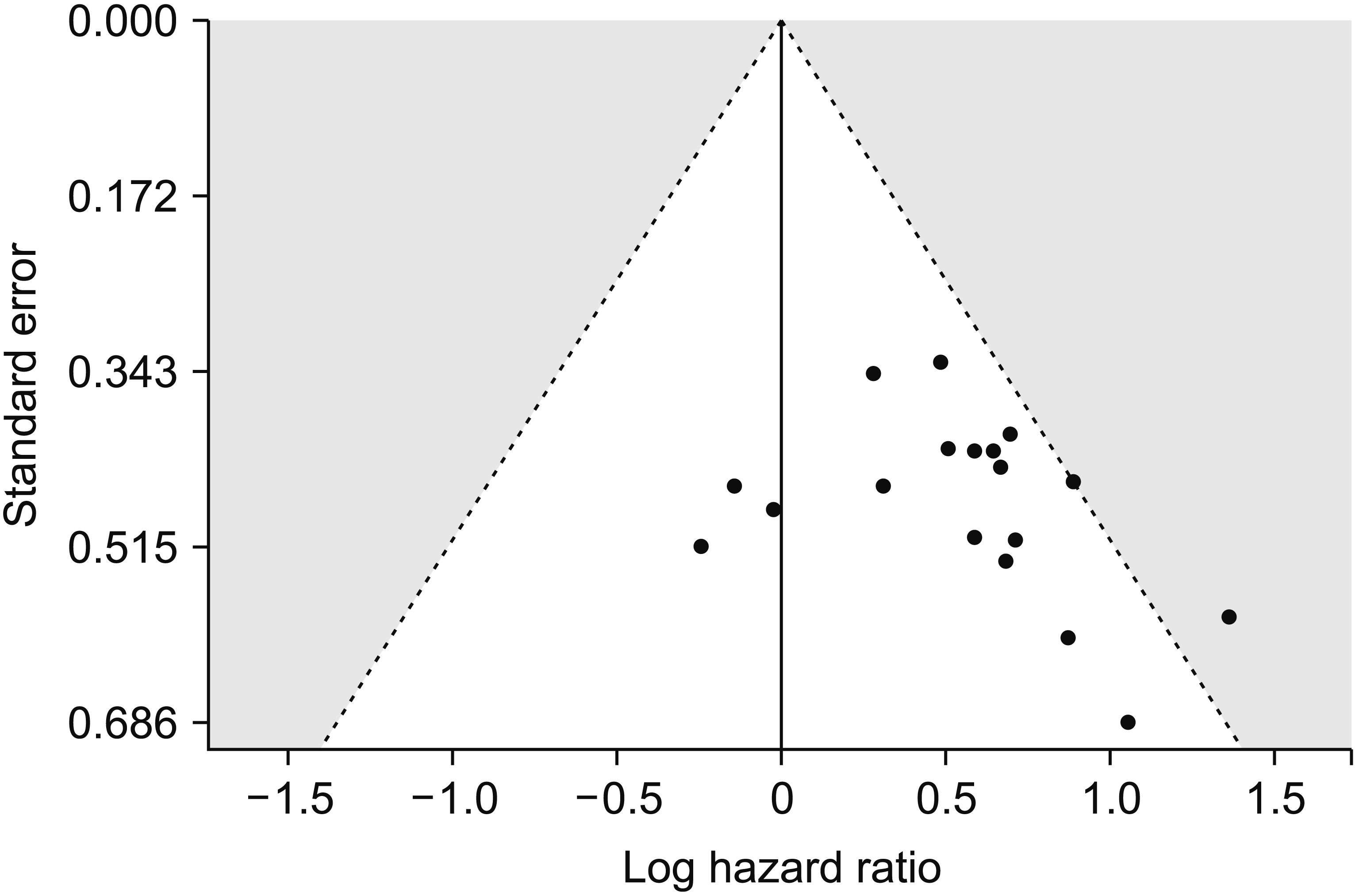
To estimate the difference of RFS between the non-cirrhosis and cirrhosis groups, HR was used. It was calculated with the fraction of the risk of tumor recurrence in the cirrhosis group compared to that in the non-cirrhosis group. Original HRs of the RFS could be extracted from ten studies [20,21,26,27,29-31,33,35,36]. However, eight studies did not show their own HRs [19,22-25,28,32,34]. The KM curve for each subject of the non-cirrhotic and cirrhotic groups was obtained separately from eight studies to secondarily calculate HR. Following the proposed algorithm [16], KM survival data were secondarily restored and used to reconstruct pseudo-KM curves and calculate the HR (Fig. 2). The cumulative HR of cirrhosis was 1.66 (95% CI, 1.43–1.93; Fig. 2) in the random-effect model. A high level of heterogeneity existed among the 18 studies (p < 0.01; I2 = 61%). This indicated a large degree of difference among effect sizes of these studies. Publication bias analysis was conducted to compare the endpoint (HR) between non-cirrhosis and cirrhosis groups. Visual evaluation of the overall funnel plot (Fig. 3) revealed an asymmetrical phenomenon. However, the Egger’s test showed no apparent publication bias (p = 0.341).
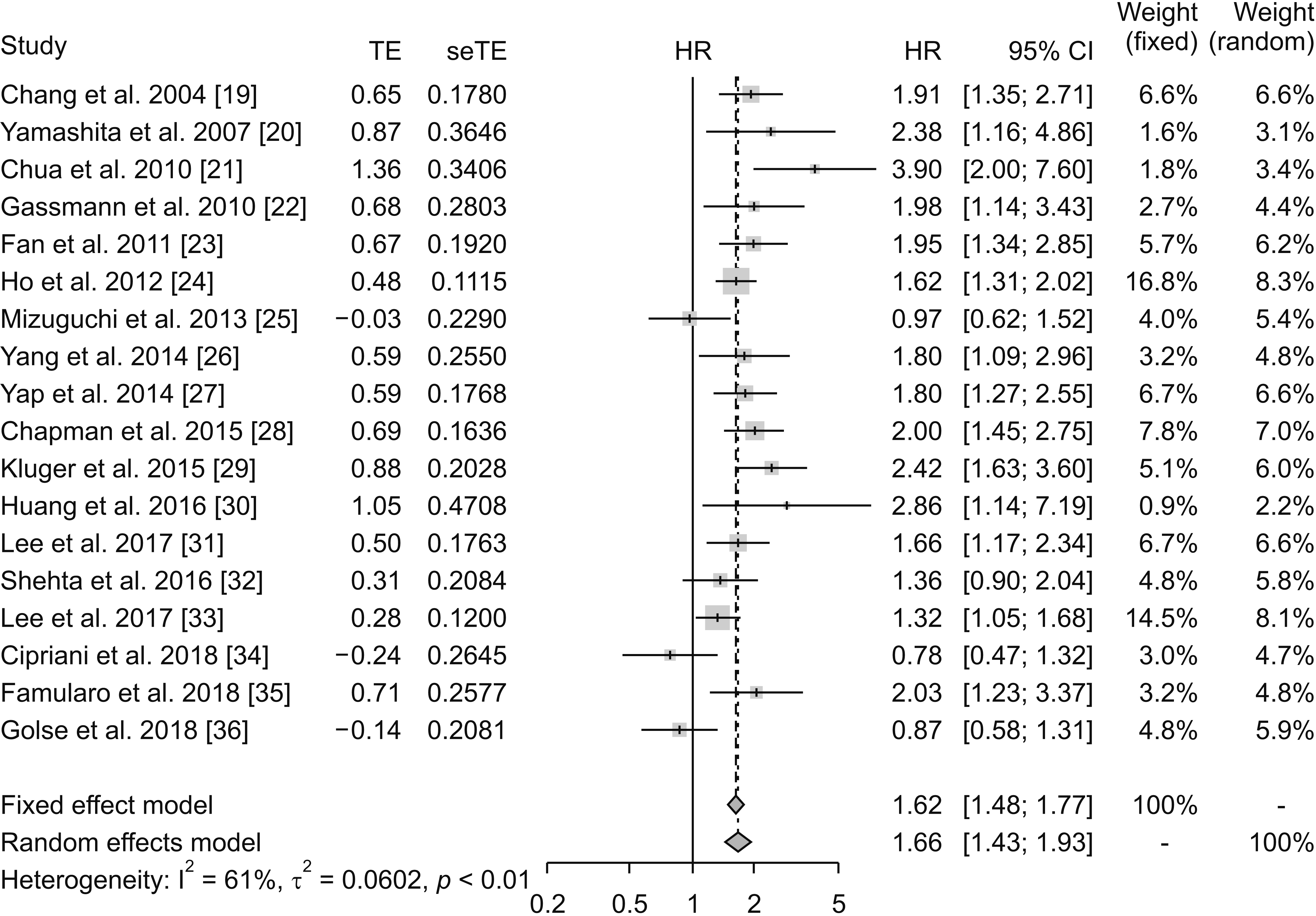
Fig. 2
Forest plot depicting hazard ratio (HR) of recurrence-free survival after partial hepatectomy in patients with hepatocellular carcinoma (HCC) accompanied by cirrhosis compared to that in patients with HCC without cirrhosis using fixed-effect and random-effect models. TE, estimated treatment effect; seTE, standard error of treatment estimate; CI, confidence interval.
![]()
Cure fractions after partial hepatectomy in each group
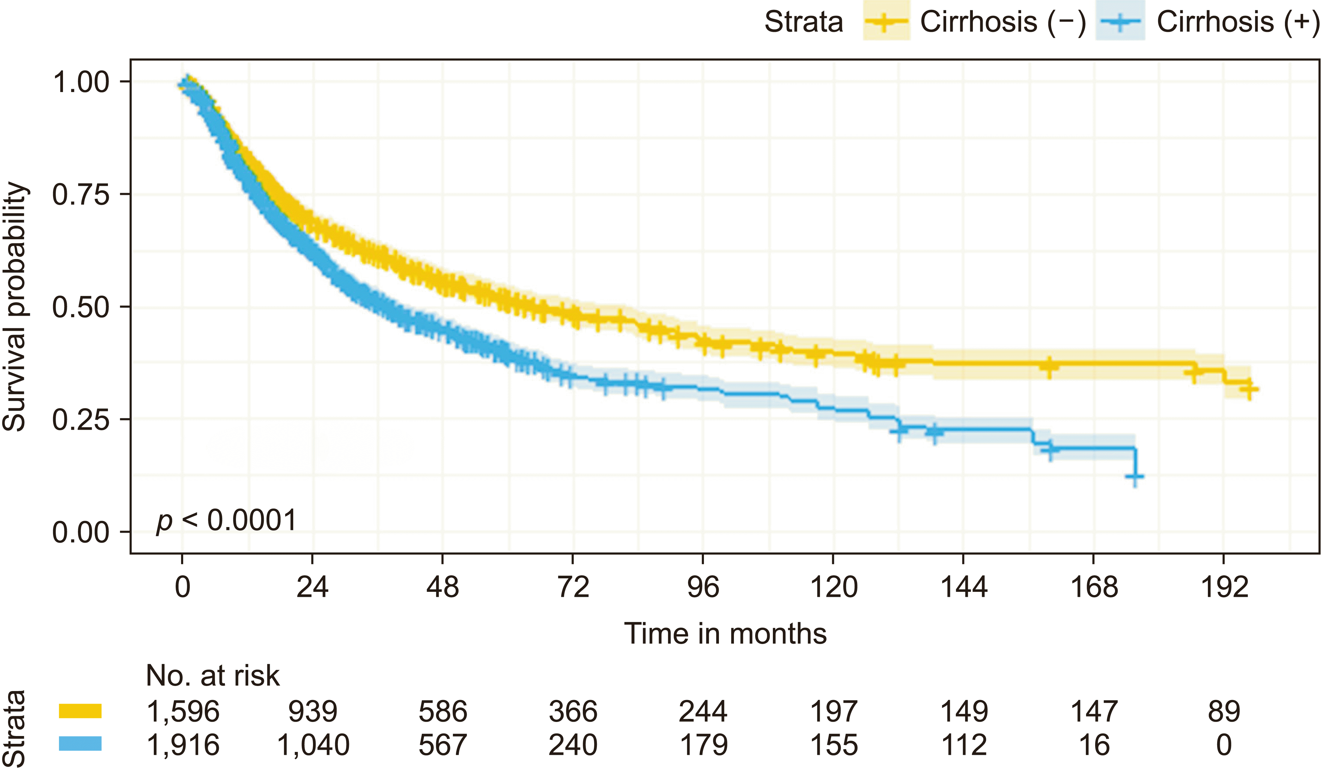
Combined patient survival data of 3,512 patients were reconstructed from KM survival curves of 13 studies: 1,596 patients in the non-cirrhosis group and 1,916 patients in the cirrhotic group [19,21-25,27,29-32,34,36]. Analysis of such data using KM methods demonstrated 1-, 3-, 5-, and 10-year survival rates of 83.3%, 62.1%, 52.2%, and 39.6% in the non-cirrhosis group and 78.8%, 51.3%, 39.7%, and 27.3% in the cirrhosis group, respectively (Fig. 4; p < 0.001). The probability of patients being cured by PH for HCC in the non-cirrhosis group was 32.5% (95% CI, 28.6%–36.4%). For patients in the cirrhosis group who underwent PH, the probability of being cured was 14.1% (95% CI, 10.6%–18.1%). From non-mixture cure model results, a larger proportion of patients were cured in the non-cirrhosis group than in the cirrhosis group (HR, 1.43; 95% CI, 1.30–1.57; p < 0.001).
DISCUSSION
Although PH is commonly used in the treatment of HCC without cirrhosis, its use in the treatment of HCC with cirrhosis remains controversial. This was attributed to the higher recurrence rate in patients with HCC accompanied by cirrhosis who underwent PH than that in patients who underwent LT [12,37]. In addition, the risk of recurrence after PH was higher in patients with HCC and cirrhosis than in those without cirrhosis [38]. Some reports suggest that PH is a viable option for treating HCC accompanied by cirrhosis by showing acceptable survival rates [38,39]. However, results derived from these studies do not reflect the curability of a certain treatment. The cure model recently discussed and applied in various diseases for its capability to predict the probability of cure is of value. In our study, we used it to examine the probability of PH curing patients with HCC and cirrhosis. We also compared the cure fraction in cirrhotic patients to that in non-cirrhotic patients. In our study, the RFS of patients with HCC accompanied by cirrhosis after PH was inferior to that of patients with HCC without cirrhosis. Nonetheless, the cure model analysis revealed that patients with HCC accompanied by cirrhosis had a meaningful cure fraction after PH (14.1%; 95% CI, 10.6%–18.1%). To the best of our knowledge, no studies have directly compared cure fractions of patients having HCC with and without cirrhosis after PH.
Recent studies have defined a “cure” as follows: survival curve reaches a plateau at the end, which occurs when the mortality rate of a patient reaches the same level as that in the general population [13,14]. This is generally seen in cancer cases. Currently, the cure model analysis is used in many clinical settings, for instance, to determine the treatment modality and the follow-up term for patients [15]. In the field of oncology, the goal of treatment is to increase a patient’s survival time or cure cancer. Application of the cure model in oncology increases with increasing number of long-term survival patients. In our study, the survival curve of patients with HCC after PH showed a plateau at the end. Thus, the cure model was used to analyze data in both patient groups that showed cure fraction. The cure fraction of patients with HCC accompanied by cirrhosis was lower than that of patients with HCC without cirrhosis. However, around 14 percent of patients with HCC accompanied by cirrhosis were cured after PH.
Cirrhosis is a major risk factor of HCC regardless of its etiology [1,40]. Many patients with HCC first suffer from repetitive hepatitis, cirrhosis, and then finally HCC [40]. Chronic liver diseases such as viral hepatitis and alcoholic liver disease can cause damage to hepatocytes, eventually leading to death. In response to liver injury, myofibroblasts are activated, which are derived from hepatic stellate cells or perivascular fibroblasts [41]. Myofibroblasts play an important role in fibrogenesis, which has a protective role in response to liver damage. However, chronic and excessive fibrosis can occur if the underlying disease is not addressed. In addition, the composition of the extracellular matrix (ECM) changes during fibrosis progression. Collagen proteins, predominantly type I collagens, can accumulate in the ECM, causing structural changes and activating growth factors that contribute to malignant changes [40]. A recent study has shown that peritumoral myofibroblasts are associated with a high recurrence rate in patients undergoing PH. These results imply that fibrosis may make the cancer more aggressive [42]. Some reports have demonstrated that the tumor in a cirrhotic liver can progress into a more aggressive form of HCC than a tumor in a non-cirrhotic liver [43,44]. One study has reported the relationship between the expression of geranylgeranyl diphosphate synthase 1 (GGPPS1) and the development of HCC from cirrhosis [43]. The expression of GGPPS1 was higher in tumor tissues than in tumor-free tissues. Moreover, cirrhotic livers showed higher expression levels of GGPPS1 than healthy livers. Interestingly, GGPPS1 also showed a close correlation with prognostic factors such as tumor stage, vessel invasion, and early recurrence [43]. Another study has suggested that 20 hub genes play an important role in HCC progression [44]. These genes were strongly expressed in patients with HCC with cirrhosis. The degree of gene expression was associated with overall survival and disease-free survival in patients with HCC. These studies unanimously demonstrate that a cirrhotic liver could lead to a more aggressive form of HCC, which in turn leads to a poor prognosis.
In patients with HCC accompanied by cirrhosis, LT is considered an ideal treatment option as it can simultaneously treat HCC and the underlying cirrhosis [7]. The overall survival rate and RFS survival rate were higher in patients who underwent LT than in those who underwent PH [11,45]. However, the postoperative mortality at 3 months was higher and hospital stay was longer in patients treated with LT than in those treated with PH [46]. LT has also been criticized for its restrictive selection criteria [8,9]. While efforts have been made to extend selection criteria for LT, only a limited number of patients have received LT. Another problem with LT is the shortage of donor organs. According to one meta-analysis, while waiting for donor organs, some patients underwent loco-regional therapy as a bridging or down-staging therapy. However, these therapeutic approaches did not significantly reduce the risk of waitlist dropout due to HCC progression. Furthermore, there were no differences in post-transplant outcomes between a locoregionally treated group and a group without receiving a loco-regional therapy [47,48]. Therefore, patients with HCC on the waitlist are at risk of disease progression and morbidity or mortality related to cirrhosis. PH can proceed without delay. It can be applied to a wider range of patients with HCC [49]. There are several additional disadvantages related to LT, including the need for immunosuppressants known to have severe side effects such as nephrotoxicity, opportunistic infections, and malignancies [50]. However, the decision on whether to perform PH or LT for HCC accompanied by cirrhosis cannot be made uniformly. It is excessively influenced by tumor characteristics (size, number, and location), presence of portal hypertension, available living donor, and patient’s performance status and underlying disease.
Recently, salvage liver transplantation (SLT) has been proposed as an alternative treatment option for HCC recurrence after primary PH. This is being discussed due to donor shortage. According to recent studies, the long-term survival outcome of SLT was comparable to that of primary LT [51,52]. Therefore, PH can be considered as an alternative to primary LT as the primary treatment for patients with HCC accompanied by cirrhosis. Our results corroborated these approaches because around 14% patients with HCC accompanied by cirrhosis could be cured after PH. Pathological data that affect prognosis such as tumor size, tumor number, and vascular invasion can also be obtained before SLT [53,54]. Consequently, after primary PH, patients who are revealed to be at high risk of recurrence could undergo “prophylactic” liver transplantation for better prognosis [55]. Furthermore, if recurrence occurs, the liver can be transplanted in the future because the majority of recurrences after PH are within the Milan criteria [56]. Thus, if applicable, PH should be initially performed for patients with HCC accompanied by cirrhosis.
The value of the cure model analysis is that the probability of cure can be calculated for patients of interest. In addition, it can provide information that may aid in making decisions in a variety of clinical settings, such as the postoperative surveillance period and decisions on treatment modalities for other combined diseases.
One limitation of this study was that it was a secondary analysis. Factors such as tumor size, alpha-fetoprotein, and vascular invasion affecting HCC recurrence were not fully identified in original articles. In addition, all included studies were retrospective studies, which might have been biased. In this study, we only compared cure fraction according to the presence or absence of cirrhosis. Future studies are needed to conduct additional subgroup cure model analyses for HCC. The impact of the extent of surgery on the prognosis of patients with HCC accompanied by cirrhosis is a matter of debate and another important research topic to be considered. Therefore, further studies about this issue are warranted.
In conclusion, according to results of our study, PH might be able to cure HCC even in patients with cirrhosis. We propose that PH is a reasonable treatment approach for patients with HCC accompanied by cirrhosis in that it could offer a chance for cure.
SUPPLEMENTARY DATA
Supplementary data related to this article can be found at https://doi.org/10.14701/ahbps.21-080.
 XML Download