Journal List > Korean J Gastroenterol > v.79(3) > 1160403

호산구성 식도염의 진단과 치료

Abstract

Eosinophilic esophagitis (EoE) is a chronic, immune/antigen-mediated esophageal disease characterized by symptoms of esophageal dysfunction and eosinophil-predominant inflammation. The diagnostic criteria for EoE have changed with our growing knowledge over the past two decades. Esophageal eosinophilia, which responds histologically to a proton pump inhibitor (PPI) is not a distinct disease but a subset of EoE. An endoscopic scoring system that relies on the assessment of exudates, rings, edema, furrows, and strictures is a useful tool for assessing endoscopic severity. PPIs are regarded as safe and effective first-line treatments for EoE. Oral topical corticosteroids or dietary therapy are also options for first-line treatment. Endoscopic dilation is effective for relieving the dysphagia symptoms of a patient with an esophageal stricture.

서 론

호산구성 식도염(eosinophilic esophagitis)은 주로 음식 항원에 대한 항진된 T help cell (Th) 2 면역반응에 의해 조직학적으로 식도 점막에 호산구의 과증식이 관찰되면서 식도장애 증상이 동반되는 만성 염증 질환이다.1,2 만성적인 염증에 의해 섬유화가 진행되면 식도 내강이 좁아져 삼킴곤란이나 음식 걸림이 발생하게 된다. 호산구성 식도염은 1977년 처음으로 호산구성 위장염의 식도 침범으로 보고되었고, 1978년 식도 이완불능증 환자에서 식도 근육층의 비후와 호산구 침착이 있는 아형으로 소개되었다.3,4 지금 개념에서 이 증례는 2차적인 원인의 식도 호산구증으로 분류되며, 현재와 같은 개별 질환으로 호산구성 식도염이 보고되기 시작한 것은 1990년대부터이다.5-7 대부분 서구에서 발표된 인구 기반 유병률 연구들은 호산구성 식도염이 꾸준히 증가하고 있다고 보고하고 있다.8 아시아 지역의 유병률 연구들은 호산구성 식도염의 유병률이 서구에 비해 낮은 것으로 보고하고 있으나 대상자 수가 적거나 증례 위주의 보고라는 한계점이 있다.9 하지만 점점 서구화 되는 식습관 등으로 최근 국내 연구에서도 유병률이 증가하는 경향을 보이는 연구 결과들이 발표되고 있다.10,11 본고에서는 이러한 호산구 식도염의 병태 생리 및 임상 증상, 정의, 내시경과 조직학적 진단, 약물 및 비약물 치료 등에 대하여 알아보고자 한다.

본 론

1. 병태생리

1) 환경적 요인

호산구성 식도염 유병률 증가의 원인으로 환경적 요인이 중요한 역할을 차지한다. 제왕절개, 조숙아, 유아기 항생제 노출, 모유 수유 실패 등이 호산구성 식도염과 연관성이 있다고 보고되었는데, 이는 면역 시스템이 형성되는 영유아기에 비정상적인 자극에 의해 질환의 발생 위험이 증가하는 것으로 추정하고 있다.12 즉, 변화된 장내 세균총이 천식이나 아토피 질환과 마찬가지로 호산구성 식도염의 발생 위험을 증가시키는 원인으로 제시되고 있다.13 실제로 호산구성 식도염에서 알레르기 질환과의 동반율을 평가한 연구에서 천식(12-38%), 아토피(70-80%), 알레르기성 비염(17-70%)의 유병률이 상당히 높게 보고되었다.14 그 외에도 여러 역학 연구에서 Helicobacter pylori 감염이 면역 기능을 조절해서 알레르기 질환의 보호 효과를 보이며, 호산구성 식도염 유병률과도 역의 상관관계가 있음을 보고하였다.15-17 하지만 이와 상반되는 연구 결과도 있어 현재로서는 Helcobacter pylori 감염과 호산구성 식도염의 연관성에 대해 결론을 내리기에는 무리가 있다.18
위식도 역류 질환도 위 음식 역류로 식도 상피에 항원 노출이 증가되고, 위산에 의한 자극으로 상피 내 세포간 공간이늘어나 점막을 통한 항원의 이동을 용이하게 만들어 호산구식도염의 병인이 될 수 있다.19,20

2) 유전적 요인

남성에서 높은 유병률, 질환의 가족력은 호산구 식도염 발생과 연관된 유전적 요인이 있음을 시사한다. 호산구성 식도염 유전자 연구에서 다양한 유전자들 중 대표적으로는 5q22에서 Th2 면역 반응을 항진시키는 thymic stromal lympho-protein (TSLP) 유전자, 2p23에서 식도 상피에 IL-13을 조절하는 물질인 calpain 14를 코딩하는 CAPN 14 유전자가 있다. 또한, 전장 유전체 상관성 분석(genome-wide association study)에서 호산구성 식도염의 염증 발생 기전과 연관된 eotaxin-3 유전자의 단일염기다형성(single nucleotide polymorphism)이 확인되었다.2 이러한 식도 장벽 기능의 손상을 일으키거나 염증 반응을 항진시키는 유전자가 발현되면 항원 물질이 점막을 통과해 진입할 수 있는 경로를 제공하므로 중요한 유전적 소인이 될 수 있다.21

3) 면역 반응

호산구성 식도염은 말초혈액 호산구 증가증이 동반되지 않는 경우도 많으며, 질환 활성도와의 연관성도 알려져 있지 않다. Th2 면역 매개성 염증으로 발생하는 호산구성 식도염에서 항원 매개성 호산구 모집(recruitment)을 유도하는 eotaxin-3가 중요한 역할을 한다.22 Eotaxin-3 수치는 질환의 활성도와 연관이 있으며 한 보고에서는 식도 조직 검사에서 89%의 진단 예측률을 보고하였다.23 호산구성 식도염 환자의 식도에서 증가된 TSLP는 Th2 면역을 촉진하여 호산구성 염증을 매개하는 여러 사이토카인들을 활성화시키는데, 그중 IL-5, IL-13, IL-15가 대표적이다. IL-5는 호산구를 활성화시켜 식도 재형성(remodeling)과 콜라겐 침착을 일으키고 IL-13과 IL-15는 eotaxin-3 발현을 증가시킨다.24,25 또한, IL-13은 desmoglein-1과 filaggrin을 억제하여 상피의 투과도를 증가시켜 식도 점막의 장벽 기능을 약화시킨다. 식도 조직에서 호산구 외에도 비만세포의 과증식도 확인이 되는데, 호산구와 더불어 이러한 비만세포의 과증식은 섬유화 인자와 혈관 생성 인자인 major basic protein, transforming growth factor 그리고 vascular cell adhesion molecule 1을 통해 식도의 염증과 재형성을 유발한다.26

2. 임상 증상

호산구성 식도염의 임상 증상은 연령에 따라 차이가 있다.8 어른이나 청소년들은 삼킴곤란 또는 음식 막힘, 흉통이 주를 이루는데 반해, 영유아에서는 잘 먹지 못하거나 성장장애로 나타나며, 3-12세 소아에서는 구토, 복통, 역류 증상이 흔하다.27 이러한 차이는 시간 경과에 따른 호산구성 식도염의 진행과 연관이 있는데, 어린 나이에는 활성화된 호산구성 염증이 증상의 주요 원인이라면 성인에서는 식도벽의 섬유화와 협착이 증상의 원인이 되기 때문이다.28 즉, 호산구성 식도염은염증 중심의 아형에서 시작하여 섬유화 중심의 아형으로 진행한다고 생각된다.29 또한 식도 근육의 기능장애도 식도 증상을일으키는 요인이다.30 그러나 삼킴곤란을 일으킬 수 있는 이러한 점막하층의 섬유화나 근육의 기능장애는 일반적인 내시경이나 조직 검사로 평가가 어렵고 임상 증상과 내시경 소견및 조직 염증 소견 사이에 불일치를 보이기도 한다.31,32
이러한 호산구성 식도염 임상 증상의 정도는 무증상부터 간헐적인 고형식 삼킴곤란, 매일 반복되는 음식 걸림까지 다양하다. 증상이 경미한 경우 병원 진료를 보지 않고 지내기 쉬운데, 치료 없이 지내며 병이 장기화되면 결국 협착 발생으로 증상이 악화되어 병원을 방문하게 된다.28 또한 삼킴곤란 증상을 완화시키기 위해 증상을 유발하였던 음식을 피하거나, 천천히 삼키고 식사 동안 물을 계속 마시는 보상 행동은 진단을 지연시킬 수 있다. 따라서 성인에서 삼킴곤란이나 음식 걸림 또는 산분비억제제 치료에도 호전되지 않는 가슴 쓰림이 지속될 때는 반드시 호산구성 식도염을 감별하기 위해 내시경 소견이 정상이더라도 식도 점막의 생검을 고려해야 하겠다.

3. 정의

호산구성 식도염은 식도 증상과 함께 조직학적으로 식도에호산구 증가증을 특징으로 하는 질환이다. 호산구성 식도염은 호산구로 인한 염증이 식도에 국한되어 있는 경우이며, 식도뿐 아니라 위나 장까지 침범되었다면 호산구성 위장염이나 호산구성 장염으로 분류할 수 있다.33 2018년 개정된 호산구성 식도염 진단에 대한 국제적인 합의에 따르면 호산구성 식도염은 1) 식도 증상(Table 1)이 있으면서, 2) 식도 조직 생검에서 고배율 시야당 15개 이상의 호산구가 관찰되고, 3) 식도 호산구증을 초래할 수 있는 2차성 원인 질환들(호산구성 위장염, 결체조직 질환, 감염, 약물 과민증, 크론병 등)이 없는 경우 진단할 수 있다(Table 2).34 과거에는 양성자펌프억제제 8주 치료에 식도 호산구증이 호전되면 양성자펌프억제제 반응성 식도 호산구증(proton-pump inhibitor responsive esophageal eosinophilia)으로 명명하고, 호산구성 식도염과는 별개의 질환으로 분류하였다.35,36 그러나 호산구성 식도염과 양성자펌프억제제 반응성 식도 호산구증의 치료 전 임상 소견, 내시경 소견 및 조직 특성에서 둘을 구분할 수 있는 차이가 없었다.37,38 게다가 아토피 질환의 동반, 알레르기와 염증 인자들의 상승, RNA 발현 검색(expression profiles) 유사성 그리고 양성자펌프억제제와 국소 스테로이드, 제한 식이요법 치료 등에 비슷한 효과를 보고하였다.39-41 또한, 양성자펌프억제제 치료가 산 억제의 효과가 아닌 알레르기성 염증 경로에 대한 직간접적인 영향으로 식도 점막의 상피 방어 기능을 개선하는 기전들이 밝혀졌다.42,43 이런 이유로 양성자펌프억제제에 대한 반응 평가가 호산구성 식도염 진단 과정에서 제외되면서 양성자펌프억제제 반응성 식도 호산구증이라는 개념은 사라지고 호산구성 식도염의 아형으로 생각되게 되었다.34

4. 진단

1) 내시경 소견

특징적인 내시경 소견은 부종(edema), 원형 주름(circular rings), 백색의 삼출물(white exudate), 종축 방향으로 선상의 골(linear furrow), 협착(stricture) 등이 있다.44 그 외에도 접촉에 의해 쉽게 출혈되고 탄성이 없는 크레페 종이 모양 (crepe-paper) 소견을 보일 수 있다. 부종은 식도 염증으로 인해 점막이 두꺼워지고 현관상의 소실로 나타난다(Fig. 1A). 동심원 모양의 주름은 횡축을 따라 나타나며, 병이 많이 경과하여 섬유화가 일어난 경우에는 치료에 반응이 있더라도 주름이 소실되지 않는다(Fig. 1B). 간혹 정상인도 내시경 검사 중구역질을 하면 원형 주름이 보일 수 있어 주의가 필요하다.백색 삼출물은 조직 검사에서 호산구가 응집되어 나타나는 미세농양(eosinophilic microabscess)이며 육안상 식도 캔디다증과 비슷하게 보이기도 한다(Fig. 1C). 선상의 골은 종축으로 관찰되며 한 메타분석에서 호산구성 식도염의 가장 흔한내시경 소견으로 보고하였다(Fig. 1D).45 하지만 내시경 소견에 대한 진단 체계가 확립되지 않아 특징적인 내시경 소견의빈도는 보고마다 다양하다.
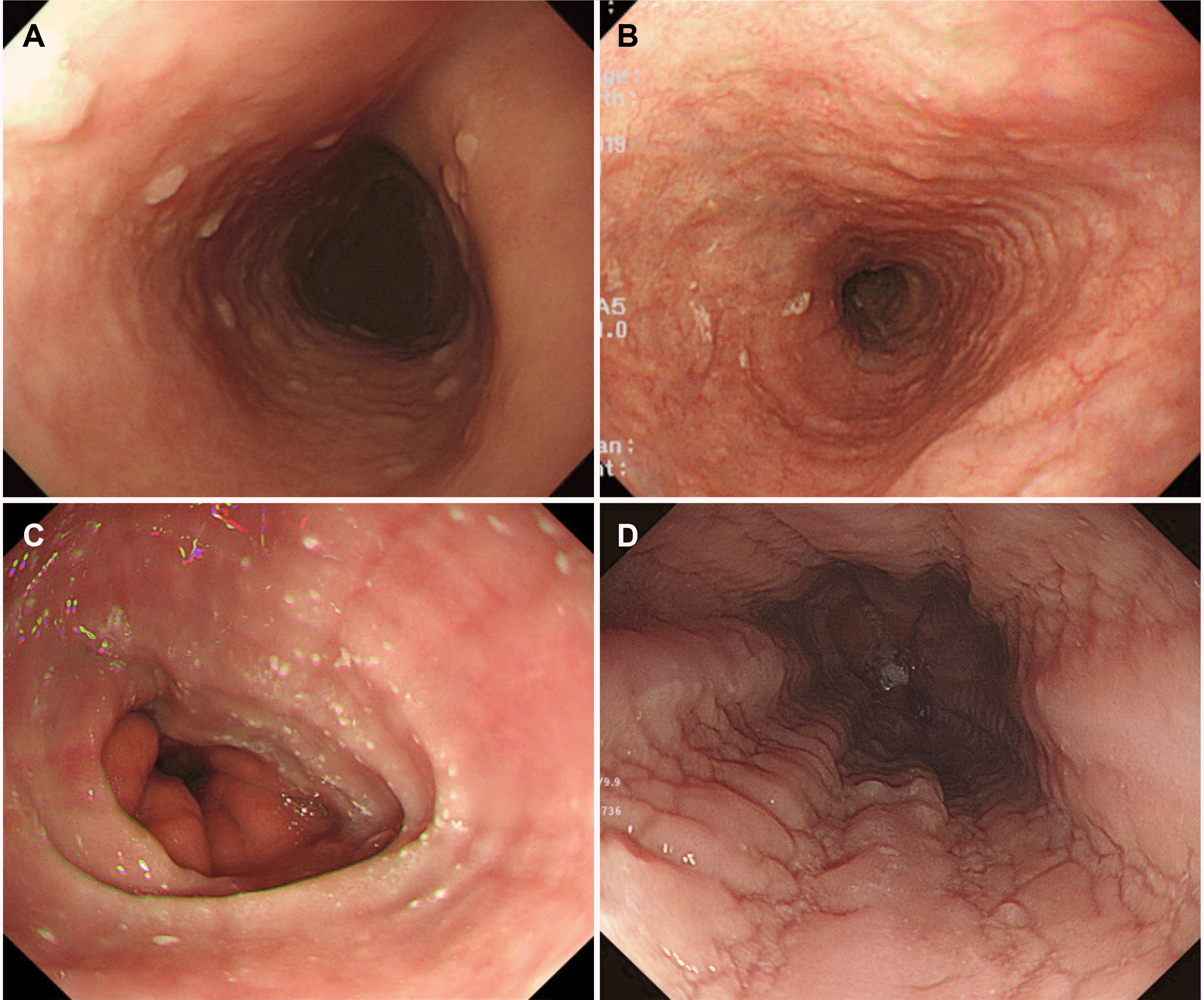
최근 부종, 원형 주름, 삼출물, 선상의 골, 협착 소견을 이용한 평가 시스템이 제안되었고 전문가 및 비전문가 평가자 간의 좋은 일치도를 보였다.44,46 새로운 EREFS (E, edema; R, rings; E, exudates; F, furrows; S, strictures) 평가 시스템은 호산구 식도염의 내시경 중증도 및 치료 반응 평가 표준화에 기여할 것으로 기대된다(Table 3). 이러한 내시경 소견 중증도와 조직학적 중증도 간에 연관성은 없지만 치료 후 EREFS 수치 감소와 조직 소견 호전과 연관성은 있다고 보고되었다.47,48
전형적인 호산구성 식도염의 내시경 소견을 보이면서 식도기능장애 증상이 있는 경우에는 호산구성 식도염으로 진단되는 빈도가 아주 높지만, 식도기능장애 소견만 있고 내시경에서 정상 소견을 보이는 경우는 조직 검사를 시행하지 않는다면 호산구 식도염을 진단할 수 없다.49 최근 메타분석에서 정상 내시경 소견을 보인 호산구성 식도염 환자가 전체 환자의 17%라고 보고하였다.45 따라서 내시경 검사자는 삼킴곤란이나 이유가 명확하지 않은 음식 걸림이 있는 환자에서 기질적 원인 평가를 위해 내시경 검사를 시행할 때 식도 점막 이상 소견이 보이지 않더라도 식도 조직 생검 시행을 적극적으로 고려해야 하겠다.

2) 조직 소견(Fig. 2)

호산구성 식도염의 진단을 위해서는 식도 조직 검사에서 고배율 시야당 15개 이상의 호산구가 관찰되어야 한다.34 하지만 호산구성 염증이 국소적으로 산발하므로 모든 조직 검사에서 이러한 소견이 확인되지 않을 수 있다. 따라서 진단율 향상을 위해 유럽 가이드라인에서는 내시경 이상 소견이 있는 부위를 중점적으로 두 군데(원위부와 근위부) 이상의 위치에서 최소 6개 이상의 생검을 권고하고 있다.36 하지만 연구자에 따라 4번의 생검으로도 높은 진단율을 보고하고 있어, 비용 효과를 고려할 때 2-4조각의 검체 채취를 권유하기도 한다.50-53 하부식도 생검 시는 식도염으로 인한 호산구 침착과 혼동되지 않도록 식도염이 관찰되는 부위를 피해 생검을 시행하고 해석에 주의를 요한다.
그 외 호산구성 식도염에서 보일 수 있는 특징적 조직 소견으로 호산구 미세 농양(eosinophil microabscesses), 상피 표층 호산구 침착(superficial layering of eosinophils), 상피하 및 고유판의 섬유화(subepithelial and lamina propria fibrosis), 호산구의 탈과립(degranulation of eosinophils) 등이 있다.54,55 기저층 과증식(basal cell hyperplasia), 유두 연장(papilla elongation), 세포간 공간 확장(dilated intercellular space), 비만세포의 증가는 호산구성 식도염과 식도 역류 질환 모두에서 관찰될 수 있다.54,55
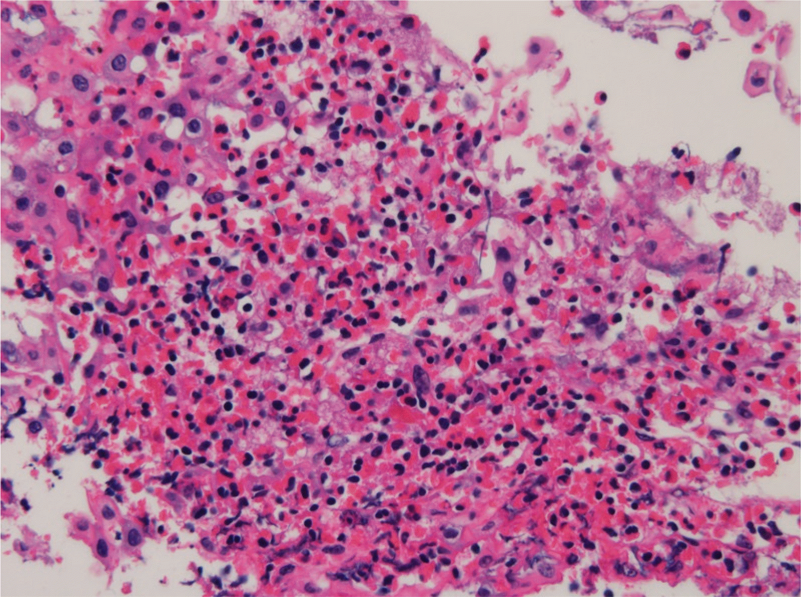
3) 영상 소견

호산구 식도염에서 복부 컴퓨터단층촬영이나 내시경 초음파에서 식도벽의 비후가 관찰될 수 있어 보조적 진단 수단으로 사용될 수 있다.56 초음파 내시경에서는 점막층부터 고유근층까지 전 층이 비후되어 관찰된다.57 일본 연구에서 호산구성 식도염 환자의 영상 검사에서 식도벽의 비후가 53%에서 관찰되었다. 이러한 식도벽의 비후와 질병 중증도의 상관관계는 아직 알려져 있지 않다.
삼킴곤란이 있는 호산구성 식도염 환자에서 상부내시경에 이상이 없더라도 식도조영술에서 식도 내강이 좁아져 있고 확장능이 많이 저하되어 있는 모습을 관찰할 수도 있다. 역류성 식도염에서 발생할 수 있는 국소적인 협착과 달리 호산구성 식도염에서는 내강이 길게 좁아진 모습을 보일 수 있는데, 특히 이런 경우 내시경 검사만으로는 진단을 못할 수 있다.58

4) 기타 검사

호산구성 식도염 환자의 약 10-50%에서 말초혈액의 호산구 증가가 관찰되며 그중 일부 환자에서는 질환의 활성도와상관관계가 있다는 보고가 있지만 이것만으로 진단적이지는않다.59 혈청 검사를 통해 음식 특이 IgE 검사나 피부 단자검사(skin prick test)는 호산구성 식도염이 IgE 비매개성 면역기전을 통해 발생하므로 원인 음식 항원을 찾는 방법으로 유용하지 못하다.8 아토피 패치 검사(atopy patch test)를 통해 확인된 음식을 제거한 식이로 호전을 보였다는 연구가 있지만, 성인에서는 예측률이 13%밖에 되지 못하였다는 보고도 있어 아토피 패치 검사 또한 유용성이 제한된다.35 따라서 호산구성 식도염의 식이 치료는 알레르기 테스트를 통한 맞춤 식이보다는 경험적 치료가 선호되고 있다. 알레르기 전문의 진료를 통해서는 흔히 동반될 수 있는 아토피 피부 질환, 천식 또는 알레르기 비염 여부를 확인하고 필요 시 치료를 받는 데 의미를 두고 있다.35
임피던스 면적측정법인 functional luminal imaging probe (EndoFLIP; Crospon Medical Devices, Galway, Ireland)를 이용하여 내강의 단면적 및 압력을 측정하여 호산구 식도염의 증상과 식도벽 확장능의 상관성을 평가한 연구들이 있다.60 호산구성 식도염 환자에서 확장능이 의미 있게 감소하고 음식 걸림의 발생과 상관관계를 보고하였으나 호산구 침윤 정도나 치료 후 호산구 감소와 식도 확장능의 관련성은 보여주지 못해 임상적 유용성이 확립되지는 않았다.
그 외에도 식도 점막 통합성(mucosal integrity)을 평가하는 mucosal impedance contour 분석, 공초점 레이저 현미내시경(endoscopic confocal laser microscopy), 식도내압검사 등을 이용한 연구들이 있으나 임상에서 사용 가능한 연구 결과는 없는 실정이다.
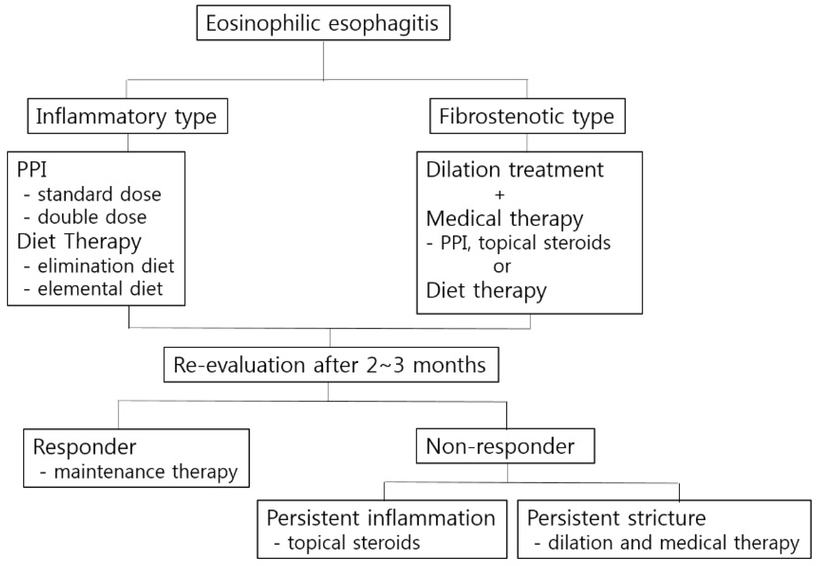
5. 치료(Fig. 3)

1) 치료 목표

이론적으로 치료의 목적은 식도에서 호산구로 인한 염증을 조절하는 것이다. 식도의 염증과 섬유화가 호산구에 의해 발생하므로 호산구를 식도 점막에서 완전히 관찰되지 않게 만드는 것이 좋으나, 이는 일부 환자에서만 가능하고 대부분 고배율 시야당 15개 미만으로 줄이는 것이 현실적인 목표가 되며 이를 위해 약물 치료 및 식이요법을 사용하고 있다.61 이차적으로 식도 협착과 그에 따르는 증상 조절도 중요한 치료 목표이며, 이를 위한 치료로 내시경 확장술이 중요한 역할을 차지하고 있다. 이러한 초기 치료로 호산구 식도염이 호전되었다 하더라도 만성적인 경과를 보이는 질환임을 기억하고 치료를 장기적으로 유지해야 하겠다.62 치료와 관련한 가장 최근 가이드라인은 미국소화기학회와 알레르기학회 공동 task force 팀에서 2020년에 발표를 하였다.63

2) 약물 치료-양성자펌프억제제

양성자펌프억제제 복용은 다른 치료에 비해 복용의 편리성 및 장기 약제 사용의 안전성이 높아 호산구성 식도염 환자에서 가장 먼저 고려되는 치료이다. 국내에서도 호산구성 식도염에 대한 다기관 및 단일기관 연구들에서 가장 흔히 사용 중인 초치료로 조사되었다.10,11 양성자펌프억제제가 호산구성 식도염에 치료 효과를 보이는 기전은 Th2 연관 사이토카인과 유전자 발현을 억제하여 호산구성 염증 경로를 억제하고, 식도 점막 상피를 산으로부터 보호하여 항원의 노출을 줄이는 것으로 추정한다.53
양성자펌프억제제 용량에 대해서는 정해지지는 않았으나 기본 용량 또는 고용량으로 2-3개월 동안 사용 후 임상 증상, 내시경 소견과 생검 소견 호전 여부를 통해 치료 효과를 평가한다.61 추적 생검에서 호산구가 보이지 않는다면 유지요법으로 양성자펌프억제제를 최소 용량으로 줄이도록 시도해야 하겠다.64 한 메타분석에서 양성자펌프억제제가 60%의 호산구성 식도염 환자에서 임상 증상 호전, 50%에서 조직학적 호전을 보였다고 보고하였다.65 하지만 양성자펌프억제제 반응성 식도 호산구증이 호산구성 식도염 진단에서 제외된 연구들도 포함되었기 때문에 해석에 주의가 필요하며 실제 치료 성적은 이보다는 높을 것으로 추정한다. 최근 국내 다기관 연구에서는 양성자펌프억제제 치료가 환자의 89%에서 임상 증상을 호전시켰다고 보고하였다.11

3) 약물 치료-국소 스테로이드

양성자펌프억제제 사용에도 호산구성 식도염이 호전되지 않는다면 스테로이드 치료를 적극적으로 고려해야 하겠다. 대부분의 호산구성 식도염 환자들에서 국소 스테로이드는 호산구 침윤을 감소시킨다.66 이러한 염증세포 감소는 이차적으로식도벽의 섬유화를 억제시킨다. 국내에서는 액상 제제인 경구점액성 제제는 시판되지 않고 분무형 budesonide와 fluticasone을 흡입하지 않고 삼켜서 투약함으로써 식도 점막에 직접적인 효과를 미칠 수 있도록 사용하고 있다.67 그렇지만 국내에서 분무형 국소 스테로이드는 호산구 식도염에 대한 사용이 식품의약품안전처 허가 사항에 없어 ‘허가범위 초과약제비급여 사용 승인’을 건강보험심사평가원에 신청하고 사용해야 한다. 유럽과 캐나다에서는 경구용 제제(orodispersible tablet budesonide)가 허가되어 사용되고 있다. 경구용 액상제제와 분무형 제제의 효과에 대한 비교 연구에서 경구용 액상 제제가 식도 점막에 약제 노출 시간이 길고 식도 호산구의수가 유의미하게 더 감소하였다.68
적절한 용량과 치료 기간에 대해서는 확립되어 있지 않지만 budesonide는 1-2 mg bid, fluticasone 440-880 μg po bid로 시작하여 2-3개월 후 평가에서 호전을 보인다면 용량을 줄이도록 시도해야 하겠다.35 꿀에 섞어서 복용하면 식도 점막과의 접촉시간을 늘리는 데 도움이 될 수 있고, 같은 이유로 약을 삼킨 후 바로 입을 헹구지 말고 30분간은 물이나 음식 섭취를 피하는 것이 좋다. 약제를 단기로 사용하고 중단하면 재발할 수 있으므로 장기적으로 사용하게 되는데, 이로 인해 경구나 식도 캔디다증, 부신기능 저하, 골밀도 저하, 성장 저하가 발생할 수 있으므로 이러한 부작용에 대한 관찰이 필요하다.

4) 비약물 치료-식이요법

특정 원인 음식을 확인하여 제거하면 장기간의 약물 치료 없이 유지할 수 있는 이상적인 치료이다. 대표적인 치료로 경험적 제거 식이는 높은 항원성을 가진 6가지 음식(유제품, 계란, 밀, 콩, 견과류, 해산물)을 제거한 식이부터 시작하여 한 가지씩 음식에 노출하면서 증상의 발생 여부를 평가하고 증상 유발 음식을 제한한 식이를 유지하는 것으로 50-75%에서 반응을 보인다.69,70 2가지 음식(유제품, 밀)을 제한한 2-foods elimination diet에서 시작하여 원인 음식을 찾지 못하면 4- 또는 6-food elimination diet로 단계를 올리는 치료도 소개되었다.71 하지만 국내 음식은 다양한 재료들을 사용하는 경우가 많아 원인 음식을 제한한 식이를 적용하기가 현실적으로어렵다. 다른 식이요법으로 성분 식이(elemental formula)가상당히 좋은 효과를 보인다. 성분 식이는 기본 영양소로 구성된 식이로 우유나 콩과 같은 물질을 포함하지 않아 알레르기반응을 피할 수 있지만 맛이 좋지 않고 비용 부담이 커서 순응도가 좋지 않다. 이러한 식이요법은 결국 장기 유지가 어렵고영양 결핍이나 음식에 대한 거부감을 만들며 사회생활의 위축을 유발할 수 있으므로 치료를 시작하기 전에 장기간의 유지가 필요하며 이로 인해 발생할 수 있는 문제점에 대해 충분한논의가 전제되어야 한다.

5) 비약물 치료-식도 확장술

호산구성 식도염 환자에서 협착이 있으면서 삼킴곤란 증상이 있다면 증상 완화를 위해 식도 확장술을 고려해야 한다. 확장술의 증상 개선 효과는 좋지만 치료 효과 유지 기간은 만족스럽지 못하다. 최근 미국의 코호트 연구에서 509명의 호산구성 식도염 환자 중 164명의 환자가 식도 확장술을 받았고, 58%에 달하는 95명이 확장술 이후 증상 재발로 1년 이내에 반복해서 확장술을 받은 것으로 보고되었다.72 확장술 방법은 부지를 이용하거나 내시경을 통한 풍선 확장술 모두 사용 가능하나 내시경으로 협착 부위를 확인하며 시술이 가능한 내시경적 풍선 확장술이 좀 더 선호된다.73 천공은 식도 확장술의 가장 큰 합병증이나, 다행히 발생률이 0.1%로 낮은 것으로 보고되었다.74 내시경 확장술은 염증 조절에는 효과가 없으므로 확장술 후 꼭 약물 치료나 식이요법을 유지해야 한다. 협착이 심하지 않다면 약물 치료나 식이요법으로 호전될 수 있으므로 치료 반응을 평가한 후에 확장술을 결정하는 것이 좋다.호산구 식도염에 대한 약물 치료나 식이요법을 선행하면서 확장술을 시행하는 것은 확장술 단독에 비해 증상 개선의 추가적인 효과는 없었다.75

6) 기타 치료

다양한 생물학 제제에 대한 임상 연구들이 시행되고 있다. 호산구성 식도염의 병리에 중요한 IL-13에 대한 단일 클론 항체를 이용한 연구에서 위약군에 비해 치료군에서 식도의 호산구 수 감소, 내시경 소견 및 조직학적 호전이 관찰되어 호산구성 식도염의 치료제로 사용될 수 있는 약제로 고려되고 있다.76 호산구 충원(recruitment)에 관여하는 IL-5에 대한 매우선택적인 인간화 항체인 mepolizumab과 reslizumab을 이용한 연구에서는 말초혈액 호산구증과 조직 호산구증 감소를 보였으나 위약군과 비교하여 증상 개선 효과의 차이가 없어 호산구성 식도염 치료제로서의 효과는 불명확한 것으로 보고되었다.77-79
다른 약제로 프로스타글란딘 D2 수용체(PTGDR2, CRTH2) 길항제(OC000459)가 호산구와 Th2 세포의 활성을 억제하는 효과를 보인다. 위약군과 비교하여 OC000459 8주 치료 후 호산구 수가 115개에서 73개로 감소되고 식도 증상과 내시경 소견 개선을 보였으나 통계적 유의성을 보이지는 못하였다.80 류코트리엔 길항제인 montelukast를 고용량으로 사용한 연구에서 증상 개선은 보였으나 식도 염증 개선 효과는 없어 호산구성 식도염 치료제로는 추천되지 않는다.81 Azathioprine과 6-mercaptopurine이 스테로이드 의존성 호산구성 식도염에효과적이었다는 증례 보고는 있으나 이후 후속 연구는 시행되지 않고 있다.82

결 론

호산구성 식도염은 과거에는 매우 드문 질환으로 인식되었으나 먼저 서구에서 발생률이 점차 증가하기 시작하여, 최근 국내 연구에서도 유병률이 증가 추세임을 보고하고 있으므로 호산구성 식도염의 진단과 치료에 대한 관심과 지식이 요구되고 있다. 삼킴곤란이나 음식 걸림, 약물 치료에 반응하지 않는 비심인성 흉통이나 가슴 쓰림 환자는 내시경을 시행하여 호산구성 식도염의 특징적인 내시경 소견 여부를 확인하고, 이상이 없더라도 식도 원위부와 근위부에서 생검을 시행하여 호산구성 식도염을 감별하려는 노력이 필요하다. 치료는 크게 약물 치료와 비약물 치료로 나눌 수 있고, 그중 양성자펌프억제제가 편리하고 부작용이 적어 첫 치료로 선호된다. 그 외에도국소 스테로이드와 식이요법을 시행해 볼 수 있다. 이미 병의경과가 오래되어 협착과 함께 삼킴곤란 증상이 있다면 내시경확장술을 시행해야 하겠다. 질환이 만성적인 경과를 보이므로증상이 개선되고 내시경 소견 및 조직 소견이 호전되더라도장기적으로 치료를 유지해야 한다.

ACKNOWLEDGEMENTS

This study was supported by a 2021 research grant from Pusan National University Yangsan Hospital.

Notes

Financial support

None.

Conflict of interest

None.

REFERENCES

1. Holvoet S, Blanchard C. 2014; Genetic and molecular mechanisms leading to eosinophilic esophagitis. Rev Esp Enferm Dig. 106:276–280. PMID: 25075658.
2. Sherrill JD, Rothenberg ME. 2011; Genetic dissection of eosinophilic esophagitis provides insight into disease pathogenesis and treatment strategies. J Allergy Clin Immunol. 128:23–34. DOI: 10.1016/j.jaci.2011.03.046. PMID: 21570716. PMCID: PMC3129465.
crossref
3. Dobbins JW, Sheahan DG, Behar J. 1977; Eosinophilic gastroenteritis with esophageal involvement. Gastroenterology. 72:1312–1316. DOI: 10.1016/S0016-5085(77)80034-6. PMID: 870380.
crossref
4. Landres RT, Kuster GG, Strum WB. 1978; Eosinophilic esophagitis in a patient with vigorous achalasia. Gastroenterology. 74:1298–1301. DOI: 10.1016/0016-5085(78)90710-2. PMID: 648822.
crossref
5. Attwood SE, Smyrk TC, Demeester TR, Jones JB. 1993; Esophageal eosinophilia with dysphagia. A distinct clinicopathologic syndrome. Dig Dis Sci. 38:109–116. DOI: 10.1007/BF01296781. PMID: 8420741.
6. Kelly KJ, Lazenby AJ, Rowe PC, Yardley JH, Perman JA, Sampson HA. 1995; Eosinophilic esophagitis attributed to gastroesophageal reflux: improvement with an amino acid-based formula. Gastroenterology. 109:1503–1512. DOI: 10.1016/0016-5085(95)90637-1. PMID: 7557132.
crossref
7. Straumann A, Spichtin HP, Bernoulli R, Loosli J, Vögtlin J. 1994; Idiopathic eosinophilic esophagitis: a frequently overlooked disease with typical clinical aspects and discrete endoscopic findings. Schweiz Med Wochenschr. 124:1419–1429. PMID: 7939509.
8. Furuta GT, Katzka DA. 2015; Eosinophilic esophagitis. N Engl J Med. 373:1640–1648. DOI: 10.1056/NEJMra1502863. PMID: 26488694. PMCID: PMC4905697.
crossref
9. Kinoshita Y, Ishimura N, Oshima N, Ishihara S. 2015; Systematic review: eosinophilic esophagitis in Asian countries. World J Gastroenterol. 21:8433–8440. DOI: 10.3748/wjg.v21.i27.8433. PMID: 26217096. PMCID: PMC4507114.
crossref
10. Kim GH, Park YS, Jung KW, et al. 2019; An increasing trend of eosinophilic esophagitis in Korea and the clinical implication of the biomarkers to determine disease activity and treatment response in eosinophilic esophagitis. J Neurogastroenterol Motil. 25:525–533. DOI: 10.5056/jnm19066. PMID: 31587544. PMCID: PMC6786448.
crossref
11. Kim SJ, Park MI, Kim GH, et al. 2021; Practice patterns for eosinophilic esophagitis patients in Busan and Gyeongnam: a Korean multicenter database study. J Neurogastroenterol Motil. 27:71–77. DOI: 10.5056/jnm20069. PMID: 32699187. PMCID: PMC7786091.
crossref
12. Jensen ET, Kappelman MD, Kim HP, Ringel-Kulka T, Dellon ES. 2013; Early life exposures as risk factors for pediatric eosinophilic esophagitis. J Pediatr Gastroenterol Nutr. 57:67–71. DOI: 10.1097/MPG.0b013e318290d15a. PMID: 23518485.
crossref
13. van Nimwegen FA, Penders J, Stobberingh EE, et al. 2011; Mode and place of delivery, gastrointestinal microbiota, and their influence on asthma and atopy. J Allergy Clin Immunol. 128:948–955. e553. DOI: 10.1016/j.jaci.2011.07.027. PMID: 21872915.
crossref
14. Liacouras CA, Furuta GT, Hirano I, et al. 2011; Eosinophilic esophagitis: updated consensus recommendations for children and adults. J Allergy Clin Immunol. 128:3–22. DOI: 10.1016/j.jaci.2011.02.040. PMID: 21477849.
15. Shah SC, Tepler A, Peek RM Jr, Colombel JF, Hirano I, Narula N. 2019; Association between Helicobacter pylori exposure and decreased odds of eosinophilic esophagitis-a systematic review and meta-analysis. Clin Gastroenterol Hepatol. 17:2185–2198. e3. DOI: 10.1016/j.cgh.2019.01.013. PMID: 30659992. PMCID: PMC7354099.
crossref
16. May M, Abrams JA. 2018; Emerging insights into the esophageal microbiome. Curr Treat Options Gastroenterol. 16:72–85. DOI: 10.1007/s11938-018-0171-5. PMID: 29350339. PMCID: PMC5843540.
crossref
17. Hooi JKY, Lai WY, Ng WK, et al. 2017; Global prevalence of Helicobacter pylori infection: systematic review and meta-analysis. Gastroenterology. 153:420–429. DOI: 10.1053/j.gastro.2017.04.022. PMID: 28456631.
crossref
18. Molina-Infante J, Gutierrez-Junquera C, Savarino E, et al. 2018; Helicobacter pylori infection does not protect against eosinophilic esophagitis: results from a large multicenter case-control study. Am J Gastroenterol. 113:972–979. DOI: 10.1038/s41395-018-0035-6. PMID: 29545632.
crossref
19. Katzka DA. 2014; The complex relationship between eosinophilic esophagitis and gastroesophageal reflux disease. Dig Dis. 32:93–97. DOI: 10.1159/000357080. PMID: 24603388.
crossref
20. Kia L, Hirano I. 2015; Distinguishing GERD from eosinophilic oesophagitis: concepts and controversies. Nat Rev Gastroenterol Hepatol. 12:379–386. DOI: 10.1038/nrgastro.2015.75. PMID: 25986303. PMCID: PMC4948861.
crossref
21. O'Shea KM, Aceves SS, Dellon ES, et al. 2018; Pathophysiology of eosinophilic esophagitis. Gastroenterology. 154:333–345. DOI: 10.1053/j.gastro.2017.06.065. PMID: 28757265. PMCID: PMC5787048.
22. Lucendo AJ, De Rezende L, Comas C, Caballero T, Bellón T. 2008; Treatment with topical steroids downregulates IL-5, eotaxin-1/CCL11, and eotaxin-3/CCL26 gene expression in eosinophilic esophagitis. Am J Gastroenterol. 103:2184–2193. DOI: 10.1111/j.1572-0241.2008.01937.x. PMID: 18844613.
crossref
23. Blanchard C, Stucke EM, Rodriguez-Jimenez B, et al. 2011; A striking local esophageal cytokine expression profile in eosinophilic esophagitis. J Allergy Clin Immunol. 127:208–217.e2177. DOI: 10.1016/j.jaci.2010.10.039. PMID: 21211656. PMCID: PMC3027004.
24. Mishra A, Wang M, Pemmaraju VR, et al. 2008; Esophageal remodeling develops as a consequence of tissue specific IL-5-induced eosinophilia. Gastroenterology. 134:204–214. DOI: 10.1053/j.gastro.2007.10.002. PMID: 18166354. PMCID: PMC2654267.
crossref
25. Blanchard C, Wang N, Stringer KF, et al. 2006; Eotaxin-3 and a uniquely conserved gene-expression profile in eosinophilic esophagitis. J Clin Invest. 116:536–547. DOI: 10.1172/JCI26679. PMID: 16453027. PMCID: PMC1359059.
crossref
26. Blanchard C. 2015; Molecular pathogenesis of eosinophilic esophagitis. Curr Opin Gastroenterol. 31:321–327. DOI: 10.1097/MOG.0000000000000186. PMID: 26039724.
crossref
27. Furuta GT, Liacouras CA, Collins MH, et al. 2007; Eosinophilic esophagitis in children and adults: a systematic review and consensus recommendations for diagnosis and treatment. Gastroenterology. 133:1342–1363. DOI: 10.1053/j.gastro.2007.08.017. PMID: 17919504.
crossref
28. Schoepfer AM, Safroneeva E, Bussmann C, et al. 2013; Delay in diagnosis of eosinophilic esophagitis increases risk for stricture formation in a time-dependent manner. Gastroenterology. 145:1230–1236.e62. DOI: 10.1053/j.gastro.2013.08.015. PMID: 23954315.
crossref
29. Dellon ES, Kim HP, Sperry SL, Rybnicek DA, Woosley JT, Shaheen NJ. 2014; A phenotypic analysis shows that eosinophilic esophagitis is a progressive fibrostenotic disease. Gastrointest Endosc. 79:577–585.e4. DOI: 10.1016/j.gie.2013.10.027. PMID: 24275329. PMCID: PMC4599711.
crossref
30. Korsapati H, Babaei A, Bhargava V, Dohil R, Quin A, Mittal RK. 2009; Dysfunction of the longitudinal muscles of the oesophagus in eosinophilic oesophagitis. Gut. 58:1056–1062. DOI: 10.1136/gut.2008.168146. PMID: 19136515.
crossref
31. Kwiatek MA, Hirano I, Kahrilas PJ, Rothe J, Luger D, Pandolfino JE. 2011; Mechanical properties of the esophagus in eosinophilic esophagitis. Gastroenterology. 140:82–90. DOI: 10.1053/j.gastro.2010.09.037. PMID: 20858491. PMCID: PMC3008315.
crossref
32. Francis DL, Foxx-Orenstein A, Arora AS, et al. 2012; Results of ambulatory pH monitoring do not reliably predict response to therapy in patients with eosinophilic oesophagitis. Aliment Pharmacol Ther. 35:300–307. DOI: 10.1111/j.1365-2036.2011.04922.x. PMID: 22111863.
crossref
33. Yan BM, Shaffer EA. 2009; Primary eosinophilic disorders of the gastrointestinal tract. Gut. 58:721–732. DOI: 10.1136/gut.2008.165894. PMID: 19052023.
crossref
34. Dellon ES, Liacouras CA, Molina-Infante J, et al. 2018; Updated international consensus diagnostic criteria for eosinophilic esophagitis: proceedings of the AGREE conference. Gastroenterology. 155:1022–1033.e10. DOI: 10.1053/j.gastro.2018.07.009. PMID: 30009819. PMCID: PMC6174113.
35. Dellon ES, Gonsalves N, Hirano I, et al. 2013; ACG clinical guideline: evidenced based approach to the diagnosis and management of esophageal eosinophilia and eosinophilic esophagitis (EoE). Am J Gastroenterol. 108:679–693. DOI: 10.1038/ajg.2013.71. PMID: 23567357.
crossref
36. Lucendo AJ, Molina-Infante J, Arias Á, et al. 2017; Guidelines on eosinophilic esophagitis: evidence-based statements and recommendations for diagnosis and management in children and adults. United European Gastroenterol J. 5:335–358. DOI: 10.1177/2050640616689525. PMID: 28507746. PMCID: PMC5415218.
crossref
37. Dellon ES, Speck O, Woodward K, et al. 2013; Clinical and endoscopic characteristics do not reliably differentiate PPI-responsive esophageal eosinophilia and eosinophilic esophagitis in patients undergoing upper endoscopy: a prospective cohort study. Am J Gastroenterol. 108:1854–1860. DOI: 10.1038/ajg.2013.363. PMID: 24145677. PMCID: PMC4574875.
crossref
38. Warners MJ, van Rhijn BD, Curvers WL, Smout AJ, Bredenoord AJ. 2015; PPI-responsive esophageal eosinophilia cannot be distinguished from eosinophilic esophagitis by endoscopic signs. Eur J Gastroenterol Hepatol. 27:506–511. DOI: 10.1097/MEG.0000000000000331. PMID: 25822858.
crossref
39. Moawad FJ, Veerappan GR, Dias JA, Baker TP, Maydonovitch CL, Wong RK. 2013; Randomized controlled trial comparing aerosolized swallowed fluticasone to esomeprazole for esophageal eosinophilia. Am J Gastroenterol. 108:366–372. DOI: 10.1038/ajg.2012.443. PMID: 23399553.
crossref
40. Dellon ES, Speck O, Woodward K, et al. 2014; Markers of eosinophilic inflammation for diagnosis of eosinophilic esophagitis and proton pump inhibitor-responsive esophageal eosinophilia: a prospective study. Clin Gastroenterol Hepatol. 12:2015–2022. DOI: 10.1016/j.cgh.2014.06.019. PMID: 24993367. PMCID: PMC4252508.
crossref
41. Shoda T, Matsuda A, Nomura I, et al. 2017; Eosinophilic esophagitis versus proton pump inhibitor-responsive esophageal eosinophilia: transcriptome analysis. J Allergy Clin Immunol. 139:2010–2013.e4. DOI: 10.1016/j.jaci.2016.11.028. PMID: 28063872.
crossref
42. Cheng E, Zhang X, Huo X, et al. 2013; Omeprazole blocks eotaxin-3 expression by oesophageal squamous cells from patients with eosinophilic oesophagitis and GORD. Gut. 62:824–832. DOI: 10.1136/gutjnl-2012-302250. PMID: 22580413. PMCID: PMC3552049.
crossref
43. van Rhijn BD, Weijenborg PW, Verheij J, et al. 2014; Proton pump inhibitors partially restore mucosal integrity in patients with proton pump inhibitor-responsive esophageal eosinophilia but not eosinophilic esophagitis. Clin Gastroenterol Hepatol. 12:1815–1823.e2. DOI: 10.1016/j.cgh.2014.02.037. PMID: 24657840.
crossref
44. Hirano I, Moy N, Heckman MG, Thomas CS, Gonsalves N, Achem SR. 2013; Endoscopic assessment of the oesophageal features of eosinophilic oesophagitis: validation of a novel classification and grading system. Gut. 62:489–495. DOI: 10.1136/gutjnl-2011-301817. PMID: 22619364.
crossref
45. Kim HP, Vance RB, Shaheen NJ, Dellon ES. 2012; The prevalence and diagnostic utility of endoscopic features of eosinophilic esophagitis: a meta-analysis. Clin Gastroenterol Hepatol. 10:988–996.e5. DOI: 10.1016/j.cgh.2012.04.019. PMID: 22610003. PMCID: PMC3424367.
crossref
46. van Rhijn BD, Warners MJ, Curvers WL, et al. 2014; Evaluating the endoscopic reference score for eosinophilic esophagitis: moderate to substantial intra- and interobserver reliability. Endoscopy. 46:1049–1055. DOI: 10.1055/s-0034-1377781. PMID: 25208033.
crossref
47. Dellon ES, Cotton CC, Gebhart JH, et al. 2016; Accuracy of the eosinophilic esophagitis Endoscopic Reference Score in diagnosis and determining response to treatment. Clin Gastroenterol Hepatol. 14:31–39. DOI: 10.1016/j.cgh.2015.08.040. PMID: 26404868. PMCID: PMC4690779.
crossref
48. van Rhijn BD, Verheij J, Smout AJ, Bredenoord AJ. 2016; The Endoscopic Reference Score shows modest accuracy to predict histologic remission in adult patients with eosinophilic esophagitis. Neurogastroenterol Motil. 28:1714–1722. DOI: 10.1111/nmo.12872. PMID: 27254480.
crossref
49. Prasad GA, Talley NJ, Romero Y, et al. 2007; Prevalence and predictive factors of eosinophilic esophagitis in patients presenting with dysphagia: a prospective study. Am J Gastroenterol. 102:2627–2632. DOI: 10.1111/j.1572-0241.2007.01512.x. PMID: 17764492.
crossref
50. Gonsalves N, Policarpio-Nicolas M, Zhang Q, Rao MS, Hirano I. 2006; Histopathologic variability and endoscopic correlates in adults with eosinophilic esophagitis. Gastrointest Endosc. 64:313–319. DOI: 10.1016/j.gie.2006.04.037. PMID: 16923475.
crossref
51. Shah A, Kagalwalla AF, Gonsalves N, Melin-Aldana H, Li BU, Hirano I. 2009; Histopathologic variability in children with eosinophilic esophagitis. Am J Gastroenterol. 104:716–721. DOI: 10.1038/ajg.2008.117. PMID: 19209168.
crossref
52. Nielsen JA, Lager DJ, Lewin M, Rendon G, Roberts CA. 2014; The optimal number of biopsy fragments to establish a morphologic diagnosis of eosinophilic esophagitis. Am J Gastroenterol. 109:515–520. DOI: 10.1038/ajg.2013.463. PMID: 24445569.
crossref
53. Abe Y, Sasaki Y, Yagi M, Yaoita T, Nishise S, Ueno Y. 2017; Diagnosis and treatment of eosinophilic esophagitis in clinical practice. Clin J Gastroenterol. 10:87–102. DOI: 10.1007/s12328-017-0725-4. PMID: 28247277.
crossref
54. Mueller S, Neureiter D, Aigner T, Stolte M. 2008; Comparison of histological parameters for the diagnosis of eosinophilic oesophagitis versus gastro-oesophageal reflux disease on oesophageal biopsy material. Histopathology. 53:676–684. DOI: 10.1111/j.1365-2559.2008.03187.x. PMID: 19076684.
crossref
55. Odze RD. 2009; Pathology of eosinophilic esophagitis: what the clinician needs to know. Am J Gastroenterol. 104:485–490. DOI: 10.1038/ajg.2008.40. PMID: 19174804.
crossref
56. Kinoshita Y, Furuta K, Ishimaura N, et al. 2013; Clinical characteristics of Japanese patients with eosinophilic esophagitis and eosinophilic gastroenteritis. J Gastroenterol. 48:333–339. DOI: 10.1007/s00535-012-0640-x. PMID: 22847555.
crossref
57. Fox VL, Nurko S, Teitelbaum JE, Badizadegan K, Furuta GT. 2003; High-resolution EUS in children with eosinophilic "allergic" esophagitis. Gastrointest Endosc. 57:30–36. DOI: 10.1067/mge.2003.33. PMID: 12518127.
crossref
58. Gentile N, Katzka D, Ravi K, et al. 2014; Oesophageal narrowing is common and frequently under-appreciated at endoscopy in patients with oesophageal eosinophilia. Aliment Pharmacol Ther. 40:1333–1340. DOI: 10.1111/apt.12977. PMID: 25287184.
crossref
59. Konikoff MR, Blanchard C, Kirby C, et al. 2006; Potential of blood eosinophils, eosinophil-derived neurotoxin, and eotaxin-3 as biomarkers of eosinophilic esophagitis. Clin Gastroenterol Hepatol. 4:1328–1336. DOI: 10.1016/j.cgh.2006.08.013. PMID: 17059896.
crossref
60. Nicodème F, Hirano I, Chen J, et al. 2013; Esophageal distensibility as a measure of disease severity in patients with eosinophilic esophagitis. Clin Gastroenterol Hepatol. 11:1101–1107.e1. DOI: 10.1016/j.cgh.2013.03.020. PMID: 23591279. PMCID: PMC3790569.
crossref
61. Straumann A, Katzka DA. 2018; Diagnosis and treatment of eosinophilic esophagitis. Gastroenterology. 154:346–359. DOI: 10.1053/j.gastro.2017.05.066. PMID: 28756235.
crossref
62. Straumann A, Conus S, Degen L, et al. 2011; Long-term budesonide maintenance treatment is partially effective for patients with eosinophilic esophagitis. Clin Gastroenterol Hepatol. 9:400–409. e1. DOI: 10.1016/j.cgh.2011.01.017. PMID: 21277394.
crossref
63. Hirano I, Chan ES, Rank MA, et al. 2020; AGA Institute and the Joint task force on allergy-immunology practice parameters clinical guidelines for the management of eosinophilic esophagitis. Gastroenterology. 158:1776–1786. DOI: 10.1053/j.gastro.2020.02.038. PMID: 32359562.
crossref
64. Gómez-Torrijos E, García-Rodríguez R, Castro-Jiménez A, Rodríguez-Sanchez J, Méndez Díaz Y, Molina-Infante J. 2016; The efficacy of step-down therapy in adult patients with proton pump inhibitor-responsive oesophageal eosinophilia. Aliment Pharmacol Ther. 43:534–540. DOI: 10.1111/apt.13496. PMID: 26662868.
crossref
65. Lucendo AJ, Arias Á, Molina-Infante J. 2016; Efficacy of proton pump inhibitor drugs for inducing clinical and histologic remission in patients with symptomatic esophageal eosinophilia: a systematic review and meta-analysis. Clin Gastroenterol Hepatol. 14:13–22.e1. DOI: 10.1016/j.cgh.2015.07.041. PMID: 26247167.
crossref
66. Alexander JA, Jung KW, Arora AS, et al. 2012; Swallowed fluticasone improves histologic but not symptomatic response of adults with eosinophilic esophagitis. Clin Gastroenterol Hepatol. 10:742–749.e1. DOI: 10.1016/j.cgh.2012.03.018. PMID: 22475741.
crossref
67. Noel RJ, Putnam PE, Collins MH, et al. 2004; Clinical and immunopathologic effects of swallowed fluticasone for eosinophilic esophagitis. Clin Gastroenterol Hepatol. 2:568–575. DOI: 10.1016/S1542-3565(04)00240-X. PMID: 15224281.
crossref
68. Dellon ES, Sheikh A, Speck O, et al. 2012; Viscous topical is more effective than nebulized steroid therapy for patients with eosinophilic esophagitis. Gastroenterology. 143:321–324.e1. DOI: 10.1053/j.gastro.2012.04.049. PMID: 22561055. PMCID: PMC3404241.
crossref
69. Kagalwalla AF, Sentongo TA, Ritz S, et al. 2006; Effect of six-food elimination diet on clinical and histologic outcomes in eosinophilic esophagitis. Clin Gastroenterol Hepatol. 4:1097–1102. DOI: 10.1016/j.cgh.2006.05.026. PMID: 16860614.
crossref
70. Arias A, González-Cervera J, Tenias JM, Lucendo AJ. 2014; Efficacy of dietary interventions for inducing histologic remission in patients with eosinophilic esophagitis: a systematic review and meta-analysis. Gastroenterology. 146:1639–1648. DOI: 10.1053/j.gastro.2014.02.006. PMID: 24534634.
crossref
71. Zhan T, Ali A, Choi JG, et al. 2018; Model to determine the optimal dietary elimination strategy for treatment of eosinophilic esophagitis. Clin Gastroenterol Hepatol. 16:1730–1737.e2. DOI: 10.1016/j.cgh.2018.04.013. PMID: 29730438.
72. Runge TM, Eluri S, Cotton CC, et al. 2016; Outcomes of esophageal dilation in eosinophilic esophagitis: safety, efficacy, and persistence of the fibrostenotic phenotype. Am J Gastroenterol. 111:206–213. DOI: 10.1038/ajg.2015.399. PMID: 26753894. PMCID: PMC4758887.
crossref
73. Dougherty M, Runge TM, Eluri S, Dellon ES. 2017; Esophageal dilation with either bougie or balloon technique as a treatment for eosinophilic esophagitis: a systematic review and meta-analysis. Gastrointest Endosc. 86:581–591.e3. DOI: 10.1016/j.gie.2017.04.028. PMID: 28461094. PMCID: PMC5601027.
crossref
74. Jacobs JW Jr, Spechler SJ. 2010; A systematic review of the risk of perforation during esophageal dilation for patients with eosinophilic esophagitis. Dig Dis Sci. 55:1512–1515. DOI: 10.1007/s10620-010-1165-x. PMID: 20238250. PMCID: PMC3121144.
crossref
75. Schoepfer AM, Gonsalves N, Bussmann C, et al. 2010; Esophageal dilation in eosinophilic esophagitis: effectiveness, safety, and impact on the underlying inflammation. Am J Gastroenterol. 105:1062–1070. DOI: 10.1038/ajg.2009.657. PMID: 19935783.
crossref
76. Hirano I, Collins MH, Assouline-Dayan Y, et al. 2019; RPC4046, a monoclonal antibody against IL13, reduces histologic and endoscopic activity in patients with eosinophilic esophagitis. Gastroenterology. 156:592–603.e10. DOI: 10.1053/j.gastro.2018.10.051. PMID: 30395812.
crossref
77. Assa'ad AH, Gupta SK, Collins MH, et al. 2011; An antibody against IL-5 reduces numbers of esophageal intraepithelial eosinophils in children with eosinophilic esophagitis. Gastroenterology. 141:1593–1604. DOI: 10.1053/j.gastro.2011.07.044. PMID: 21835135.
78. Spergel JM, Rothenberg ME, Collins MH, et al. 2012; Reslizumab in children and adolescents with eosinophilic esophagitis: results of a double-blind, randomized, placebo-controlled trial. J Allergy Clin Immunol. 129:456–463.e4633. DOI: 10.1016/j.jaci.2011.11.044. PMID: 22206777.
crossref
79. Straumann A, Conus S, Grzonka P, et al. 2010; Anti-interleukin-5 antibody treatment (mepolizumab) in active eosinophilic oesophagitis: a randomised, placebo-controlled, double-blind trial. Gut. 59:21–30. DOI: 10.1136/gut.2009.178558. PMID: 19828470.
crossref
80. Straumann A, Hoesli S, Bussmann Ch, et al. 2013; Anti-eosinophil activity and clinical efficacy of the CRTH2 antagonist OC000459 in eosinophilic esophagitis. Allergy. 68:375–385. DOI: 10.1111/all.12096. PMID: 23379537.
crossref
81. Attwood SE, Lewis CJ, Bronder CS, Morris CD, Armstrong GR, Whittam J. 2003; Eosinophilic oesophagitis: a novel treatment using Montelukast. Gut. 52:181–185. DOI: 10.1136/gut.52.2.181. PMID: 12524397. PMCID: PMC1774975.
crossref
82. Netzer P, Gschossmann JM, Straumann A, Sendensky A, Weimann R, Schoepfer AM. 2007; Corticosteroid-dependent eosinophilic oesophagitis: azathioprine and 6-mercaptopurine can induce and maintain long-term remission. Eur J Gastroenterol Hepatol. 19:865–869. DOI: 10.1097/MEG.0b013e32825a6ab4. PMID: 17873610.
crossref

Fig. 1
Endoscopic findings in patients with eosinophilic esophagitis. (A) Decreased vascularity is present. (B) Cocentric rings are present. (C) Granular white exudates are present in the lower esophagus. (D) Linear furrows run along the longitudinal axis of the esophagus.
kjg-79-3-99-f1.tif
Fig. 2
Histologic findings of eosinophilic esophagitis (hematoxylin and eosin, ×400). Surface sloughing of squamous cells with abundant eosinophils was present and is one of the important features that can distinguish eosinophilic esophagitis from gastroesophageal reflux disease or eosinophilic gastroenteritis.
kjg-79-3-99-f2.tif
Fig. 3
Treatment algorithm for patients with eosinophilic esophagitis.
kjg-79-3-99-f3.tif
Table 1
Common Clinical Manifestations in Patients with Eosinophilic Esophagitis
Adults Children
Esophageal symptoms Dysphagia Feeding dysfunction (median age 2 years)
Food impaction Vomiting (median age 8 years)
Chest pain Abdominal pain (median age 12 years)
Heartburn Dysphagia (median age 13 years)
Upper abdominal pain Food impaction (median age 17 years)
Table 2
Eosinophilic Esophagitis Diagnostic Criteria (Must Include All of the Following)
• Clinical symptoms of esophageal dysfunction
• ≥15 eosinophils in a high-power field on esophageal biopsy
• Exclusion of secondary cause of esophageal eosinophilia
Table 3
Modified Classification and Grading System for Endoscopic Assessment. Eosinophilic Esophagitis Endoscopic Reference Score (EREFS; E, edema; R, rings; E, exudates; F, furrows; S, strictures)
Major features
Fixed rings
Grade 0: none
Grade 1: mild (subtle circumferential ridges)
Grade 2: moderate (distinct rings that permit passage of a endoscope)
Grade 3: severe (distinct ring that do not permit passage of a endoscope)
Exudates
Grade 0: none
Grade 1: mild (lesions involving <10% of the esophageal surface)
Grade 2: severe (lesions involving >10% of the esophageal surface)
Furrows
Grade 0: absent
Grade 1: present
Edema
Grade 0: absent
Grade 1: present (loss of clarity or absence of vascular marking)
Stricture
Grade 0: absent
Grade 1: present
Minor features
Crepe paper esophagus
Grade 0: absent
Grade 1: present (mucosal fragility or laceration upon passage of endoscope)
TOOLS
Similar articles