Abstract
The stomach temporarily stores food and secretes gastric juices to break down and digest food. The normal process is the movement of food digested from the stomach to the duodenum, with the pylorus as a passageway. This paper reports the case of a patient with an ectopic gastric pylorus who presented with gastrointestinal bleeding. A 62-year-old man complained of melena with mild dizziness and nausea. An endoscopic examination revealed a gastric ulcer, approximately 1 cm in diameter, and exposed blood vessels on the posterior wall of the upper body. No normal pyloric structure was observed in the distal antrum, and an opening leading to the duodenum was noted in the posterior wall of the upper body adjacent to the ulcer. This case presents a congenital pyloric ectopic opening in the upper body of the stomach, not in the distal antrum, suggesting a rare gastric morphological variation.
Go to : 
The stomach temporarily stores food that enters the esophagus and secretes gastric juice containing digestive juices to break down the food and digest it. The movement of food digested from the stomach to the duodenum, with the pylorus as a passageway, is a normal process.
The gastric pylorus exists as a hole approximately 5-10 mm and secretes gastric juice containing digestive juices to break down the food and digest it. The movement of food digested from the stomach to the duodenum, with the pylorus as a passageway, is a normal process. The gastric pylorus exists as a hole approximately 5-10 mm in the distal antrum. Anatomical variations of the pylorus are often reported, with the most common being congenital pyloric stenosis and double pylorus.1,2 Case reports of ectopic pylorus in the distal antrum with blind ducts are rare. This paper reports a patient with ectopic gastric pylorus who presented with gastrointestinal bleeding.
Go to : 
A 62-year-old man presented to the Dong-A University Hospital with melena. The symptoms started the morning of the day of the visit. The patient complained of mild dizziness and nausea accompanying the symptoms. His vital signs were as follows: blood pressure 140/90 mmHg, body temperature 36.5℃, pulse 107 beats/min, respiration rate 22 beats/min, and oxygen saturation 99%. There were no specific findings other than hemoglobin 8.3 g/dL in the laboratory blood test.
The patient was admitted to the gastroenterology depart ment on April 29, 2021. He had a history of endoscopic hemostasis for gastric ulcer bleeding one year earlier and was taking medications for hypertension (amlodipine 5 mg, losartan 100 mg, and hydrochlorothiazide 12.5 mg). He had a history of an appendectomy at 17 years of age. He had never undergone any abdominal surgery other than the appendectomy that could have affected the gastroduodenal anatomy. He had never smoked and occasionally drank alcohol.
The physical examination revealed no specific findings other than anemic conjunctiva. There was no abdominal tenderness or palpable mass, and melena was confirmed on the digital rectal examination.
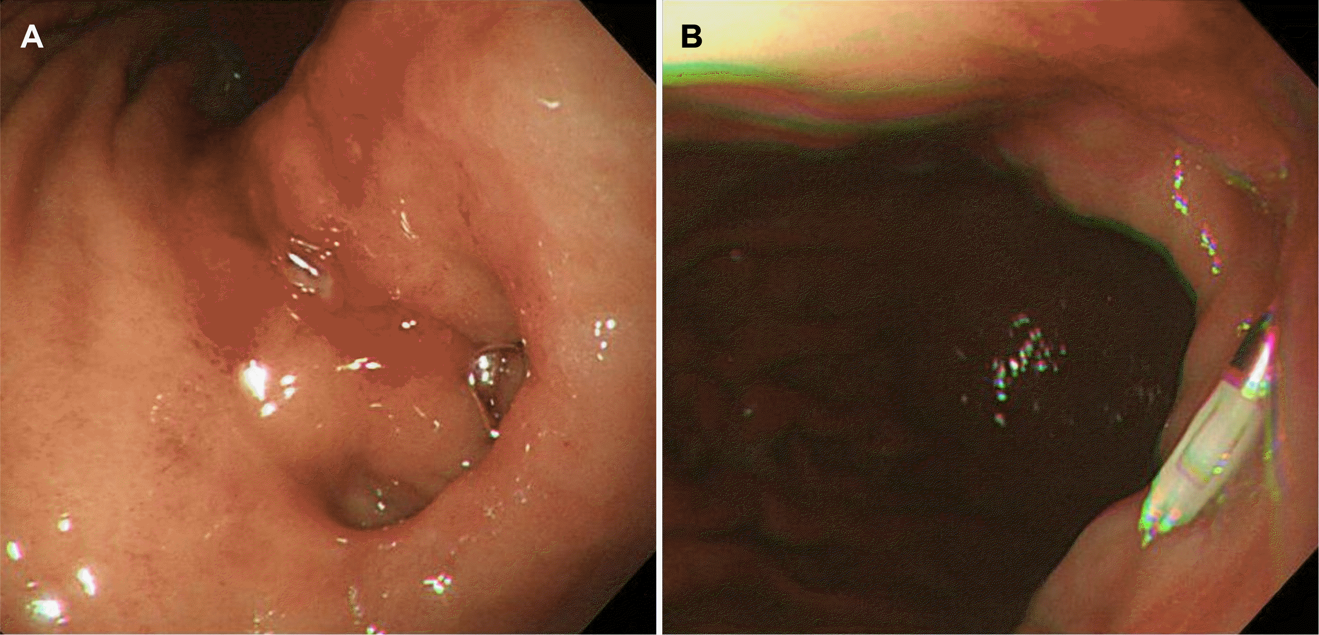
Emergency gastrointestinal endoscopy was performed 3 hours after the visit. The endoscopic examination revealed a gastric ulcer with a diameter of approximately 1 cm and exposed blood vessels on the posterior wall of the upper body. Endoscopic hemostasis was performed with hemoclips and epinephrine injection (Fig. 1). The patient’s endoscopic findings were unusual. No normal pyloric structure was observed in the distal antrum, and an opening leading to the duodenum was found in the posterior wall of the upper body adjacent to the ulcer. When the endoscope entered the opening, a mucous membrane presumed to be the duodenum was observed. There were no obvious abnormalities in the duodenum or near the ampulla of Vater.
After admission, the patient received intravenous nutritional support in the fasting state, and a high-dose proton pump inhibitor was administered intravenously. Follow-up endoscopy was performed 5 days after endoscopic hemostasis. The previously observed gastric ulcer improved, and regenerated epithelium was observed at the margin of the ulcer. On the other hand, some food remained in the stomach even after 5 days of fasting.
A histological examination of the gastric ulcer confirmed the presence of chronic superficial gastritis. There were no Helicobacter pylori (H. pylori) on Giemsa staining, and the rapid urease test was also negative. Hence, it is unlikely to be related to a H. pylori infection.
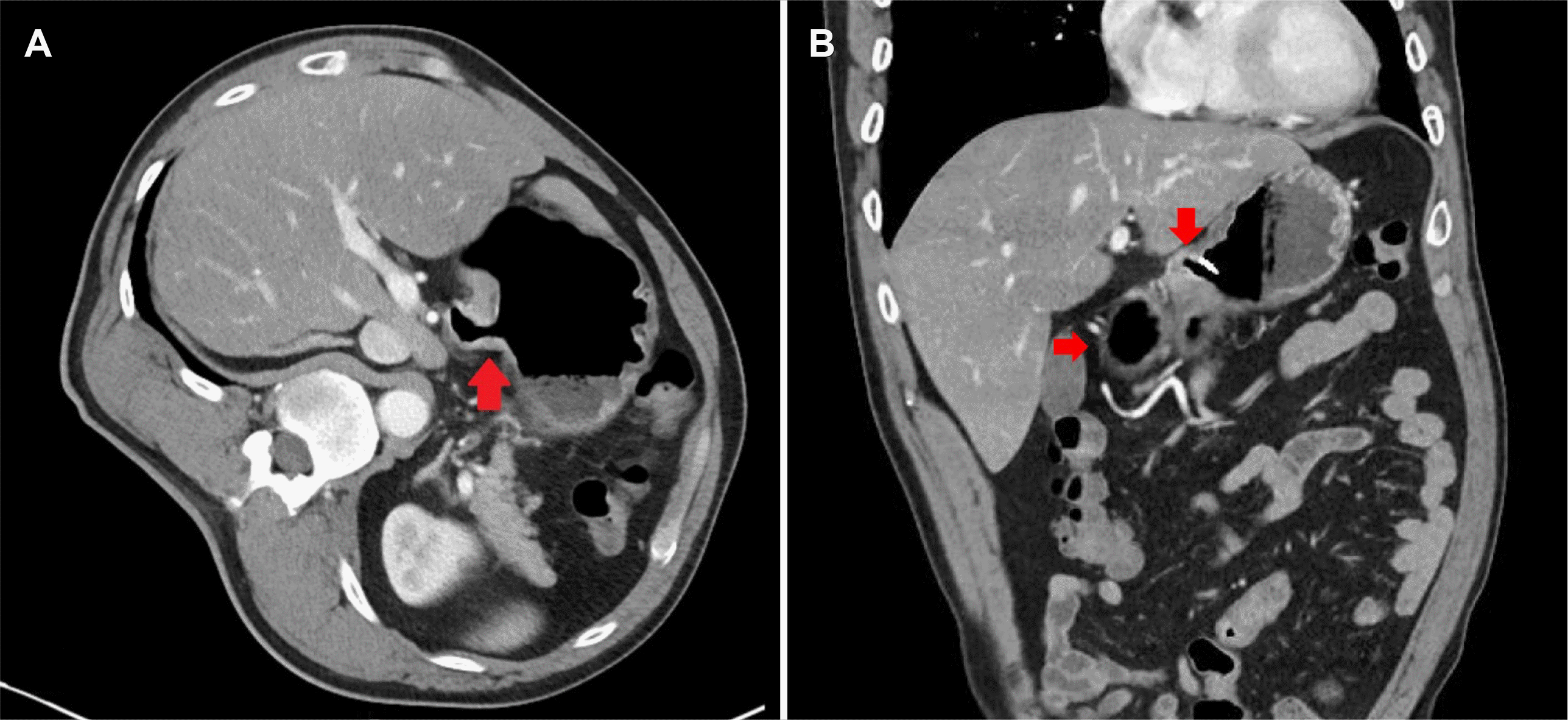
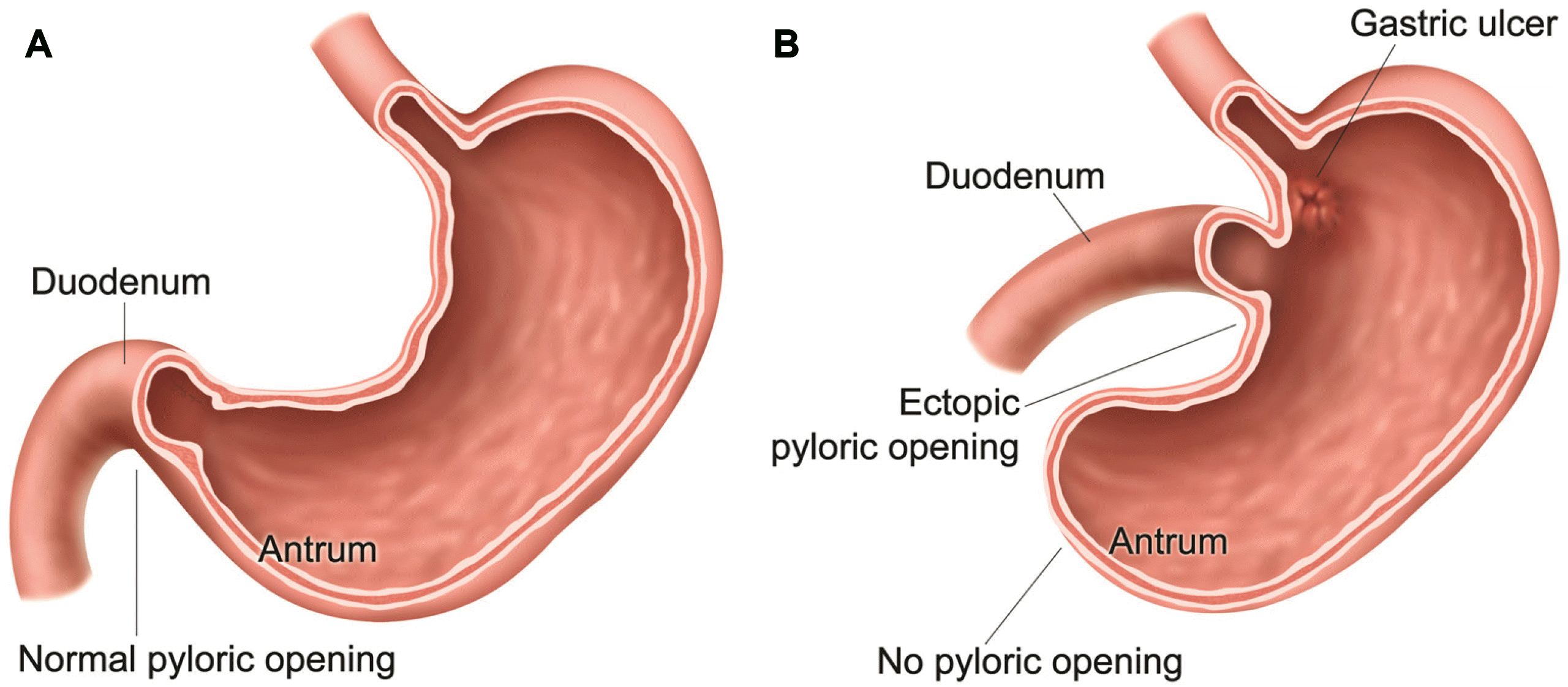
An abdominal CT scan was performed to confirm the anatomical variation in the patient’s stomach, and a CT scan confirmed the anatomical abnormalities in the pylorus (Fig. 2). This paper describes these anatomical abnormalities based on the endoscopy and CT findings (Fig. 3). The diet was continued, and there was no bleeding. Finally, the patient was safely discharged.
Go to : 
This case report presents a patient with gastric ulcer bleeding with ectopic pylorus. The anatomical structure of the stomach could vary with or without clinical symptoms. According to previous reports, the anatomical variations of the stomach can be divided into five types: (I) abnormal position along longitudinal and (II)horizontal axis, as well as (III) an abnormal shape and (IV) stomach connections, and (V) mixed forms.3 In addition, they were classified as hernias, abnormal rotations, and congenital mutations.4
The causes of gastric anatomical abnormalities include congenital anomalies and acquired deformities. For congenital anomalies, pyloric abnormalities, including double pylorus and congenital pyloric stenosis, have been reported.5,6 Acquired anomalies can occur due to the repeated occurrence and healing of peptic ulcers. In some cases, surgery may lead to gastric deformities.
In general, the gastric pylorus exists as an opening approximately 5-10 mm in the distal antrum of the stomach. This patient did not have a normal pyloric opening in the distal antrum but showed an ectopic pyloric opening in the posterior wall of the upper body. The patient had previously undergone an appendectomy, but it is unlikely related to the ectopic pylorus considering the anatomical location and surgical technique.
The patient may also have unusual features on CT imaging. Lu and Yang7 reported a 72-year-old man with gastric ectopic pylorus. The patient complained of discomfort in his upper left abdomen. An endoscopic and radiologic examination revealed a ‘hammer’-shaped stomach and gastric ulcer.7 Lu and Yang7 suggested that these anatomical gastric abnormalities would cause food retention and mechanical stimulation, eventually leading to delayed gastric emptying, gastric discomfort, or gastric ulcer. In the case of the present patient, food was retained in the stomach in the follow-up endoscopy performed after 5 days of fasting. Hence, continuous mechanical stimulation due to delayed gastric emptying may be related to a gastric ulcer. In a stomach with a normal anatomy, food passes from the antrum through the pylorus to the duodenum. In contrast, gastric emptying was expected to be delayed in this patient with a blind duct at the end of the antrum. On the other hand, the patient did not complain of any discomfort due to delayed gastric emptying. Therefore, additional endoscopic or surgical treatment for the patient was not considered.
As the gastric ulcer heals, the shape of the stomach changes, and the antrum is shortened. Therefore, the normal pylorus can be mistaken for a gastric ectopic pylorus. Although this patient had a history of gastric ulcer bleeding, it is not appropriate to assume that the position of the pylorus had shifted due to gastric shortening. In this patient, the pylorus was adjacent to the gastroesophageal junction, and there was a portion of the gastric angle in the distal portion. Therefore, it was not considered to be related to the shifting of the pyloric opening position because of the shortening of the antrum due to previous ulcer healing.
Studies have shown that a H. pylori infection can delay gastric emptying and lead to complications. Conversely, delayed gastric emptying can worsen H. pylori infections. Nevertheless, the present patient showed no evidence of H. pylori infection, and delayed gastric emptying was confirmed endoscopically.8
Although it is difficult to confirm because of the few case reports, the treatment for the ectopic gastric pylorus itself is unnecessary. Hence, only the treatment of gastric ulcers caused by delayed gastric emptying is sufficient. This paper describes this rare case with a review of the literature.
Go to : 
REFERENCES
1. Ladd WE, Ware PF, Pickett LK. 1946; Congenital hypertrophic pyloric stenosis. J Am Med Assoc. 131:647–651. DOI: 10.1001/jama.1946.02870250001001. PMID: 20984883.

2. Einhorn RI, Grace ND, Banks PA. 1984; The clinical significance and natural history of the double pylorus. Dig Dis Sci. 29:213–218. DOI: 10.1007/BF01296254. PMID: 6697860.

3. Burdan F, Zinkiewicz K, Szumiło J, et al. 2012; Anatomical classification of the shape and topography of the operated stomach. Folia Morphol (Warsz). 71:129–135. PMID: 22936546.
4. Burdan F, Rozylo-Kalinowska I, Szumilo J, et al. 2012; Anatomical classification of the shape and topography of the stomach. Surg Radiol Anat. 34:171–178. DOI: 10.1007/s00276-011-0893-8. PMID: 22057798. PMCID: PMC3284679.

5. Naidoo R, Singh B. 2012; Congenital double pylorus. Case Rep Gastrointest Med. 2012:537697. DOI: 10.1155/2012/537697. PMID: 22924137. PMCID: PMC3423775.

6. Teele RL, Smith EH. 1977; Ultrasound in the diagnosis of idiopathic hypertrophic pyloric stenosis. N Engl J Med. 296:1149–1150. DOI: 10.1056/NEJM197705192962006. PMID: 854046.

7. Lu B, Yang L. 2019; Gastric ectopic pyloric opening: an unusual case. Surg Radiol Anat. 41:1395–1398. DOI: 10.1007/s00276-019-02276-x. PMID: 31264000. PMCID: PMC6841747.

8. Chang CS, Chen GH, Kao CH, Wang SJ, Peng SN, Huang CK. 1996; The effect of Helicobacter pylori infection on gastric emptying of digestible and indigestible solids in patients with nonulcer dyspepsia. Am J Gastroenterol. 91:474–479. PMID: 8633494.
Go to : 


 PDF
PDF Citation
 Print
Print


 XML Download