

 PDF
PDF Citation
 Print
Print


I. Introduction
Temporomandibular joint (TMJ) hypermobility is defined as the hypertranslation of the mandibular condyle towards the anterior and superior aspects of the articular eminence as the mouth opens1. In the aetiology of TMJ hypermobility, factors such as the morphology of mandibular condyle, articular eminence and glenoid fossa, lateral pterygoid muscle activity, long anaesthesia intake, loss of tightness of joint ligaments, and trauma may play a role2.
TMJ hypermobility, which gives clinical symptoms such as pain, inability to perform mandibular movements, and open locking, has various treatments, ranging from minimally invasive methods such as medication, Botox injection or intracapsular injection of sclerosed solutions, to advanced surgeries such as myotomy of the lateral pterygoid muscle1,3,4.
Prolotherapy is a method that has gained popularity in recent years and has been reported to have positive short-term and long-term clinical results in maxillofacial surgery, especially TMJ hypermobility5,6. Prolotherapy, also called regenerative injection therapy or growth factor stimulation injection therapy, was first defined by Schultz in 19371,7. Various non-pharmacological proliferants, such as glycerin, dextrose, and phenol, have been used for proliferation purposes8. Among these, dextrose is the most preferred. The mechanism of prolotherapy in TMJ has not been clearly defined. However, hypertonic dextrose applied to tendons and ligaments that have lost their flexibility for various reasons is considered to increase the repair process by affecting the non-inflammatory and inflammatory processes5,7. In the treatment of TMJ hypermobility, dextrose is used at concentrations of 10%-50% and in a single session or multiple sessions2,5,9. Most studies on this subject are clinically oriented. Studies showing the effect of prolotherapy on the hard tissue components of the joint are limited. The effect of prolotherapy using hypertonic dextrose on hard tissue can be examined by fractal dimension (FD) analysis.
FD analysis is a statistical method based on fractal mathematics used to describe complex shapes and structures10. Fractals consist of geometric shapes, such as curves, lines and points11. FD analysis is a proven and effective method for evaluating the microarchitecture structures of bones12. It has been confirmed to show early bone changes in the medical field13,14.
This study investigated FD change in the mandibular condyle in patients who had hypertonic dextrose prolotherapy for one, two, or three sessions due to TMJ hypermobility.
Go to : 
II. Materials and Methods
1. Patients
In this retrospective study, patients who received prolotherapy treatment from the author at Department of Oral and Maxillofacial Surgery of Bolu Abant Izzet Baysal University (Bolu, Turkey) between June 2017 and January 2020 were examined.
The inclusion criteria were one or more injections of bilateral dextrose prolotherapy due to TMJ hypermobility, having panoramic radiography taken before and six months after the end of the treatment, not having a previous operation in the TMJ area, no trauma history, and not having a systemic disease that could affect bone structure. Records of clinical examination findings and radiological examination were considered in the diagnosis of TMJ hypermobility. In all patients included, the TMJ radiographs taken before the procedure with the mouth open showed that the mandibular condyle was located beyond the articular eminence.
All patients except the control group received 20% dextrose prolotherapy through at least one injection into the TMJ area. The patients received follow-up for at least six months. The patients were divided into three groups according to the number of prolotherapy injections they received; Group 1: one injection, Group 2: two injections, and Group 3: three injections. The control group was formed from patients with TMJ hypermobility who did not receive any treatment.
2. Prolotherapy procedure
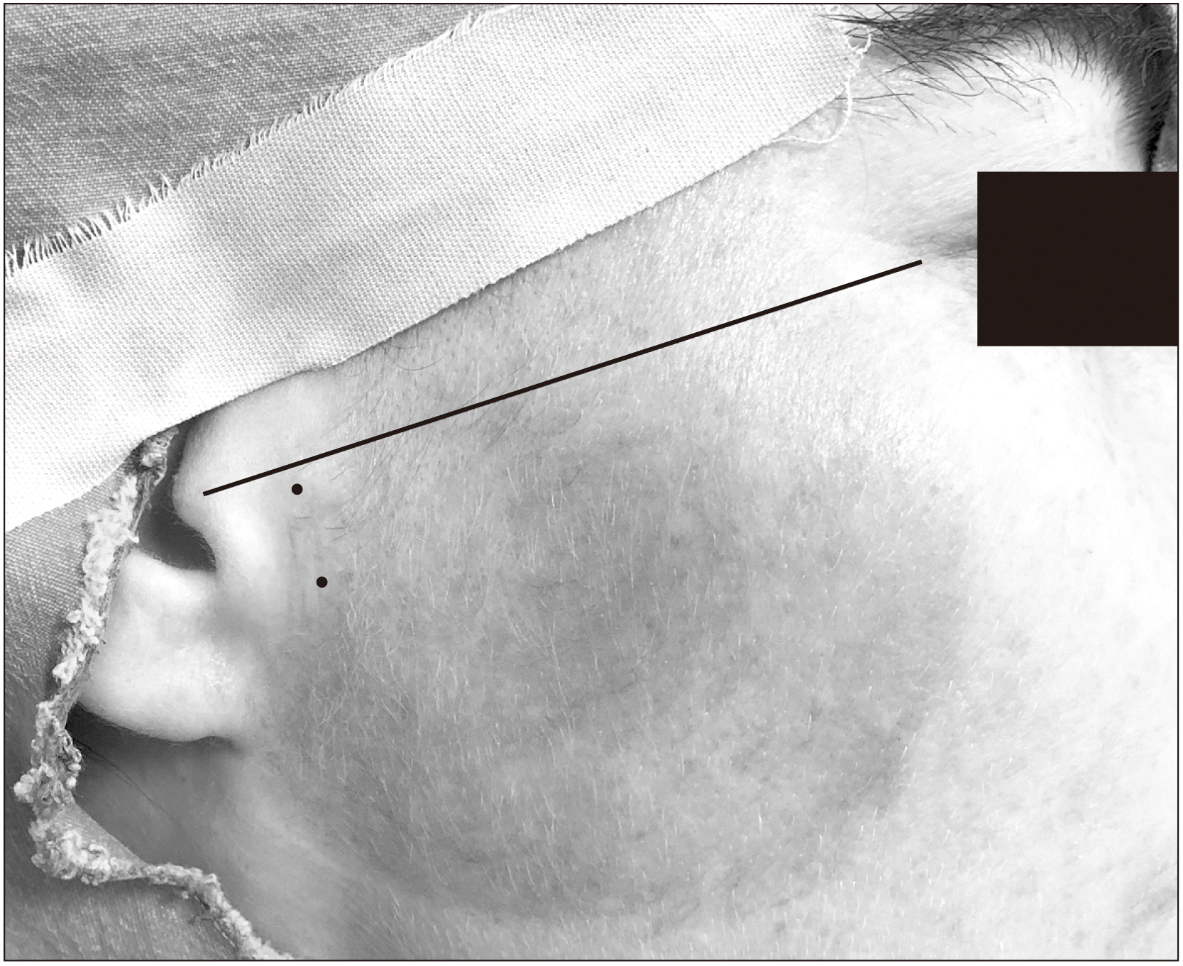
The prolotherapy procedures applied to all patients were performed under local operating room conditions. A 27-gauge needle injector was used for each injection. The syringe to be applied to each addition was prepared as 3 mL dextrose solution (2 mL of 20% dextrose solution and 1:200,000 epinephrine with 1 mL of articaine). In the preauricular area, the skin surface was disinfected with povidone iodine solution. With reference to the cantal tragus line, the first entry point was 1 cm anterior and 2 mm inferior from the mid-tragus point; 1 cm below this point, the second entry point was marked with a surgical marker pen.(Fig. 1) The condyle was palpated by giving commands for mouth opening and closing to the patient. With the mouth open, 0.75 mL of solution was injected into the upper joint cavity from the upper entry point. Without removing the injector, it was directed upward, and the needle was removed by injecting 0.75 mL of solution into the area where the joint capsule was attached to the lateral margin of the glenoid fossa. The patient was instructed to close his/her mouth, and 0.75 mL of the solution was injected through the second needle entry located below, where the capsule was attached to the condyle neck from below. Then, the needle was directed superiorly, and 0.75 mL of the solution was injected into the surface of the TMJ capsule. The needle was then removed. The patients were advised to take paracetamol if they felt pain after the procedure. They were also instructed not to use different analgesics and anti-inflammatories to eliminate possible negative effects on prolotherapy.
The use of single or multiple injections of prolotherapy was decided based on the clinical findings of the patients, such as mouth opening measurements, pain status, and inability to perform jaw movements comfortably. Prolotherapy was not performed again on the patients whose clinical complaints resolved. In patients who received more than one injection, an interval of six weeks was given between procedures.
3. Radiography
All patients’ X-rays were obtained from the same archive. All radiographs were taken using the same panoramic machine (Soredex; Cranex Novus, Tuusula, Finland) at 70 kVp and 10 mA for an 8 seconds exposure time. The patients were positioned such that the Frankfurt plane was parallel to the floor and the sagittal plane was parallel to the vertical plane of the dental panoramic machine. All patients included in the study had panoramic radiographs taken before the procedure (T0) and six months after the procedure (T1).
4. Fractal analysis
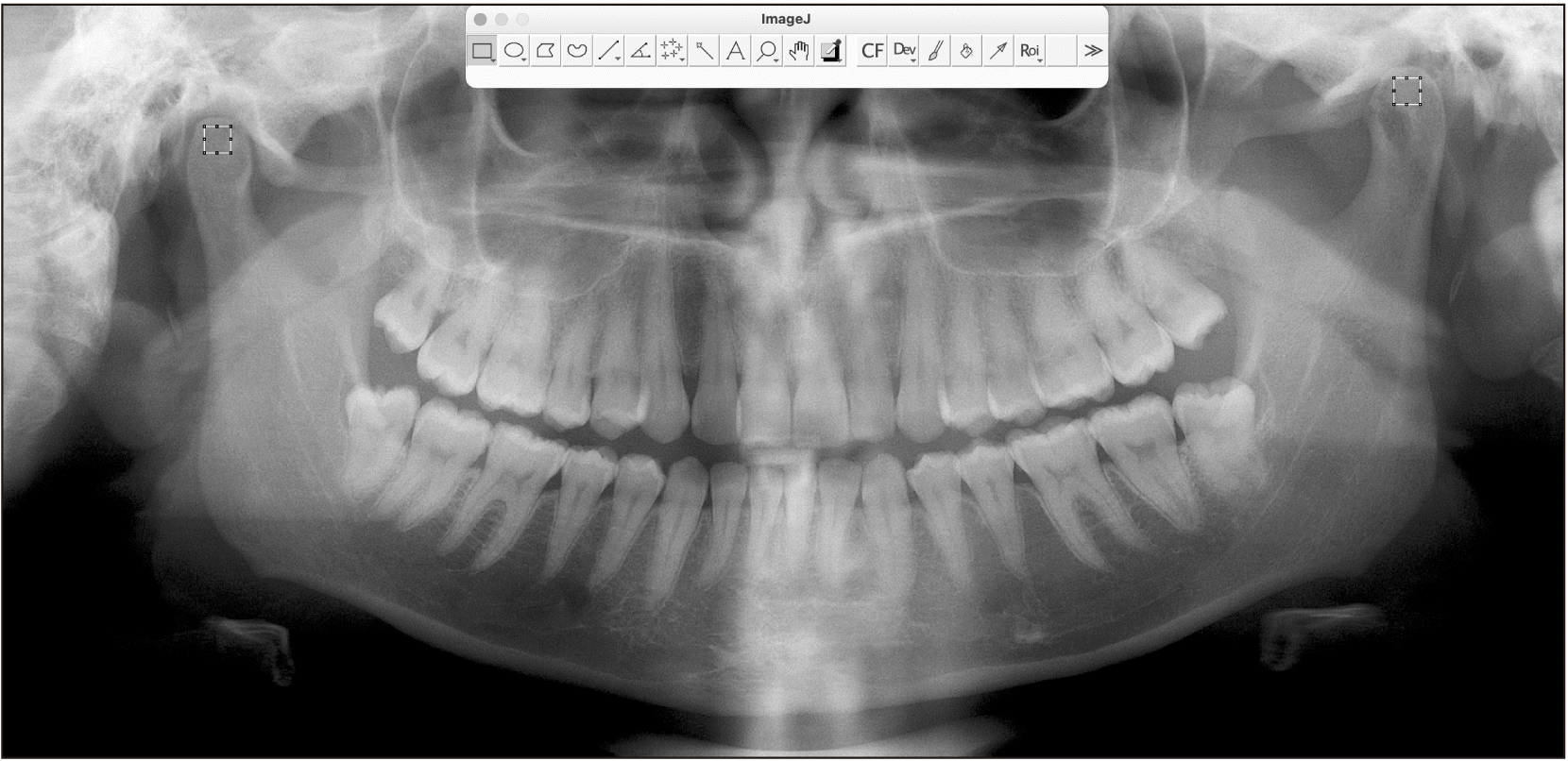
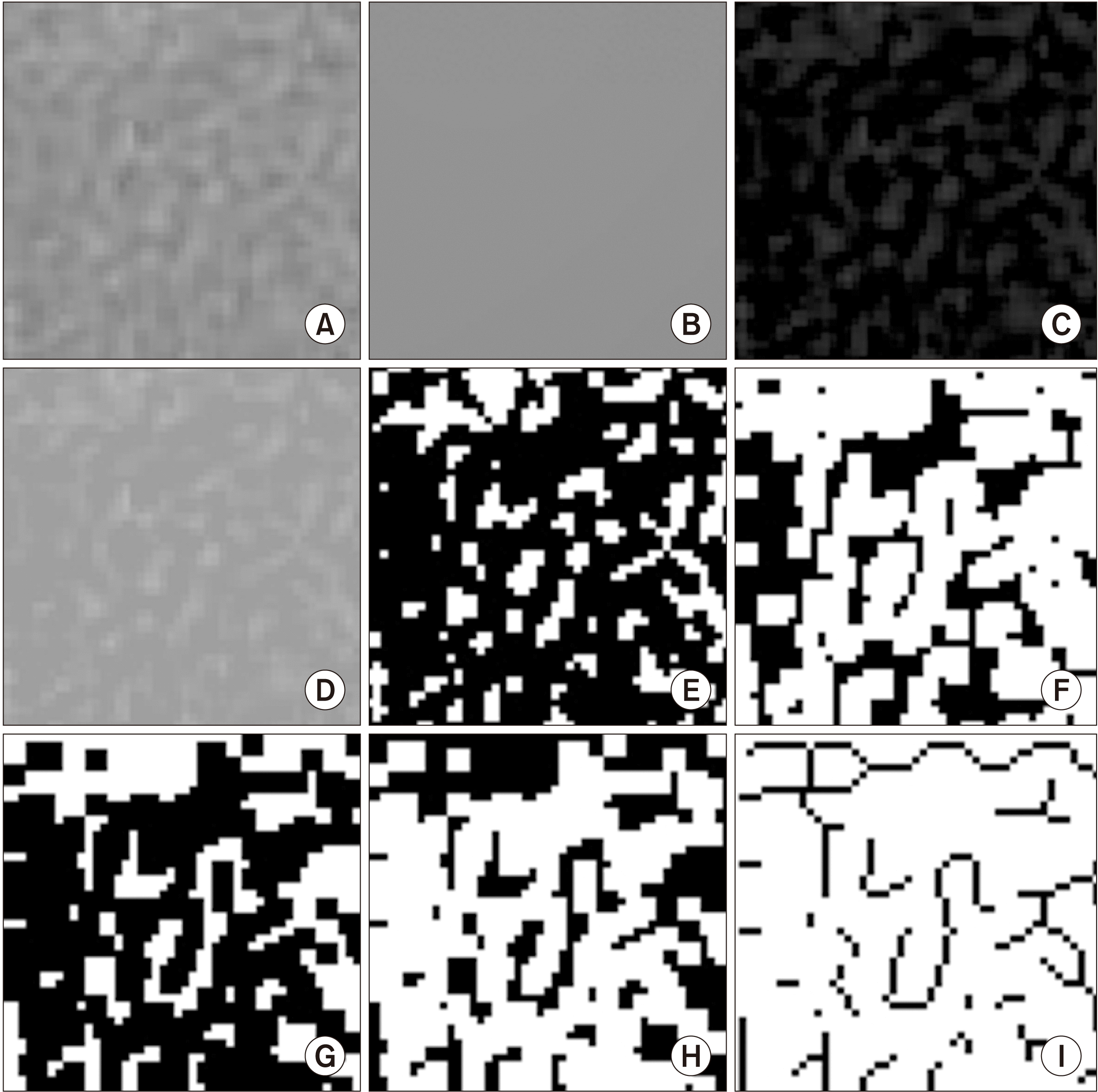
The FD values were calculated for the right and left sides of the TMJ on each panoramic radiograph using the box counting method described by White and Rudolph15. The ImageJ version 1.53g (National Institutes of Health, Bethesda, MN, USA; http://rsb.info.nih.gov/nih-image) software was used for this process. Panoramic radiographs were opened using the ImageJ software. All containing bone tissue, the regions of interest (ROIs) with 50×50 pixels were selected from the mandibular condyles close to the articular surfaces.(Fig. 2) The ROIs were duplicated. The image was blurred using the Gaussian filter. The blurry image was removed from the original image using the subtraction process. A total of 128 shades of grey were added to each pixel of the image. The image was turned into black and white using the threshold process, and erode, dilate, invert and skeletonization procedures were then applied to the image. Fractal box counting was performed in the resulting image.(Fig. 3) The FD values were calculated and recorded separately for panoramic radiographs taken at T0 and T1 times for both condyles of each patient.
 | Fig. 2Fractal dimension analysis process. A. Region of interest. B. Blurred image of the cropped and duplicated region of interest. C. Subtracted blurred image from the original image. D. Addition of a grey value of 128 to each pixel location. E. Binarization. F. Erosion. G. Dilatation. H. Inversion. I. Skeletonization.
|
All images were reviewed by the same researcher. The images of the patients were re-evaluated after 30 days to eliminate any errors and inconsistencies that may occur due to the researcher. Pearson correlation coefficients were calculated to determine the relationship between measures. As a result of the correlation analysis, there was no significant difference between the two measurements (P>0.05).
5. Statistical analysis
As the data were distributed homogeneously according to the results of the Levene statistic test of homogeneity of variances, a two-way repeated measures ANOVA was used to evaluate the FDs. Gabriel’s post hoc test was used for the paired comparison of differences between groups. The differences within groups were evaluated using a paired sample t-test. The significance level was set to P≤0.05. Statistical analysis was performed using IBM SPSS Statistics software for Mac (ver. 25.0; IBM, Armonk, NY, USA).
Go to :
III. Results
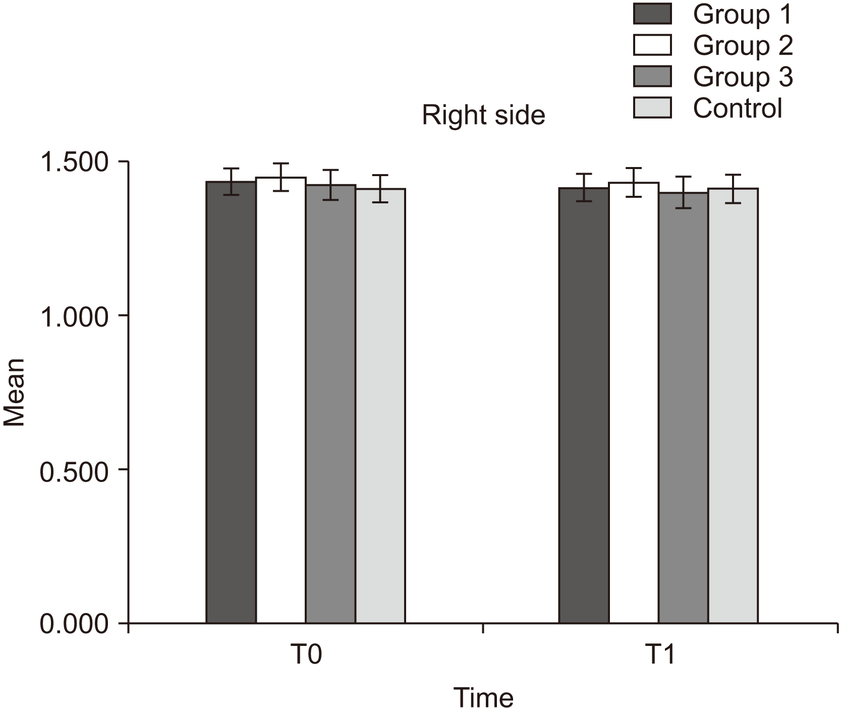
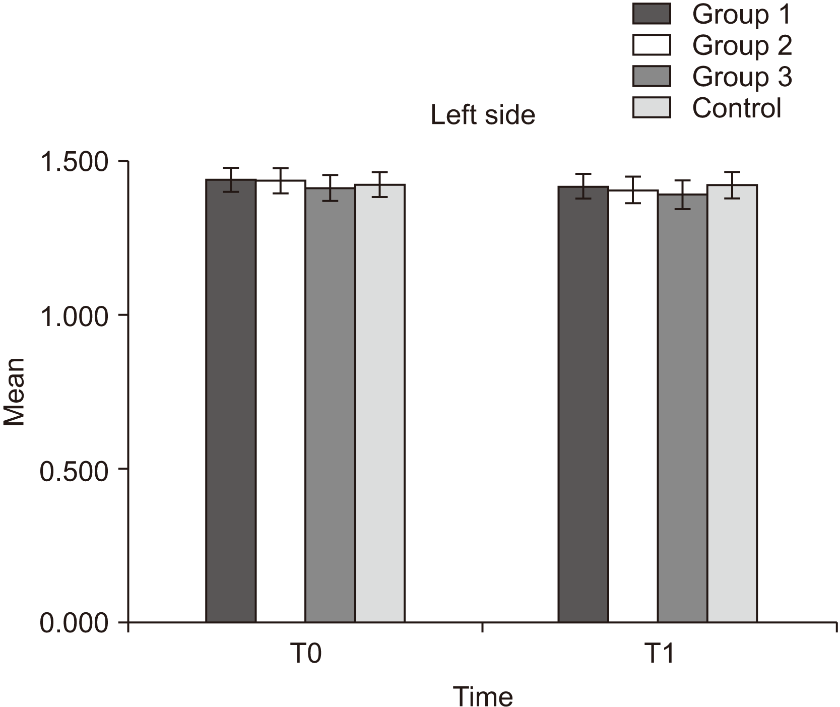
A total of 40 females and 20 males aged 18-68, with a mean of 30.95±12.38 years, were included in the study. The number of patients in the groups was as follows: Group 1, n=17; Group 2, n=15; Group 3, n=13, and the Control group, n=15. The measurements by group are presented in Table 1, considering the right and left sides.
Table 1
Fractal dimension measurements by group
(SD: standard deviation, T0: before treatment, T1: six months after treatment, RCFD T0: fractal dimension value of right condyle at time T0, RCFD T1: fractal dimension value of right condyle at time T1, LCFD T0: fractal dimension value of left condyle at time T0, LCFD T1: fractal dimension value of left condyle at time T1)
![]()
There was no significant effect of sex on the FD variable [F (1, 58)=0.766, P=0.385]. The main effect of side on the FD value was not significant [F (1, 56)=0.041, P=0.840]. The effect of side×group interaction was not significant [F (3, 56)=0.440, P=0.726]. The main effect of time on the FD value was significant [F (1, 56)=86.176, P<0.001]. This effect was qualified by a significant time×group interaction effect [F (3, 56)=9.023, P<0.001]. The effect of side×time interaction effect was not significant [F (1, 56)=0.740, P=0.393]. The effect of side×time×group interaction was not significant [F (3, 56)=0.924, P=0.435].(Table 2)
The main effect of the group on the mean FD value across time was not significant [F (3, 56)=0.295, P=0.829]. No significant difference was found between the pairwise comparisons of the groups (P=0.947).(Table 3)
Table 3
Results of multiple comparisons
![]()
In addition, the within-group differences in all groups were evaluated at T0 and T1 times using the paired t-test. The decreases in FD values in treatment groups between T0 and T1 times were significant (P=0.004). However, changes in FD values were not significant in the control group (P=0.728).(Table 4)
Table 4
Results of evaluating the within-group differences between two different times
| Group | Mean | SE | P-value | |
|---|---|---|---|---|
| Group 1 | RCFD T0–RCFD T1 | 0.019235 | 0.005102 | 0.002* |
| LCFD T0–LCFD T1 | 0.021882 | 0.004013 | 0.000* | |
| Group 2 | RCFD T0–RCFD T1 | 0.018267 | 0.005321 | 0.004* |
| LCFD T0–LCFD T1 | 0.029333 | 0.006657 | 0.001* | |
| Group 3 | RCFD T0–RCFD T1 | 0.023769 | 0.006634 | 0.004* |
| LCFD T0–LCFD T1 | 0.021385 | 0.004474 | 0.000* | |
| Control | RCFD T0–RCFD T1 | 0.000467 | 0.001316 | 0.728 |
| LCFD T0–LCFD T1 | –0.000267 | 0.001449 | 0.857 | |
(SE: standard error, T0: before treatment, T1: six months after treatment, RCFD T0: fractal dimension value of right condyle at time T0, RCFD T1: fractal dimension value of right condyle at time T1, LCFD T0: fractal dimension value of left condyle at time T0, LCFD T1: fractal dimension value of left condyle at time T1)
![]()
The means of the FD values at T0 and T1 times are plotted graphically in Fig. 4 and 5.
Go to :
IV. Discussion
In patients with bilateral TMJ hypermobility, the FD values calculated in the mandibular condyles of patients who received hypertonic dextrose prolotherapy decreased depending on time (P<0.001). No interactions of the decrease in FD values with side, number of injections, or sex were found. FD analysis has been used previously to evaluate bone healing after endodontic surgery, orthognathic surgery, and implant treatments and to evaluate bone structures in TMJ disorders11,16-20. Higher FD values indicate more complex structures. However, structures with the same FD value may not show the same texture feature10,21. FD value reduction in surgical sites is associated with decreased bone complexity. Arsan et al.11 reported that erosive and sclerotic changes in the condyle in TMJ patients could be seen as a decrease in FD values.
Various proliferants are used in prolotherapy. Dextrose is the most widely used proliferant because it is inexpensive, accessible, and reliable. The techniques used in the treatment of TMJ hypermobility and dysfunction with the purpose of prolotherapy with a single injection or multiple injections and using dextrose in different concentrations have been described5,6,9,22. There is no definite protocol for prolotherapy in TMJ hypermobility. Dextrose concentrations used in prolotherapy range from 10% to 50%2,6,9,22-24. In this study, dextrose at a concentration of 20%, which gives positive clinical results on TMJ hypermobility in the literature, was used, and positive clinical results were obtained for all patients25,26.
Although the mechanism of prolotherapy has not been fully discovered, it is believed that its inflammatory and non-inflammatory mechanisms play a role9. Dextrose in concentrations above 10% is known to cause partial inflammation1,9. In histopathological examinations performed after a dextrose injection, first-day hemorrhage, inflammation and necrosis in the soft tissues and ligaments were reported, followed by fibrosis, repair tissue, and regeneration symptoms9,27. Histopathologically, the strengthening and thickening of the tendons and ligaments were demonstrated after prolotherapy28. Studies on changes in hard tissues are limited. There is a need for histopathological studies on the effect of inflammation after prolotherapy and subsequent regeneration of the hard tissue components of the joint at different concentrations and numbers of injections.
Studies on prolotherapy in TMJ have generally focused on clinical findings, such as pain, maximal interincisal opening (MIO), locking episode frequency, and sound in TMJ6,29. Studies have reported different results on clinical findings after prolotherapy in TMJ hypermobility patients. Ungor et al.6 reported a decrease in pain on function after prolotherapy in patients with TMJ dislocation. Taskesen and Cezairli29 found a reduction in pain after prolotherapy in patients with TMJ hypermobility. Cömert Kiliç and Güngörmüş2 reported that prolotherapy caused a decrease in MIO in TMJ hypermobility, but there was no difference in the placebo group in other clinical findings, such as joint noise and pain.
Unlike other studies, this study evaluated the change in the trabecular structure in the long-term areas of the condyles close to the articular surface after prolotherapy using the FD analysis method. There was a decrease in the obtained FD values at T1 time compared with T0 time. This can be interpreted as the inflammation that occurs in this area and affecting the hard tissues over a period of six months. Longer follow-up studies are recommended at dextrose concentrations different from those used in this study.
In the literature review, dextrose prolotherapy is mostly performed as three or four injections in TMJ5,6,30. In some studies, prolotherapy was performed as a single injection in the TMJ and obtained positive results7,9,25. In the current study, three groups of patients who received positive clinical results after one injection, two injections, and three injections of prolotherapy were formed, and their results were compared. The number of injections was found to have no effect on the change in FD value in the condyles when dextrose was used at a concentration of 20%. However, to relieve the clinical symptoms of patients, re-injection/injections are required.
Prolotherapy is widely used, especially in diseases of the musculoskeletal system. Prolotherapy, which is used in the regeneration of ligaments and tendons, has also been reported to provide effective results in osteoarthritis24,28,31. In a disease such as osteoarthritis in which bone tissue is affected, the effectiveness of dextrose suggests that it may also affect hard tissues. During prolotherapy application, not only intra-articular injection but also injection around the joint may affect the hard tissue. Due to the slower regeneration times of hard tissues compared with soft tissues, a decrease in FD value in bone may be observed in the sixth month.
The changes in FD values observed in the treatment groups may not indicate large structural changes. Since the jaw movements and muscle functions of the patients in the treatment groups may change after the treatment, FD changes can be considered not only as a result of dextrose injections, but also as an adaptation response in the condyles to these mechanical changes. The fact that FD changes were not significant in the control group may be attributed to the absence of changes in the TMJ movements and chewing mechanisms of the patients.
Go to :
 XML Download