

 PDF
PDF Citation
 Print
Print


Introduction
Anti-N-methyl-D-aspartate receptor (NMDAR) encephalitis is a rare autoimmune disorder that predominantly affects young adults. The clinical course begins with prominent psychosis or memory deficits leading to seizures, movement abnormalities, or autonomic instability [1-5]. It is a paraneoplastic syndrome accompanied by ovarian neoplasms, mostly mature teratoma. Once this fatal encephalitis is clinically suspected, the ovarian tumor is removed and immunosuppressive therapy is initiated. The diagnosis is confirmed by the presence of antibodies in the cerebrospinal fluid (CSF) or serum. This is a case report of anti-NMDAR encephalitis associated with ovarian neoplasms, including immature teratoma, in Korea.
Cases
1. Case 1
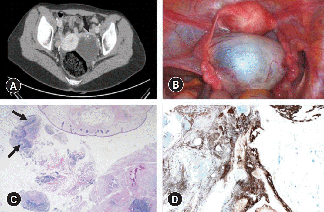
An 18-year-old woman presented with abdominal pain associated with a 6 cm left ovarian teratoma observed by an abdominal computed tomography (CT) scan (Fig. 1A). Surgical removal was planned. However, on the day of admission, she experienced severe insomnia, followed by global amnesia, aggressive behavior, and suicidal ideation. The patient had headaches and memory disturbance for 2 weeks before admission (Table 1). As a result, the planned operation was postponed. Electroencephalogram (EEG) and brain magnetic resonance imaging (MRI) were unremarkable. Intensive antipsychotic treatment did not improve the symptoms. Under the suspicion of anti-NMDAR encephalitis, removal of the ovarian teratoma was performed (Fig. 1B). Both serum and CSF samples were found to be positive for antibodies against NMDAR. The histopathological diagnosis of the ovarian tumor was an immature teratoma (Fig. 1C, 1D). Immunoglobulin treatment and four cycles of rituximab were administered. The symptoms progressively improved, and at the 9-month follow-up, whole-body positron emission tomography CT and transvaginal sonography were normal.
2. Case 2
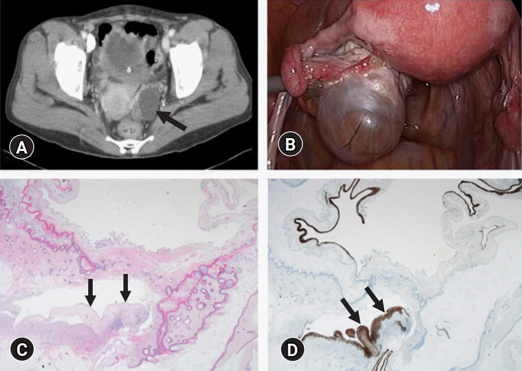
A 41-year-old woman was admitted to the hospital with fever and headache. The serum and CSF examinations showed no signs of inflammation. The patient complained about episodes of self-talking and disorientation for the last few days before admission. Brain MRI and EEG were normal. Despite intensive treatment, the clinical symptoms did not improve. Under the impression of anti-NMDAR encephalitis, an abdominal CT scan was performed (Fig. 2A). It showed a 4 cm ovarian teratoma that was immediately removed, and the patient received corticosteroids and immunoglobulin treatment (Fig. 2B). Anti-NMDAR antibodies were detected in serum and CSF samples. The pathologic diagnosis of the ovarian tumor was a mature teratoma (Fig. 2C, 2D). There was no evidence of recurrence at the 2-month follow-up.
3. Case 3
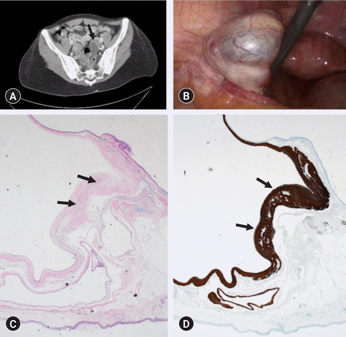
A 29-year-old woman experienced confusion, agitation, and auditory hallucinations. The serum blood test, CSF examination, and brain MRI showed no abnormalities. Only the EEG test revealed cerebral dysfunction. Empirical treatment with high-dose immunoglobulin, methylprednisolone, and plasma exchange was started; however, symptoms did not improve. Anti-NMDAR encephalitis was considered, and a 3 cm left ovarian teratoma was detected on an abdominal CT scan (Fig. 3A). The serum and CSF tested positive for anti-NMDAR antibodies. The tumor was removed surgically, and the patient received corticosteroids, plasmapheresis, and rituximab thereafter (Fig 3B). The pathologic diagnosis of the ovarian tumor was a mature teratoma (Fig. 3C, 3D). She completely recovered within 3 months.
Discussion
Anti-NMDAR encephalitis is an autoimmune encephalitis first reported by Dalmau et al. [4,5] in 2007. It typically begins with prodromal symptoms such as nonspecific fever, diarrhea, vomiting, headache, or upper-respiratory symptoms before the onset of psychiatric and neurological symptoms, which are often accompanied by persistent amnesia. It is known that age, sex, and ethnicity are important factors related to the disease, as most patients are women, especially of Asian ethnicity [6,7]. NMDAR hypofunction is related to cognitive defects, whereas overstimulation causes excitotoxicity and subsequent neurodegeneration [8].
The clinical features that distinguish anti-NMDAR encephalitis from other neurological or psychiatric disorders are acute onset of mood and behavioral changes with no prior history of seizures or abnormal movements. The differential diagnoses include primary psychiatric disorder, drug abuse, neuroleptic malignant syndrome, migraine, antipsychotic medications, or infectious encephalitis. Anti-NMDAR encephalitis should be considered once the above conditions are excluded [9]. The detection of immunoglobulin G antibodies in the serum or CSF confirms the diagnosis. In the advanced stages, the CSF antibodies usually remain elevated, while serum antibodies substantially decline with treatment [10]. There is limited evidence regarding the relationship between the size of the adnexal tumor and encephalitis. However, ovarian teratomas in anti-NMDAR encephalitis are usually not large, and an ovarian teratoma as small as 6 mm in diameter has been reported in association with anti-NMDAR encephalitis [11].
Treatment is focused on resection of the tumor followed by immunotherapy, including glucocorticoids, immunoglobulin, and plasma exchange, which collectively improves the symptoms within 4 weeks. For patients who do not respond to first-line immunotherapy, rituximab or cyclophosphamide may be used. Immunotherapy is applied to treat encephalitis regardless of the type of ovarian teratoma.
This is a case report of a rare autoimmune anti-NMDAR encephalitis associated with ovarian tumors, including immature teratoma, in Korea. Patients showed a wide spectrum of initial symptoms, and immediate surgical removal of the adnexa tumor was performed in all patients, followed by intensive immunotherapy. The course of the disease correlated with previous findings in that neurological symptoms progressively improved following the operation. Consciousness improved first, and then, autonomic function recovered. The frequency of epileptic seizures was reduced, and the social ability returned to the state before the onset of the disease.
Although we were not able to compare the clinical symptoms among anti-NMDAR encephalitis patients with or without ovarian teratoma, according to the study by Zhang et al. [12], there may be differences in laboratory and clinical findings between anti-NMDAR encephalitis with or without ovarian teratoma [12]. Anti-NMDAR encephalitis patients without ovarian teratoma experience more viral prodromal symptoms, whereas patients with teratoma present with milder neurological symptoms. In our study, two patients presented with fever and myalgia, mimicking viral infection, whereas one patient complained about memory disturbance. Moreover, the prevalence of immature teratomas, which generally comprise less than 1% of ovarian teratomas, is much higher among patients with anti-NMDAR encephalitis compared to the general population [13]. Therefore, it is important to inform patients about the possibility of malignancy in cases of anti-NMDAR encephalitis with ovarian teratoma.
In conclusion, anti-NMDAR encephalitis is a rare yet serious disorder that must be considered in patients with ovarian teratoma and neurological symptoms. Anti-NMDAR encephalitis may be speculated in patients who present with psychotic symptoms that are not improved by typical antipsychotic treatments, and the presence of an ovarian tumor detected on imaging studies. A comprehensive evaluation, including blood tests, CSF examination, brain and abdomen imaging, and antibody screening, is required to exclude other causes before making the diagnosis.
Once the disease is suspected, a combination of surgical removal of adnexal tumors and immunotherapy should be applied. The disorder can be fatal without appropriate treatment. Therefore, it should be included in the differential diagnosis in patients with ovarian teratoma and neurological symptoms.
 XML Download