

 PDF
PDF Citation
 Print
Print


Introduction
Infections of the heart primarily manifest as endocarditis, which affects 7.6 to 80/100,000 adult admissions [1]. Prosthetic materials, including valve replacements, grafts, implantable devices, and related materials, such as leads, can lead to the development of endocarditis [2]. Moreover, cardiac implantable electronic device (CIED) infections occur in approximately 1.8/1,000 pacemakers a year according to a nationwide cohort study [3]. The diagnosis of such infections can be difficult, and the clinical presentation, as well as microbiological and imaging approaches have been used to reach an accurate diagnosis.
The diagnosis of infective endocarditis (IE) is largely based on the modified Duke criteria (mDC), which has an overall sensitivity of 80%. The mDC comprises causative pathogen detection and echocardiographic features of endocardial involvement [4]. Echocardiography is useful in diagnosing and managing patients with IE, by showing the presence of a swinging intracardiac mass or vegetation, prosthetic valve partial dehiscence, annular abscesses, and new valve regurgitation, which are categorized as major criteria in the diagnosis of IE [4]. Furthermore, the diagnostic sensitivity for vegetation in native and prosthetic valves is better for transesophageal echocardiography (TEE) than transthoracic echocardiography (96% and 92% vs. 70% and 50%, respectively) [5]. Echocardiography has superior accuracy as a primary imaging tool for native valve imaging but has disadvantages such as acoustic shadowing and noise when imaging implanted material. Moreover, blood cultures often lead to indeterminate results for confirming suspected IE, and in up to 24% of patients with pathologically proven IE, it is misclassified as a “possible” IE based on the mDC alone [6]. Recently, the addition of F-18 fluorodeoxyglucose (FDG) positron emission tomography/computed tomography (PET/CT) to the mDC has improved the diagnostic accuracy in patients suspected with prosthetic valve endocarditis (PVE) or intracardiac device infection. FDG PET/CT is also useful to evaluate the extent of valve and device infections, as well as the detection of extracardiac infections, such as septic embolism [6,7].
Unlike TEE, for which numerous well-designed prospective randomized studies have been conducted, knowledge on FDG PET/CT is largely from observational case studies and retrospective data reviews. Despite this limitation, published data for cardiac-device related infections are generally consistent and support its judicious application in the workup of IE [8,9]. Furthermore, FDG PET/CT may have unique advantages over TEE in the following aspects: (1) to provide confirmatory information when TEE findings are inconclusive; (2) to diagnose IE earlier than TEE before morphological damage ensues; (3) to detect unexpected sources of infection and embolisms; and (4) to potentially guide clinical management [10]. The European Society of Cardiology (ESC) recommends adding abnormal FDG uptake as the major criterion for PVE in patients with suspected PVEs classified as “possible” or “denied” in initial mDC [5]. In the 2017 appropriate use criteria, FDG PET/CT was included as ‘may be appropriate’ for PVE and CIEDs [11]. However, the American Heart Association guideline states that more studies are needed to determine the role of FDG PET/CT in the diagnosis and management of patients with IE, although it is stated to be useful for detecting extracardiac complications [12].
Early adoption of a new technique in clinical care based on recommendations from a panel of experts may improve patient outcomes, even when scientific evidence is lacking. In this article, the general concepts and available evidence for FDG PET/CT use in heart infections are reviewed, and recommendations for image acquisition, interpretation, and pitfalls are given.
Go to : 
Prosthetic valve endocarditis
Several studies have examined the usefulness of FDG PET/CT for PVE. Saby et al. [7] reported that FDG PET/CT had a sensitivity of 73% and a specificity of 80% in a cohort of 72 patients. Furthermore, the sensitivity has significantly increased from 70% with the mDC to 93% with the addition of abnormal FDG uptake around the prosthetic valve as a new main criterion. A meta-analysis of 13 studies by Mahmood et al. [13] showed a pooled sensitivity of 80.5% and specificity of 73.1% and supported the utility of FDG PET/CT as an ancillary diagnostic tool in challenging IE cases. Recently, a large retrospective multicenter cohort study reported FDG PET/CT had a sensitivity of 74% and a specificity of 91%. In addition, if confusing factors, such as low inflammatory activity (defined as C-reactive protein [CRP] levels <40 mg/dL), and the use of surgical adhesives during transplantation, are excluded, the sensitivity and specificity of FDG PET/CT in PVE diagnosis would increase to 91% and 95%, respectively. Moreover, FDG PET/CT is performed at the early stage of PVE diagnosis, positive results can be confirmed even if the existing diagnostic tests, such as blood culture and echocardiography, are negative. Another interesting finding is that by using FDG PET/CT with echocardiography for diagnosis of IE, the sensitivity increases from 65% to 96% compared to echocardiography alone; this facilitates early diagnosis before structural damage, and consequently reduces the incidence of serious complications, such as valve dehiscence or perivalvular abscesses [14].
Another single center prospective study of 151 patients with suspected PVE reported a sensitivity of 60% for echocardiography alone and 42% for mDC, which increased to 91% when focal uptake in PET/CT was included in the mDC. This study also confirmed that possible IE could be reduced from 33% to 8% if the diagnosis included FDG PET/CT findings [15]. Recent research has indicated that positive FDG PET/CT results are related to major cardiac events such as, death, recurrence of IE, acute heart failure, unsuspected cardiovascular hospitalization, and new embolic events [16].
Go to :
Native valve endocarditis
In contrast to PVE, there is limited research for the use of FDG PET/CT in suspected native valve endocarditis (NVE). de Camargo et al. [15] reported on 115 patients with NVE, the sensitivity, specificity, positive predictive value (PPV), and negative predictive value (NPV) were 22%, 100%, 100%, and 66%, respectively. Furthermore, Kouijzer et al. [17] reported that although FDG PET/CT has a low sensitivity of 45% in NVE, its ability to diagnose NVE increases when FDG PET/CT is added to mDC, which is particularly useful in cases that are difficult to diagnosis using conventional techniques.
A recently published pathological study showed that a higher FDG uptake was related to higher inflammatory infiltration, higher fibrin, lower fibrosis, and a predominance of polymorphonuclear cells in tissues. The study also reported that polymorphonuclear cell infiltration was significantly increased and fibrosis was reduced in PVE, compared to NVE [15]. Therefore, the sensitivity of FDG PET/CT is expected to be lower in NVE than in PVE.
FDG PET/CT may influence the clinical management of patients with NVE by identifying the source of primary extracardiac infection or infective emboli. A prospective study showed that FDG PET/CT identified additional infection sites, such as the lungs, skeleton, brain, and other organs, in 74.5% of patients with diagnosed NVE. Based on these extracardiac findings on FDG PET/CT, the incidence of IE relapse decreased by two-fold as a result of appropriate treatment [18]. In line with this, moderate to intense FDG uptake in the perivalvular area is associated with worse prognosis, such as increased new embolic events (hazard ratio [HR], 8.8) and rehospitalization (HR, 3.57) [16].
Go to :
Cardiac implantable electronic device infection
CIEDs, including pacemakers, implantable cardioverter-defibrillators, and cardiac resynchronization therapy devices (with or without defibrillation capacity), consist of pulse generators to provide the electrical stimulus and transvenous or epicardial leads to deliver the stimulus to the heart. Approximately 1.2 million to 1.4 million CIEDs are implanted each year worldwide [19]. CIED infections have an incidence of 1.37/1,000 device-years for pocket infection alone and 1.14/1,000 device-years for device-related endocarditis. Among a cohort of 2,760 patients with definite IE, 177 cases (6.4%) involved CIED [20]. CIED infections are generally considered in two categories; pocket infection and systemic infection such as lead and/or valvular infection [21]. However, these categories are not exclusive, and the two forms can coexist (Fig. 1). Antibiotics alone can be useful for managing superficial soft tissue infections, but deep pocket infections or lead infections require complete removal of the device, which has important implications for patient care. Thus, fast and accurate diagnosis and, rapid treatment are of supreme importance; however, difficulties remain, especially if the symptoms are delayed or mild [19].
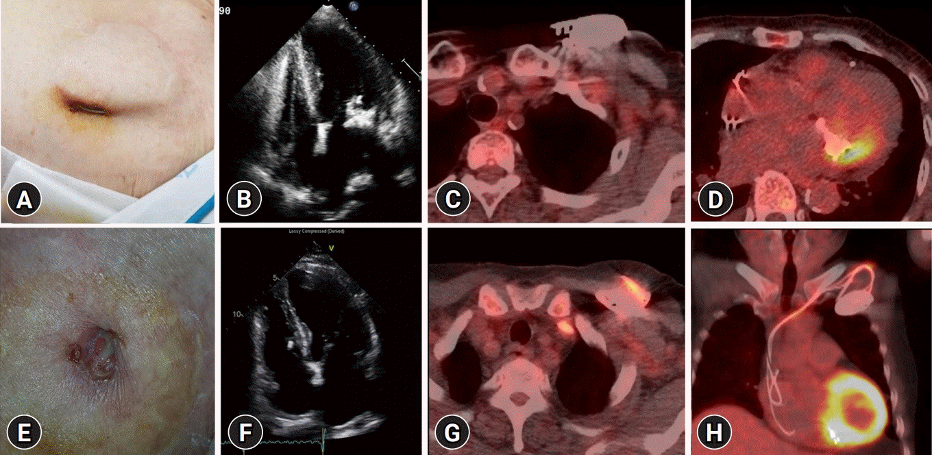 | Fig. 1.Two patients (A–D and E–H) with suspected cardiac implantable electronic device (CIED) infection. (A) Gross photo showing CIED exposure with redness in a 79-year-old woman. (B) Echocardiography reveals vegetation in the mitral valve. (C, D) Axial FDG PET/CT shows no FDG uptake in the CIED pocket, but shows high uptake in the mitral valve. The patient did not undergo extraction of the CIED, and instead, was treated with vancomycin for CIED-infective endocarditis with positive blood culture for Staphylococcus epidermidis. (E) Gross photo showing pus drainage from the pacemaker insertion site in a 73-year-old man with positive wound culture for Serratia marcescens. (F) Echocardiography confirming absence of abnormal findings in the heart. (G, H) Axial and coronal FDG PET/CT shows increased FDG uptake in the pocket and along the lead. The patient underwent pacemaker removal. FDG, F-18 fluorodeoxyglucose; PET/CT, positron emission tomography/computed tomography.
|
The role of FDG PET/CT in CIED infection has received increasing interest in recent years, and it is considered to be especially useful when a diagnosis of pocket or lead infection is unclear with other imaging techniques, such as TEE. Sarrazin et al. [22] reported that PET/CT could distinguish skin infections from pocket, lead, or intravascular infections in cases of unclear diagnosis or extension. They also proposed that PET/CT can help differentiate an active cardiac device infection from residual normal postoperative inflammation. In addition, Juneau et al. [23] presented a 93% sensitivity and 98% specificity for pocket infection, but 88% and 65% for lead/IE, respectively. Furthermore, a large meta-analysis involving 14 studies with a total of 492 patients showed that FDG PET/CT had a high sensitivity and specificity (96% and 97%, respectively) for pocket infection, but a relatively low sensitivity and specificity (76% and 83%, respectively) for lead infections or CIED-IE [24]. The reason for the poor diagnostic ability for lead infection or IE was only speculated. Of the included studies, only four involved physiological myocardial suppression, while lead infection was difficult to interpret in the other studies [25]. The vegetation in the lead or valve was too minimal and leukocyte induction was not sufficient for visualization of the FDG accumulation. Moreover, many patients were investigated after initiation of antibiotic therapy.
A recent study involving 105 patients with confirmed CIED infection demonstrated the ability of FDG PET/CT to predict the outcomes of post transvenous lead extraction. Patients with positive findings of CIED pockets on FDG PET/CT, with or without systemic involvement, had better survival rates (HR, 0.493). However, patients with CIED infection with no pocket infection in skin lesions or on PET/CT images showed poor long-term survival. The results can be explained by the hypothesis that CIED infection can arise from two mechanisms, which are associated with different long-term outcomes: (1) CIED infection can originate in a CIED pocket and later spread to the bloodstream; and (2) primary bloodstream infection (transient or recurrent bacteremia) can cause metastatic infection of the lead. The latter may explain why patients with no pocket infection in skin lesions or on PET/CT images have a poor outcome. In addition, approximately 25% of patients diagnosed with CIED infection can be restratified with the presence/absence of CIED-IE by incorporating the FDG PET/CT results [26]. The ESC guideline and appropriate use criteria refer to the usefulness of FDG PET/CT in CIED infection as “may be considered” [5,11].
Go to :
Left ventricular assist device infection
A left ventricular assist device (LVAD) is mechanical circulatory support device that is surgically implanted in patients with acute or chronic refractory heart failure and serves as a bridge for transplant or destination therapy [27]. LVADs typically consist of a pump with an inflow conduit from the left ventricular apex and an outflow conduit to the ascending aorta. The pump is placed into a pocket, and a driveline, tunneled from the pump, is connected to an external power source through an exit site on the lower abdominal wall. The driveline is particular at risk of infection, with an infection incidence of between 17% to 30% and a mortality rate of 9.8% at 6 months and 31% at 12 months [28]. Early treatment can improve the prognosis, but proper diagnosis is difficult at the time of infection. Although the use of FDG PET/CT has shown good diagnostic results for driveline infection, the number of patients included in each study was small.
Bernhardt et al. [29] reported the sensitivity of FDG PET/CT in LVAD infections as 87.5%, the specificity as 100%, the PPV as 100%, and the NPV as 86.7%. They also confirmed the utility of FDG PET/CT in the location and quantification of the extent of infection. Moreover, a recent case series and meta-analysis of four studies showed a pooled sensitivity of 92% and specificity of 83% for the diagnosis of LVAD infection, but the specificity varied considerably between studies (25% to 100%) [30]. Akin et al. [31] showed that FDG PET/CT imaging provided accurate information on the location and extent of LVAD-related infections 3 weeks after implantation. Although data are limited, preliminary studies have shown that FDG PET/CT can differentiate and localize the site and extension of infection within the central portion of the device, or along the peripheral driveline. This finding has clinical significance, in that patients with infection involving the central portion of a LVAD (including the pump and cannula) have a poorer survival rate than those with an infection involving the peripheral driveline and exit site [32].
de Vaugelade et al. [33] compared the diagnostic performance of FDG PET/CT and leucocyte labeled scintigraphy and demonstrated that FDG PET/CT showed greater sensitivity (95.2% vs. 71.4%, respectively). A recent analysis of 57 patients who underwent 85 PET/CT scans showed that a threshold of peak standardized uptake value (SUVpeak) of 2.5 could accurately diagnose driveline infections. On dividing the LVAD infection into four components on FDG PET/CT, patients with three or more components showed lower survival. Moreover, the presence of thoracic lymph nodes with FDG avidity was also associated with lower survival. Patients who underwent early surgical revision after PET/CT had a shorter hospital stay [34].
Although visual analysis by FDG PET/CT is highly associated with LVAD infection, quantitative parameters provide greater sensitivity and specificity than visual grading alone. A retrospective study that evaluated both visual and semiquantitative approaches reported that FDG uptake along the driveline is rarely an artifact; however, this depends on the reader’s experience and is not suitable for inter-examination or patient-to-patient comparison. In a semiquantitative analysis of FDG PET/CT, the diagnostic ability could be further improved with a sensitivity and specificity of 100%, and the maximum SUV (SUVmax) increased by 3.88 or more compared to the basal scan [35]. Dell’Aquila et al. [36] reported that quantitative FDG PET/CT analysis using SUVmax was accurate in diagnosing superficial and deep driveline infections, but had limited use in pump housing infections, in which visual analysis was better. Furthermore, Avramovic et al. [37] proposed that metabolic volume had more diagnostic capability than visual score or SUVmax for LVAD driveline infection. The results demonstrated that the sensitivity, specificity, PPV, and NPV were 87.5%, 79%, 81%, and 86% for visual score; 87.5%, 87.5%, 87.5%, and 87.5% for SUVmax; and 96%, 87.5%, 88.5%, and 95.5% for metabolic volume, respectively.
The use of surgical adhesives to strengthen inflow and outflow cannulas can lead to false-positive findings. Careful review of each patient’s surgical report with regard to the implantation procedure could improve the discriminant power for LVAD infection. Moreover, the large, dense structures of the pump housing are susceptible to beam hardening and scatter in the low-dose CT images used for attenuation correction (AC), which can lead to false uptake. In addition, FDG uptake by a foreign body reaction around the LVAD, physiologic FDG uptake by the adjacent left ventricular myocardium, presence of chronic fistula, and the possibility of internal surface infection of pump housing are all possible reasons for false uptake in FDG PET/CT [36].
Go to :
Extracardiac complications of infective endocarditis
Extracardiac complications following IE and CIED infection occur in 22% to 43% of patients within the first 2 weeks of treatment [38]. Metastatic infection, spondylodiscitis, osteomyelitis, septic arthritis, and metastatic soft tissue abscess can occur. Previous studies have shown that FDG PET/CT is useful to detect and localize these complications before clinical suspicion in patients with NVE [17,38-40] (Fig. 2). As such, the ESC guidelines recommend the use of FDG PET/CT in combination with other imaging studies, such as whole-body CT and brain magnetic resonance imaging, for the examination of embolic events [5]. Orvin et al. [40] reported on the use of FDG PET/CT in patients with confirmed IE and demonstrated that pertinent extracardiac findings on FDG PET/CT were present in 75% of patients. Consequently, treatment plans were changed in up to 35% of patients and included antibiotic treatment prolongation, referral to surgical procedures, and avoidance of unnecessary device extraction.
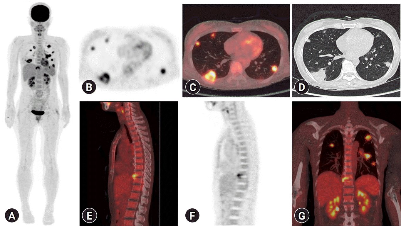 | Fig. 2.(A) Extracardiac metastatic infection in a 51-year-old woman demonstrating bilateral lung infection and spondylodiscitis resulting from Staphylococcus aureus. (B) Axial FDG PET, (C) axial FDG PET/CT, and (D) axial lung CT showing extensive lung septic emboli. (E) Sagittal FDG PET/CT, (F) sagittal FDG PET, and (G) coronal FDG PET/CT showing infective spondylodiscitis in T10 to T11. FDG, F-18 fluorodeoxyglucose; PET, positron emission tomography; CT, computed tomography.
|
Go to :
Monitoring response to antimicrobial therapy
For treatment of IE, the duration of antimicrobial therapy depends on the causative organism and the site of infection (NVE, PVE, or CIED infection). The recommended duration of treatment in the 2015 ESC guidelines [5] and the Northern American guidelines [41] is based on early randomized studies conducted in the 1990s or expert opinion, and there are very few recent comparative studies on the treatment duration [42]. An encouraging role of FDG PET/CT in evaluating the effectiveness of antimicrobial therapy in IE was recently proposed in a small observational study [43]. Moreover, García et al. [44] reported the usefulness of determining a need for continued therapy in cases with remnants of infection at the end of standard treatment; thus, continuation of therapy is needed until increased metabolism is no longer observed on FDG PET/CT. Further large-scale studies are necessary to utilize and prove the usefulness of this application.
Go to :
Practical consideration in clinics
The main limitation of FDG PET/CT in the diagnosis of cardiac infections is the absence of current standardized protocols, which have varied across previous studies. FDG PET/CT guidelines for diagnosis and monitoring in IE should include information on patient preparation, such as the diet protocol, FDG dosage, duration of uptake, acquisition time, and imaging processing including motion correction of cardiac and respiratory gating and image interpretation.
1. Patient preparation
In an oncological setting, myocardial FDG uptake varies from patient-to-patient and even for serial examinations of the same patient. This variation is considered to be due to nonspecific FDG uptake patterns in which diffuse myocardial activity is higher than liver activity or uptake in the lateral wall and/or ring shaped/circumferential basal uptake. Since this physiological uptake can mask pathological activities, patient preparation methods to inhibit physiological uptake have been proposed [45,46]. The degree of myocardial glucose metabolism varies greatly depending on the patient’s overall metabolic status, while the fasting myocardium uses free fatty acids (FFAs) as the main energy source (90%) [47]. Following dietary carbohydrate intake, myocardial metabolism shifts to glucose, following the Randle cycle [47,48]. The myocardium metabolizes glucose when blood sugar and insulin are elevated and FFAs are decreased. Conversely, as glucose and insulin levels decrease and FFAs increase in the fasting state, FFAs are used as the primary source of myocardial energy. The glucose metabolism in inflammatory cells is regulated by glucose transporter 1 (GLUT1) and GLUT3, unlike in the myocardium, where it is regulated via GLUT 4 and is independent of insulin effects [49,50]. Therefore, a patient preparation method that enables myocardial FFA metabolism, while simultaneously suppressing physiologic glucose uptake, is essential for successful FDG PET cardiac infection/inflammation imaging.
Most previous studies have implemented a high-fat, low-carbohydrate (HFLC) diet prior to a prolonged fast, or prolonged fast alone, while some studies have also used additional intravenous heparin administration. Although many preparation methods have been proposed to suppress physiologic myocardial glucose uptake, most studies included a small number of patients, and the preparation strategies were heterogeneous. Therefore, no clear consensus has been reached on the optimal method [51]. The guidelines of Society of Nuclear Medicine and Molecular Imaging, American Society of Nuclear Cardiology, and Society of Cardiovascular CT (SNMMI/ASNC/SCCT guideline) recommend a preparation regimen that incorporates a fat-rich and low-carbohydrate diet for 12 to 24 hours before scanning, and fasting 12 to 18 hours before and/or the infusion of intravenous heparin approximately 15 minutes before FDG injection [52]. More recently, the Japanese Society of Nuclear Cardiology (JSNC) performed an analysis of previous research and suggested a more detailed method, which involved fasting for 12 to 18 hours, and a low-carbohydrate diet, with a total carbohydrate content of less than 5 g for dinner the day before the scan. In addition, they suggested that patients with diabetes underwent same preparation as those without diabetes, albeit with particular attention to sugar control [53].
Regarding the effect of heparin preadministration on inhibition of physiologic uptake, Osborne et al. [51] reviewed 31 dietary preparation studies and found that the myocardium appropriately suppressed FDG uptake in 87% to 93% of patients who fasted for 4 hours after two HFLC meals. In addition, the same effect was reported when unfractionated heparin was injected 15 minutes before FDG injection after at least one HFLC meal and overnight fasting. Moreover, the authors did not recommend the fasting-only methods, food or drink intake, unrestricted diets, and high-fat supplements within 4 hours prior to scanning [51]. Furthermore, heparin showed an anticoagulant effect at doses above 10 U/kg, and the majority of published studies used unfractionated heparin 50 U/kg [54]. In order to lower the risk of heparin-induced thrombocytopenia (HIT), an uncommon but potentially life-threatening risk, low molecular weight heparin has been suggested; however, low molecular weight heparin undergoes approximately 60% lesser lipolysis than unfractionated heparin [55]. The aforementioned SNMMI/ASNC/SCCT guidelines suggest the use of 15 to 50 U/kg heparin, while the JSNC guideline does not recommend the use of heparin. By way of explanation, the JSNC explained that when fasting for more than 18 hours after a low carbohydrate diet, the inhibition of physiological myocardial uptake by heparin was limited, and the risk of HIT following unfractionated heparin is not negligible. However, only a small number of patients were included in the studies cited in these guidelines or review, and studies may provide varying suggestions owing to different fasting times, dietary methods, and frequency; therefore, one is not absolutely correct. The JSNC performed an analysis of fasting over 18 hours by dividing 82 patients into two groups [53], while Scholtens et al. [56] performed an analysis by dividing 150 people into three groups, and found that heparin additionally induced a significant decrease in myocardial intake during 12 hours of fasting.
Intracellular calcium is known to stimulate glucose uptake, and Gaeta et al. [57] reported a significant decrease in myocardial FDG uptake in mice treated with verapamil prior to FDG injection. However, these findings have not been replicated in humans [58].
Studies to date have failed to establish a single preparation technique that is far superior to all others, and metabolic profiles vary among patients; therefore, each center must optimize its own protocols that adhere to the principles described above.
2. Imaging acquisition
Hyperglycemia has been shown to impair the inflammatory cell uptake of FDG, as a result of competition with endogenous blood glucose; thus, it is recommended to perform scanning when the patient’s blood glucose is below 200 mg/dL. While most studies perform imaging acquisition 1 hour after FDG injection, Caldarella et al. [59] reported that an improved target-to-back-ground ratio is possible if imaging is delayed by 2 to 3 hours. This finding may have additional value for CIED infections, especially in lead infections with low diagnostic sensitivity. However, a patient series study comparing 1- and 2.5-hour post-injection images in PVE patients reported a false-positive interpretation trend for late images, requiring attention to interpretation [60].
Whole-body (head to feet) FDG PET/CT scanning has an advantage with regard to assessing localization of extracardiac infection, including clinically unpredicted distant foci [40,61]. In addition, treatment can be modified depending on the presence or location of the lesions [40]. Although physiological uptake leads to difficulty in visualizing small intracranial lesions, additional infectious lesions have been found on whole-body PET/CT in 17% of patients [13].
Gated cardiac PET imaging can improve spatial resolution and enhance the detection of small moving lesions with the heartbeat, and comparison with CT angiography (CTA) images is easier [62]. However, gated cardiac PET requires a surplus scan time, and there are currently no publications that have examined the additional value of gated cardiac PET for the diagnostic performance of endocarditis.
Pizzi et al. [63] suggested that a combination of FDG PET with CTA could improve the sensitivity in PVE and CIED infection. In their study, they demonstrated that the sensitivity, specificity, PPV, and NPV were 54.5%, 93.8%, 92%, and 60.9% for mDC; 86.4%, 87.5%, 90.2%, and 82.9% for PET/nonenhanced CT; and 91%, 90.6%, 92.8%, and 88.3% for PET/CTA. They also demonstrated that the use of PET/CTA significantly reduced the proportion of suspicious cases to 8%, from 20% in PET/nonenhanced CT. Furthermore, the high sensitivity of FDG PET for diagnosing infections, combined with the high spatial resolution of cardiac CTA, which delineate structural damage, was able to the nine possible cases in PET/nonenhanced CT to be reclassified into eight rejects and one definite case [6]. Moreover, in a study of adult patients with congenital heart disease and suspected IE and/or CIED infection, FDG PET/CTA enhanced the diagnostic sensitivity from 39.1% to 89% and confirmed the diagnosis in 92% of cases [63].
It has been advised that FDG PET/CT is untrustworthy in the 2 months after surgery [64]. Moreover, the ESC guidelines recommended that it should be used with caution in interpreting FDG PET/CT results in patients <3 months after cardiac surgery, as postoperative inflammatory responses may cause nonspecific FDG uptake; thus, in these cases, radio-labeled leucocyte single photon-emission CT (SPECT)/CT can be considered as an alternative [5]. However, recent studies refuted that scanning for an early after the surgery can be correctly displayed true-negative and the waiting may not resolve potential misidentification problem [14,65,66].
3. Image interpretation
Most studies used visual image analysis, to distinguish diseased states from physiologic uptake. The visual evaluations have been reported to have 74%, 91%, 89%, and 78% sensitivity, specificity, PPV, and NPV for PVE, respectively; and were significantly improved by excluding confounders, as described above (91%, 95%, 95%, and 91%) [14]. The most typical finding in infection is an increased localized uptake in the valve annuli, near the valve leaflets, or in prosthetic materials, where FDG uptake is not observed physiologically. In contrast, a mild homogeneous uptake near mechanical prosthetic valves should be considered as a physiologic uptake. However, a high uptake near prosthetic valves, particularly in cases with clinically high suspicion of infection without other infectious foci, should be considered as possible infection, even if homogeneous.
FDG PET/CT can overlook small vegetations, and endocarditis can be ruled out if there is no FDG uptake in the heart. However, if FDG avid metastatic infection lesions, such as septic pulmonary emboli, fungal aneurysms, or brain abscesses, are found, they may be considered as evidence of endocarditis, even in the absence of cardiac abnormalities [67]. This guidance is especially important when FDG PET/CT is less sensitive at the primary focus, such as in NVE or lead endocarditis, as mentioned earlier.
AC artifacts occur when high-density structures produce beam hardening or scattering artifacts in low-dose CT used for AC, and can result in false uptake. Non-AC PET images are difficult to evaluate quantitatively, but if the lesion uptake is less than two-fold that of the liver uptake, the artifacts should be excluded by comparing the non-AC images with the corresponding AC images to reduce the possibility of false positives (Fig. 3) [25]. In addition, metal artifact reduction algorithms can improve the confidence of AC image analysis in patients with metallic cardiac devices or valves [68,69].
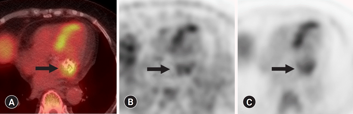 | Fig. 3.(A) Axial FDG ETP/CT, (B) non-attenuation, and (C) AC PET images of a case of definite infective endocarditis (IE) resulting from Escherichia coli in an 89-year-old patient with a mitral native valve. She suffered from fever of unknown origin, and FDG PET/CT was performed to evaluate the source of the fever. FDG PET/CT shows focal areas of enhanced glycolytic metabolism (arrow) around the calcified mitral valve, in which the standard uptake value was 5.8. Dense calcification can result in false uptake through AC, and therefore, readers should check for non-AC PET. FDG, F-18 fluorodeoxyglucose; PET, positron emission tomography; CT, computed tomography.
|
Semiquantitative measurements of the metabolic activity of lesions using SUVs are less subjective and provide a more objective threshold for determining infection. For this purpose, Scholten et al. [70] analyzed studies on the SUV value of PVE. However, intercenter exchanges were not possible due to the lack of standardized protocols between studies; and because the threshold for rejected PVEs (0.5 to 4.9) and definite PVEs (4.2 to 7.4) showed wide ranges. A semiquantitative measure of FDG uptake, the European Association of Nuclear Medicine Research Ltd.-standardized SUV value ratio (affected valve/blood pool) of ≥2.0, was a 100% sensitive and 91% specific interpreter of PVE [14]. It has been proposed that grading of valvular FDG uptake as intense, moderate, mild, or absent is more useful than a simple grading of positive or negative. Similar to the mDC for echocardiography, it is also possible to set up a registry to predict the probability of PVE for each uptake intensity [71].
False-positive findings may occur in areas where surgical adhesives have been applied, which may last for 2 or 3 months [14,45]. False-negative findings may be due to low inflammatory activity at the time of imaging or as a result of prolonged antibiotic treatment [14]. Swart et al. [14] reported that a CRP value less than four-fold the upper normal limit (<40 mg/L) was a remarkable false-negative factor. Although PET is highly sensitive to detecting disease activity, it has lower spatial resolution than CT. To visualize sites with an abnormal uptake by PET, the target structure should have a volume larger than 1 cm3, with significant accumulation of the administered radio-tracer [72]. In addition, PET requires data collection for a relatively longer time compared to CT, and motion from the heart beat and breathing during data acquisition can degrade the spatial resolution of PET. However, the detrimental effect caused by motion cannot be noticeably overcome by physiological gating during data collection. Therefore, the use of PET is not optimal for detecting and characterizing small lesions [73]. Overall, clinicians face considerable challenges in diagnosing and characterizing infectious heart diseases with structural or functional imaging, and further improvement in imaging technology will be needed.
Go to :
Conclusion
FDG PET/CT imaging is a valuable diagnostic modality for patients with suspected IE with prosthetic valves or intracardiac devices. Proper use of PET/CT imaging increases the diagnostic capacity of the mDC. Although the role of FDG PET/CT in NVE is limited, adding the results of FDG PET/CT to the mDC is helpful in diagnosis, particularly if the decision is difficult with conventional approaches. FDG PET/CT can help determine treatment regimens related to CIED infection and the monitoring of antimicrobial therapy in IE. In LVAD, FDG PET/CT is considered to be accurate in diagnosing driveline infections, although it has limited value in evaluating pump housing infections. For all cases of IE, whole-body FDG PET/CT is advantageous in the early detection of metastatic infection and embolic events. The patient should be carefully prepared using a HFLC diet, heparin infusion prior to imaging, and increasing the specificity through AC and non-AC images to reduce impact on metal artifacts. The benefits of using FDG PET/CT for cardiac infection will become more apparent through the development of standardized protocols and large-scale prospective studies.
Go to :
 XML Download