

 PDF
PDF Citation
 Print
Print


Introduction
Intussusception rarely causes intestinal obstruction in adults and accounts for approximately 1%–5% of all cases of bowel obstruction [1]. In contrast to pediatric intussusception, most cases of adult intussusception are secondary to an underlying pathology, such as primary/metastatic malignancies or benign tumors that serve as the leading point of intussusception [2].
An inflammatory fibroid polyp (IFP) is a benign tumor usually localized to the gastric antrum, although it can occur throughout the gastrointestinal tract [3]. Meckel’s diverticulum (MD) is a remnant of the omphalomesenteric duct occurring in approximately 2% of the population [4]. Both IFP and MD can act as leading points in adult intussusception. We present a rare case of synchronous ileal IFP and MD noted during laparoscopic surgery for adult intussusception.
Go to : 
Case
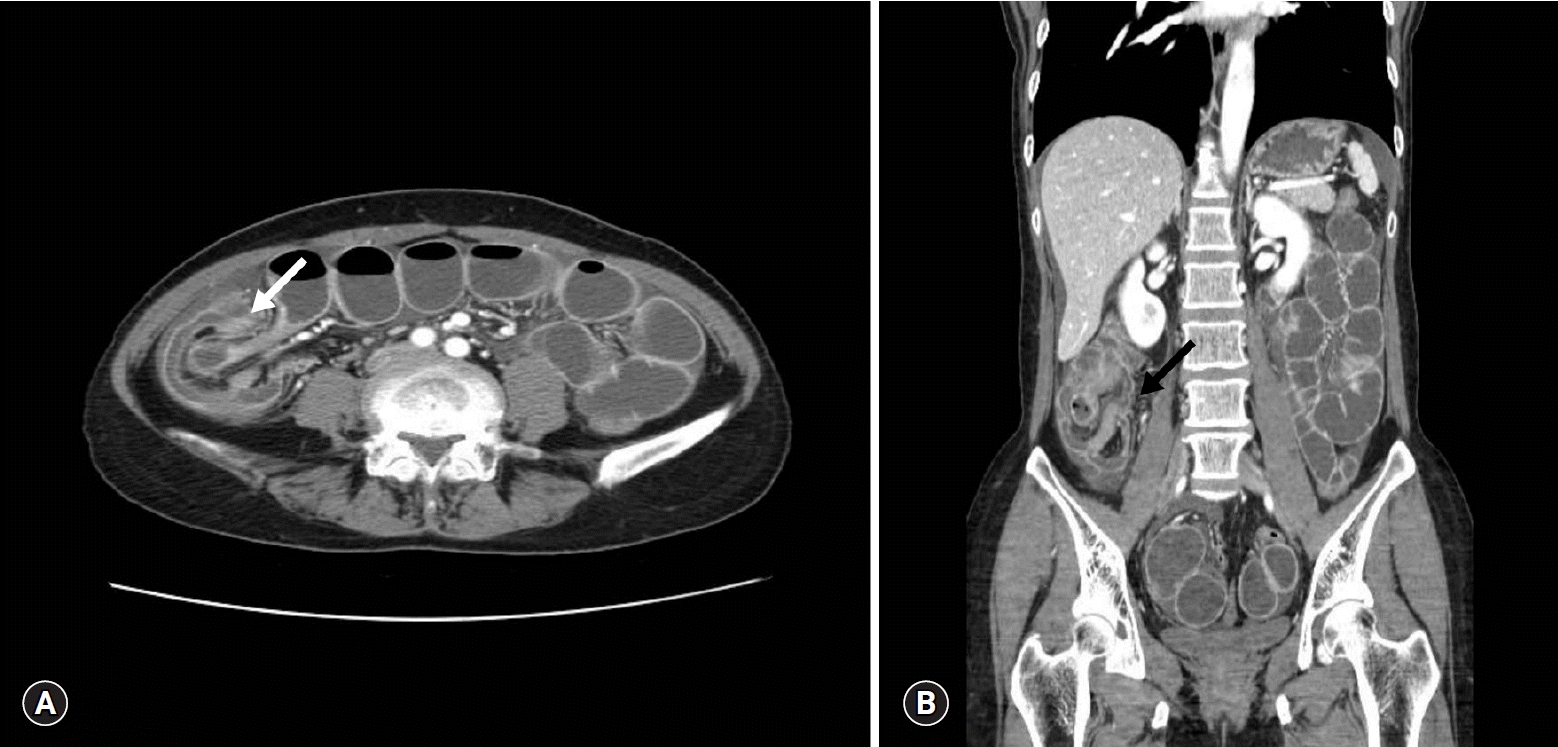
A 48-year-old woman was referred to our emergency department due to a sudden onset of severe abdominal pain. Her vital signs were stable on physical examination, and direct tenderness in the right lower quadrant without rigidity or distention was observed on abdominal examination. The patient reported a history of total laparoscopic hysterectomy for early-stage endometrial cancer, 4 years prior to the presentation. Abdominal radiography revealed a stepladder sign with several small bowel loops. Blood tests were within normal limits. Abdominal computed tomography (CT) revealed ileocecal intussusception with proximal small bowel obstruction (Fig. 1).
Urgent laparoscopic exploration was performed using 3 trocars (a 12-mm supraumbilical port for the camera and two 5-mm ports placed in the left middle and lower quadrants of the abdomen), which revealed spontaneous resolution of intussusception. However, an intraluminal mass was found in the distal ileum, located approximately 30 cm from the ileocecal valve (Fig. 2A). Further bowel exploration revealed an outpouching closed luminal structure on the antimesenteric side wall of the ileum, located approximately 70 cm from the ileocecal valve (Fig. 2B). Umbilical mini-laparotomy, approximately 4 cm in length including the supraumbilical port site, was performed, and the affected ileum was retracted (Fig. 2C). The intraluminal tumor was resected via enterotomy followed by primary closure. The ileal outpouching was resected, and ileal side-to-side anastomosis was performed. The total operative time and estimated blood loss were 90 minutes and 20 mL, respectively.
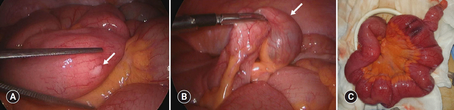 | Fig. 2.Intraoperative laparoscopic images. (A) An intraluminal mass (arrow) is observed in the distal ileum approximately 30 cm from the ileocecal valve. (B) An outpouching closed luminal structure (arrow) is observed on the anti-mesenteric side wall of the ileum, located approximately 70 cm from the ileocecal valve. (C) The affected ileum is retracted via mini-laparotomy.
|
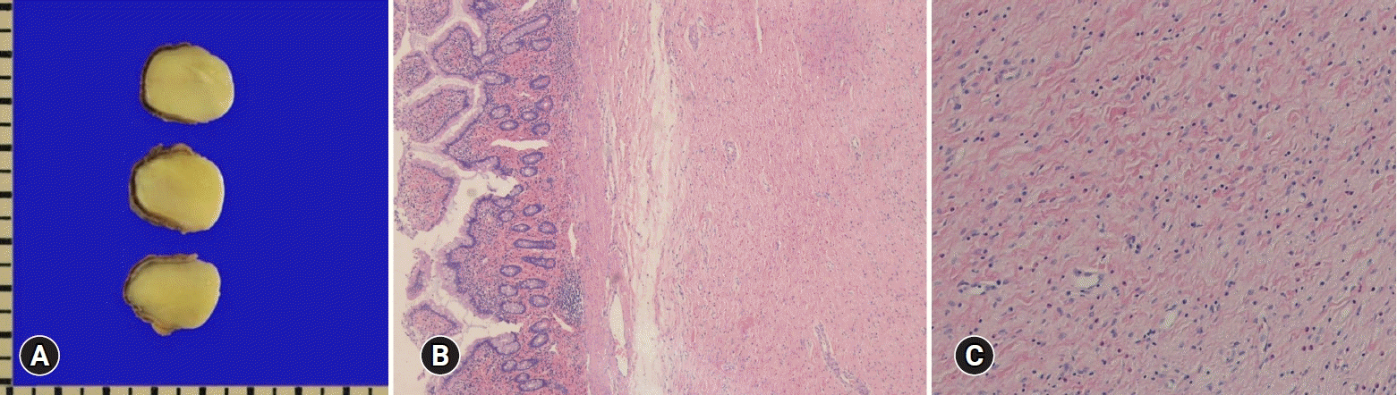
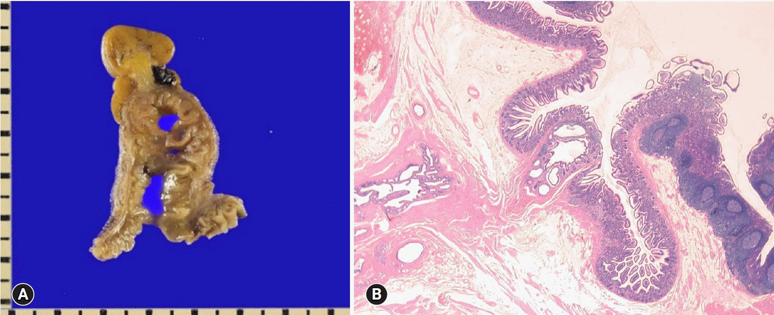
The patient was discharged on the 4th postoperative day following an uneventful recovery. Histopathological examination confirmed an IFP measuring 2.5×1.5 cm (Fig. 3) as well as MD with ectopic pancreatic tissues measuring 5.5×2.5 cm (Fig. 4).
Go to :
Discussion
IFP is a benign gastrointestinal tract lesion and usually asymptomatic. It develops in the submucosa and is composed of mononuclear spindle-shaped cells and prominent eosinophils [1,3]. However, small bowel IFPs tend to be symptomatic and present with bleeding or abdominal pain secondary to bowel obstruction, but less commonly occur as compared with gastric IFP [5-7]. Although MD is usually asymptomatic in adults, abdominal pain secondary to obstruction, stricture, or MD-induced intussusception is the predominant characteristic of symptomatic cases [3].
The incidence of adult intussusception secondary to IFP and MD is extremely low. Notably, <100 cases of intussusception secondary to ileal IFP and 4%–14% of adult intussusception attributed to intussusception are reported in the existing literature [1,8,9]. Moreover, to our knowledge, no report has described the synchronous occurrence of IFP and MD discovered during a surgery for adult intussusception to date.
Intussusception is usually diagnosed by imaging studies because its clinical manifestations and history were nonspecific. CT is preferred over ultrasonography for the differential diagnosis of intussusception because adult intussusception usually present with tumors (including malignancies). In our case, ileocecal intussusception was diagnosed with CT. However, because of the overlapping ileocolic wall, as well as edematous and dilated small bowel loops, CT did not reveal any apparent lesions that might have served as a leading point in this case.
Surgical resection is the primary treatment for adult intussusception, and some patients require radical surgery due to the malignant potential of the tumor within the leading point [10]. Laparoscopic surgery for adult intussusception has been increasingly performed in recent years due to its proven advantages such as faster recovery, less pain, and minimal scarring [10-12]. In addition, laparoscopic exploration is effective even for the confirmative diagnosis of adult intussusception to avoid unnecessary incision.
Therefore, diagnostic laparoscopy was performed first, because no pathologic data has confirmed whether the tumor was malignant or benign. In addition, a leading point that caused the ileocolic intussusception was not identified in radiologic examinations. During the laparoscopic exploration, a small ileal mass and MD were detected. However, we could not determine whether one of them was the leading point of intussusception because the ileocolic intussusception resolved spontaneously. However, surgical resection of the two abdominal lesions was performed because both of them were considered as leading points of intussusception.
In conclusion, we report a rare case of synchronous occurrence of ileal IFP and MD detected during laparoscopic surgery for adult intussusception, which was successfully treated with a laparoscopic approach.
Go to :
 XML Download