

 PDF
PDF Citation
 Print
Print


Introduction
Benign adrenal vascular pathologies, including tumors, cysts, and pseudocysts, are relatively rare entities that are mostly right-sided and predominantly observed in females [1]. These lesions may occur concomitantly with other adrenal tumors associated with hormone secretion. Cysts of the adrenal glands are rare and often detected incidentally during radiological examinations or at autopsy. Their reported incidence ranges 0.06%–0.18% [2-4].
Adrenal cysts are classified into the following four main groups: endothelial cysts (45%), pseudocyst (39%), epithelial cysts (9%), and parasitic cysts (7%) [5]. Endothelial cysts include hemangioma and lymphangioma [6]. Adrenal cystic lymphangiomas do not exhibit pathognomonic clinical or radiological presentation. Certain features of these entities raise the suspicion of malignancy, including a heterogeneous appearance in imaging analyses, the presence of necrosis in the center of the mass accompanied by calcification, and the size of the adrenal mass [7].
We present a case of a cystic lesion on the right adrenal gland in an elderly man and discuss its common and distinct features.
Go to : 
Case
We found multiloculated cysts on the right adrenal gland in the cadaver of a 75-year-old Korean man, whose cause of death was pneumonia, during routine educational dissection.
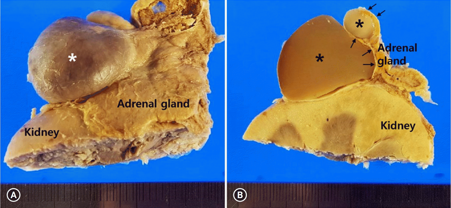
An ovoid cystic mass protruded from the outer surface of the adrenal gland (Fig. 1A). Dissection revealed a multilocular cystic lesion measuring 3.3×3.0 cm (longest and transverse diameters in external dimension, respectively) within the adrenal gland. The cystic walls were thin, and the cavities contained a pale to tan-colored gelatinous material (Fig. 1B).
Microscopy analysis revealed that the cystic structures were surrounded by flattened adrenal parenchyma (Figs. 2A, 2B) and the inner surfaces were lined by a single layer of flattened cells with bland nuclei (Fig. 2C). The lining cells were diffusely positive for cluster of differentiation 31 (CD31) (Fig. 2D) and podoplanin (D2-40) (Fig. 2E), and negative for pan-cytokeratin (Fig. 2F). The pathological diagnosis confirmed a cystic lymphangioma arising from the adrenal gland.
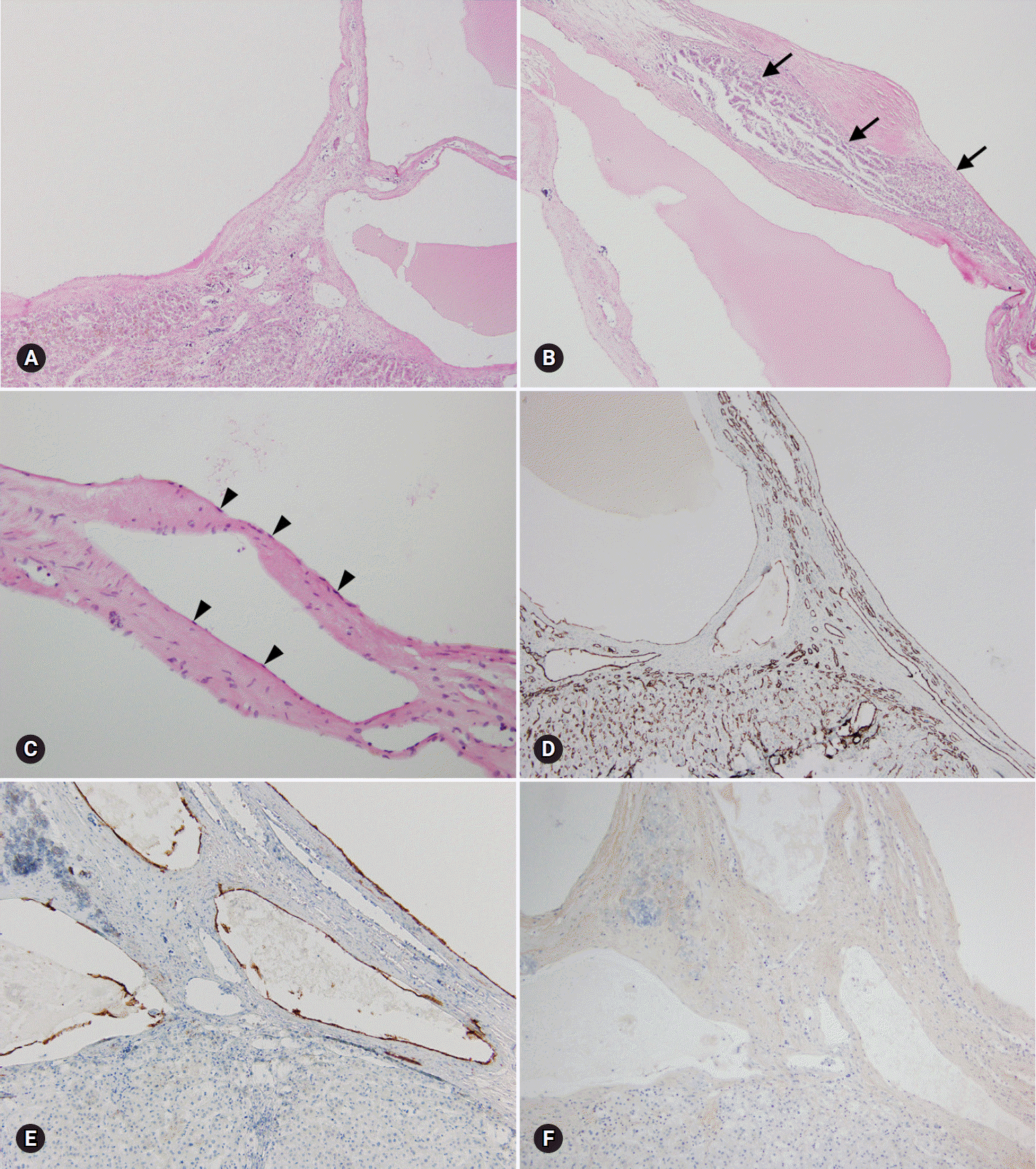 | Fig. 2.Microscopic findings of the right-sided adrenal cystic lesion. (A) Multilocular cystic spaces are present. (B) Entrapped adrenal parenchyma (arrows) is present. (C) The cystic wall is lined by a single layer of flattened endothelial cells (arrowheads) (hematoxylin and eosin stain, ×40 [A and B] and ×200 [C]). The lining endothelial cells are positive for cluster of differentiation 31 (D) and podoplanin (E), and negative for pan-cytokeratin (F) (immunohistochemical stain, ×40 [D] and ×100 [E and F]).
|
Go to :
Discussion
Lymphangiomas, benign malformations of lymphatic vessels, are most frequently noted in children aged less than 2 years [8,9]. Although the exact cause has not been established yet, these entities are generally regarded as congenital malformations in which obstruction or agenesis of lymphatic tissue results in lymphangiectasia due to a lack of normal communication within the lymphatic system [8,10]. The majority of lymphangiomas are detected in the neck, axillary region and mediastinum (95%), and the remaining 5% are found in the abdominal cavity [8,11]. Adrenal lymphangiomas account for less than 1% of all abdominal lymphangiomas [12]. To date, less than 60 adrenal lymphangioma cases have been described [13], with only 7 cases reported in Korean [14].
While lymphangiomas are typically detected in childhood and are rare in adults, adrenal lymphangiomas are predominantly noted in women in aged approximately 40 years, mostly right-sided, and unilateral (mean size 8.9 cm; range 2.0–35.0 cm). Histologically, all adrenal lymphangiomas show a typical multicystic lesion composed of irregular dilated spaces that are lined by flattened bland simple endothelial cells [12] and have potential malignancy incidence of about 4.1% [10,15]. Adrenal cystic lymphangiomas are usually asymptomatic because of their size and location [8]. The most common complaint is pain, which is localized in back, the right upper quadrant of abdomen, or is generalized as abdominal pain [10]. Transabdominal adrenalectomy is the treatment of choice. Recent recommendations entail the aspiration of material from the adrenal cysts instead of their surgical excision both for diagnosis and management, in cases where malignancy is not suspected or the lesion is non-functional and asymptomatic [6,8,10,12,16].
In conclusion, small asymptomatic adrenal cystic lymphangiomas without malignant transformation were incidentally detected in an elderly Korean male cadaver and the diagnosis was confirmed by histopathological staining for CD31 and D2-40. Unlike other adrenal cystic lymphangiomas, the adrenal lymphangioma in the present case was detected in an elderly male cadaver due to their size during postmortem investigation. Therefore, careful inspection of the adrenal cysts by clinicians and anatomists is warranted, despite the patient’s age and sex.
Go to :
 XML Download