

 PDF
PDF Citation
 Print
Print


Introduction
Forefoot disorders are extremely prevalent in the general population and can hinder patients’ activities of daily living. In cases where severe pain exists during gait, patients are reluctant to walk around, which can lead to poor physical function [1,2]. In many cases, forefoot disorders are caused by wearing ill-fitting or high-heeled shoes, altered foot alignment, and foot arthropathy [3,4]. Although there are several forefoot disorders, metatarsalgia, hallux valgus, hallux limitus/rigidus, lesser toe deformities (hammer, claw, and mallet toes), and Morton’s (interdigital) neuroma are common. For the management of these disorders, conservative treatment is usually attempted prior to surgical intervention. Conservative treatments include corrective shoes, the application of insoles or orthoses such as pads or supports, oral medications, and steroid injections [5]. Despite the high incidence of forefoot disorders, their conservative treatments have rarely been studied. Thus, in this review, we aim to summarize common forefoot disorders and present conservative treatments by focusing on shoe modifications and the application of insoles or orthoses (Table 1).
Table 1.
Summary of conservative treatment for forefoot disorders (other than shoe modification)
![]()
Go to : 
Metatarsalgia
Metatarsalgia refers to pain in the plantar aspect of the foot in the region of the metatarsal (MT) head, particularly in the second, third, and fourth rays [6]. During the toe-off phase of gait, most of the pressure is concentrated in this area. A deteriorated biomechanical condition of the foot can increase pressure on the MT head during weight bearing, resulting in metatarsalgia. Metatarsalgia can be divided into three types: primary, secondary, and iatrogenic [6]. The incidence of metatarsalgia is approximately 5–36% [7].
Primary metatarsalgia is caused by anatomical abnormalities of the MT and the relationships between the MT and other parts of the foot. The common causes of primary metatarsalgia include first ray insufficiency [8], a long second MT [9], and increased MT declination caused by pes cavus [8]. First ray insufficiency is caused by inability of the first ray to accept loads during weight bearing, leading to transfer pressure to the lesser MT [8]. First ray insufficiency occurs due to several conditions, such as hallux valgus, pes planus, and hypermobility of the first metatarsophalangeal (MTP) joint [10]. The long second MT shifts loads during weight bearing from the first to the second ray [9]. Moreover, in pes cavus, MT declination increases and pressure with heel strike focuses on the MT head and heel without adequate lateral plantar midfoot support [8]. These conditions increase lesser MT load and cause metatarsalgia.
Secondary metatarsalgia is induced by indirect overloading of the forefoot [6]. Foot trauma can alter foot alignment, causing angular or rotational MT displacement. Fracture or injury to the supporting structures of the MTP joint (plantar plate and collateral ligaments) deteriorates the foot’s biomechanical alignment, leading to forefoot instability and pain [6]. Other conditions, including hallux rigidus, Morton’s neuroma, tarsal tunnel syndrome, Freiberg infarction, and chronic inflammatory diseases can increase forefoot load without direct load on the MT [8,11-13].
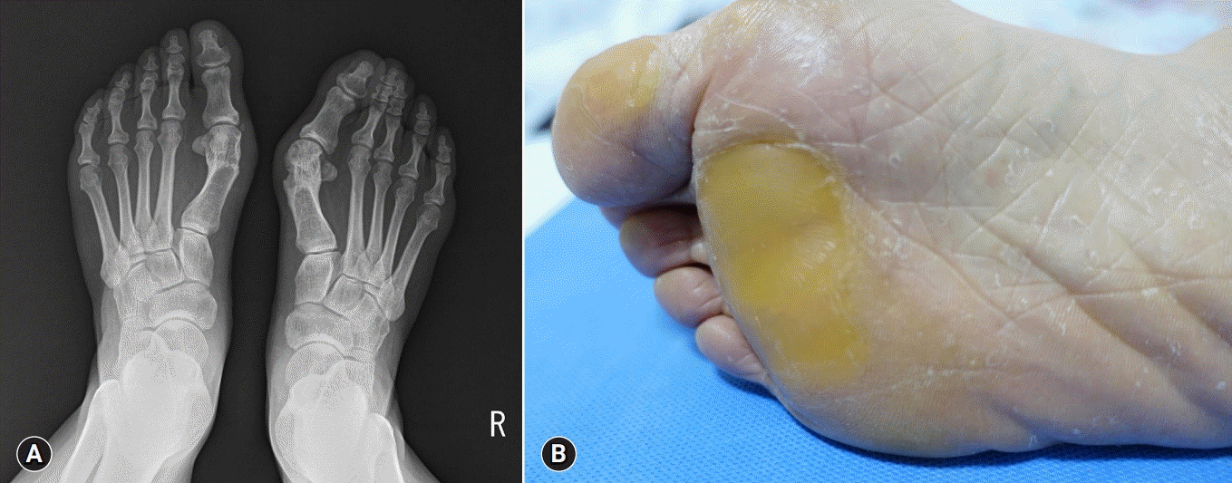
Iatrogenic metatarsalgia arises as a sequela of prior forefoot surgery, such as altered MT position after proximal MT osteotomy and excessive MT shortening after MT osteotomy or hallux valgus surgery (Fig. 1) [14-16]. Complications after forefoot surgery, including nonunion, malunion, or avascular necrosis, can cause iatrogenic metatarsalgia [14-16].
To effectively treat metatarsalgia, clinicians should diagnose its causal factor and focus on solving it. For symptomatic relief, an MT pad made of rubber, polyurethane, or silicone can be applied. The pad reduces pressure under painful MT heads by spreading it to a larger area, improving functional ability [17-19]. In 2005, Hsi et al. [18] reported that the optimal method is to apply an MT pad just proximal to the MT head. It also elevates the horizontal arch of the forefoot, which can widen the space between MT heads, reducing interdigital nerve compression and irritation [18]. Use of an MT bar or forefoot cushion is also effective for controlling metatarsalgia [20].
Go to :
Hallux valgus
Hallux valgus, the most common foot deformity, is characterized by medial deviation of the first MT and lateral deviation of the hallux. The incidence of hallux valgus is approximately 23% in 18–65-year-olds and 35% in people over 65 years of age [21]. When the hallux valgus angle, defined as the angle between the shaft axis of the first MT and the proximal phalanx of the hallux, is greater than 15°, patients are diagnosed with hallux valgus. Hallux valgus can be categorized as mild (15–20°), moderate (21–39°), and severe (≤40°) [22]. In severe cases, subluxation of the first MTP joint can be involved. Pain in the MTP joint, especially during weight bearing, is routinely seen in patients with hallux valgus [22].
The first ray bears a significant amount of weight because it maintains the position of the medial arch [23]. Several factors that deteriorate the integrity of the first ray, such as restrictive footwear, foot deformities, and pes planus, can be ascribed to the occurrence of hallux valgus [24]. The much higher prevalence of hallux valgus in women (women:men=15:1) is associated with the differential use of tight-fitting and high-heeled shoes [25]. Moreover, the fact that women have higher rates of ligamentous laxity, which disrupts the integrity of the first ray, seems to contribute to the different incidence in men and women [26].
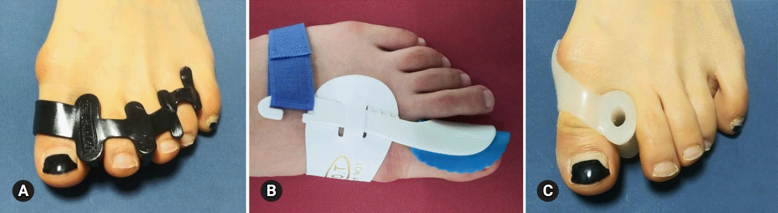
Prior to surgical correction, conservative treatment should be initiated. Patients should avoid tight-fitting or high-heeled shoes and wear soft and wide-toed shoes instead [22]. A toe spreader, valgus splint, and bunion shield are suggested treatments for hallux valgus (Fig. 2) [22]. The toe spreader separates the first toe from the second toe and reduces pain caused by the bunion, reducing protrusion of the MT head [27]. Many types of ready-made toe spreaders are being applied in patients with hallux valgus. In 2008, Tehraninasr et al. [27] recruited 30 patients with hallux valgus and compared the effects of a toe separator made of plastazote material with a semi-rigid total contact insole and those of a valgus splint. After 3 months, pain intensity was significantly reduced in 15 patients who were managed with the toe separator, whereas the change in pain was not significant in the other 15 patients treated with the valgus splint. On the other hand, the hallux valgus angle was not significantly reduced in either group. In addition, ready-made toe spreaders do not accurately fit the toe spaces of each patient; thus, the toe axis cannot be accurately corrected. In 2018, Cha et al. [28] designed personalized toe spreaders with three-dimensional scanning and printing and successfully applied them in eight patients with hallux valgus.
Go to :
Hallux limitus/rigidus
Hallux limitus refers to restricted sagittal range of motion (ROM) of the first MTP joint. Also, hallux rigidus is defined as a condition in which sagittal ROM is completely absent [29-31]. Hallux limitus/rigidus occurs in 35–60% of peoples over 65 years old [32-34]. In addition, a functional reduction in ROM of the first MTP joint that occurs during walking with no structural limitations is referred to as functional hallux limitus [34].
The predisposing factors of hallux limitus/rigidus include, osteoarthritis, trauma, rheumatoid arthritis, age, female sex, hypermobile first ray, hallux valgus, and pes planus [33,35-37]. The first MTP joint bears about 60% of one’s body weight [38]. Pes planus leads to excess weight load on the first MTP joint, which causes arthritic changes in the first MTP joint.
A kinetic wedge foot orthosis can be applied to treat hallux limitus or rigidus [39,40]. This involves the use of a cut out under the first MT head, which seems to increase dorsiflexion at the first MTP joint and allow the first ray to plantarflex more freely. The kinetic wedge foot orthosis reduces plantar pressure under the first MTP joint. In 2003, Rambarran et al. [40] reported that plantar pressure under the first MTP joint during gait was significantly reduced after the application of a kinetic wedge foot orthosis. The addition of a rocker sole also helps relieve symptoms [41].
Go to :
Hammer, claw, and mallet toes
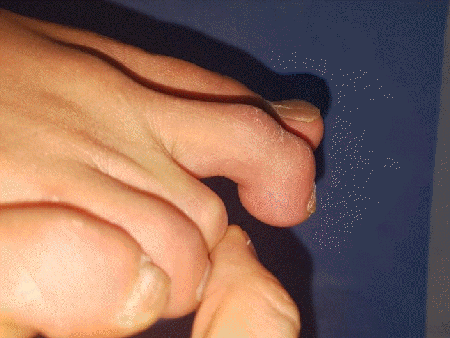
Hammer, claw, and mallet toes are sagittal plane deformities of the lesser toes. The incidence of these deformities is approximately 30% [42]. Hammer toe refers to a flexion deformity at the proximal interphalangeal (PIP) joint of the toe accompanied by a slight MTP joint extension deformity (Fig. 3) [5]. Claw toe is defined as a hyperextension deformity at the MTP joint and secondarily having flexion deformity in the PIP and distal interphalangeal (DIP) joints (Fig. 4) [5]. Mallet toe is defined as a flexion deformity at the DIP joint. Difficult in wearing shoes (impingement of the toes on the shoe box) and metatarsalgia can occur during these deformities (Fig. 5) [5]. These deformities of the lesser toe are associated with ill-fitting or high-heeled shoes [43]. Hallux valgus can also contribute to the formation of lesser toe deformities [44]. Shortening of the first ray by hallux valgus slackens the plantar fascia and weakens the windlass effect on the first toe, which leads to greater strain on the lesser toes [44]. This makes the supporting structures of the lesser toe more likely to fail. Pathologies such as diabetes mellitus, neuromuscular disorders, and inflammatory arthritis can also cause lesser toe deformities [43]. Clinicians should recommend that patients wear wider shoes with a larger toe box [45]. A toe sleeve or padding can be applied over high-pressure areas of the PIP or DIP joints or under the MT heads [46]. MT off-loading insoles can also be used to alleviate symptoms following lesser toe deformities [47]. However, there is paucity of clinical data on the effect of conservative management of lesser toe deformities; thus, a clinical trial should be performed on this theme in the future.
 | Fig. 3.Hammer toe with a flexion deformity at the proximal interphalangeal joint of the toe accompanied by a slight metatarsophalangeal joint extension deformity.
|
Go to :
Morton’s (interdigital) neuroma
Morton’s neuroma is a compression neuropathy of the plantar digital nerve with associated perineural fibrosis [48]. Morton’s neuroma reportedly affects 30% of the population and is predominant in the female sex (female:male=4:1) [49]. It most commonly affects the third space (66% of cases), followed by the second (32%) and fourth spaces (2%) [50]. Its symptoms are a burning and tingling sensation in the forefoot, with occasional numbness in the affected toe [48]. Morton’s neuroma, a thickening of the interdigital nerve, occurs just distal to the MT transverse ligament and before bifurcation of the digital nerves. Entrapment of the interdigital nerve between the intermetatarsal ligaments is considered the key factor in the occurrence of Morton’s neuroma [51]. It is related to various overload mechanisms and the use of inadequate footwear [52]. Morton’s neuroma can be diagnosed by a physical examination. The application of plantar pressure on the area between and just proximal to the MT heads should replicate the patient’s usual pain [48]. Moreover, concomitant lateral, dorsal, and plantar compression of the MT heads can produce an audible and painful Mulder click [48].
For treatment, patients should favor shoes with a lower heel and wider toe box [49]. An MT bar on the sole of the shoe reduces pressure loading on the space between MT heads and transfers it more proximally [53]. Plantar pads placed to the insole proximal to the neuroma elevate the MT head and decrease interspace pressure [53]. The use of a more cushioned thicker insole can decrease the pressure and impact on the intermetatarsal space [53].
Go to :
Conclusion
In clinical practice, many patients complain of forefoot pain or deformity. The modification of shoes and application of insoles or orthoses can be beneficial for managing forefoot disorder. However, clinical data or evidence of these conservative treatment methods are insufficient. To clarify the utility of these treatments in forefoot disorders, further studies are needed. This review will help clinicians consider various tools for treating forefoot disorders.
Go to :
 XML Download