

 PDF
PDF Citation
 Print
Print


Information
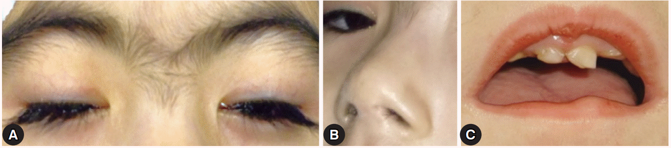
Cornelia de Lange syndrome (CdLS) is a rare multisystemic disorder that is characterized by mental retardation, prenatal and postnatal growth retardation, limb anomalies, and distinctive facial features, which include arched eyebrows that often meet in the middle (synophrys), long eyelashes, low-set ears, small and widely spaced teeth, and a small and upturned nose [1-3]. The clinical features vary widely among affected individuals and range from relatively mild to severe. Ophthalmic manifestations include long eyelashes, nasolacrimal duct obstruction, myopia, ptosis, and strabismus [4,5]. There has been no report of surgical treatment for esotropia and unilateral ptosis in patients with CdLS in Korea. I report a patient with CdLS who underwent surgical treatment for esotropia and unilateral ptosis with a good surgical outcome.
Case
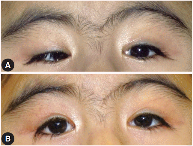
Informed consent was obtained from the patient’s parent. A 5-year-old girl was referred to our ophthalmology department for surgical treatment of esotropia and unilateral ptosis of the right eye. She had been previously diagnosed with CdLS at another clinic and had undergone regular follow-up. She had been born at 36 weeks of gestation with a birth weight of 1,900 g. She had the typical facial features of CdLS, psychomotor delay, and intellectual disability (Fig. 1). We were unable to measure the visual acuity due to poor cooperation. The fixation of the right eye was weaker compared with that of the left eye. She also had ptosis, which covered the visual axis of the right eye. The extraocular examination showed 50 prism diopters of esotropia with abduction limitation of the right eye (Fig. 2A). Nystagmus was not observed. The refractive errors were -0.50 diopters (D) in both eyes. The fundus examination revealed peripapillary pigmentation of the right eye and a normal appearance of the left eye. The forced duction test was performed under general anesthesia; it did not reveal any restriction of the medial rectus in the right eye. Exploration showed that the lateral rectus muscle had a normal appearance, and it did not have any restriction. A 7-mm resection of the lateral rectus and 6-mm recession of the medial rectus were performed on the right eye without augmentation. The frontalis sling procedure was performed for the treatment of right ptosis. Four months later, the patient had stable ocular alignment with good eyelid position (Fig. 2B).
Discussion
CdLS occurs sporadically in most patients [2,5]. The exact etiology of CdLS is still unknown, but five genes have been reported to be associated with CdLS; the mutations of these genes comprise the underlying defect in 70% of the patients [2,6]. The initial diagnosis of CdLS is based on the clinical features, with specific signs, symptoms, and developmental characteristics forming the main criteria. Clinical diagnostic criteria have been proposed and include long eyelashes, ptosis, tear duct malformation or blepharitis, myopia ≥ -6.00 D, and a major eye malformation or peripapillary pigmentation as ophthalmic features [2]. Therefore, an ophthalmic examination can be helpful in the diagnosis of CdLS in an atypical case. In the present case, the patient had large-angle esotropia and severe unilateral ptosis. Compared with strabismus, ptosis is a relatively common clinical feature. Wygnanski-Jaffe et al. have reported the ophthalmic features in 120 patients with CdLS [7]. In their largest series, 10% of patients (11/113) had esotropia and 44% of patients (41/117) had varying degrees of ptosis, ranging from mild cosmetic changes to functional obstruction of the visual axis. Nallasamy et al. have evaluated the genotype-phenotype correlation with regard to the severity of ophthalmic findings in patients who had been previously screened for mutations in the Nipped-B-like gene [5]. In their study, nearly one-half of the included patients (25/54) had ptosis, and its severity was increased in patients who had truncating mutations compared to those who had missense mutations. Only 2 of the 15 patients with strabismus required surgical treatment. Because patients with CdLS have short horizontal palpebral fissures [7], it is sometimes difficult to get a clear surgical field during strabismus surgery, especially during medial rectus muscle surgery. A surgeon should take these into consideration during surgical planning.
Patients with CdLS have many ophthalmic problems, such as ptosis, strabismus, refractive errors, and nasolacrimal duct obstruction [5,7,8]. The treatment strategy should be based on each patient’s symptoms. The surgical correction of ptosis and strabismus can improve the quality of the life of an affected patient. This report should help surgeons to consider surgical treatment of ptosis and strabismus in patients with CdLS.
 XML Download