

 PDF
PDF Citation
 Print
Print


INTRODUCTION
Congenital cholesteatoma is composed of a white mass located in the middle ear cavity and medial to the normal tympanic membrane. This condition can be diagnosed in the absence of otorrhea, tympanic membrane perforation, and a history of otologic surgery [1]. In patients with cholesteatomas, a growth of keratinized squamous epithelium is present that induces the bony destruction of nearby tissue. This bony destruction leads to hearing impairment, dizziness, facial palsy, and intracranial complications [2]. Therefore, the main purpose of cholesteatoma treatment is to surgically remove the lesion and preserve the patient’s hearing. Congenital cholesteatoma located within the tympanic membrane is referred to as congenital intratympanic membrane cholesteatoma (ITMC). Its progression can also cause several complications, similar to any other cholesteatoma. In most case reports, the treatment is surgical removal [3-10]. We recently observed a case of ITMC in a child, wherein we originally planned to execute surgical treatment. However, this case developed into an external auditory canal cholesteatoma after 2 months. In this report, we describe this unusual case.
CASE
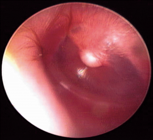
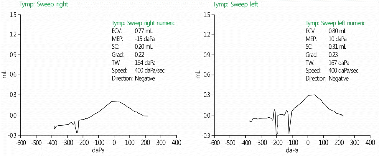
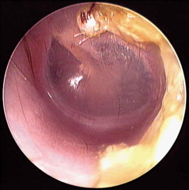
A 16-month-old female patient with a history of frequent otitis media visited our department. She had no history of otologic surgery. Otoscopic examination showed that her right tympanic membrane and right external auditory canal were normal, but there was a pearly white mass located at the end of the malleus handle, in the center of her left tympanic membrane (Fig. 1). A presumed diagnosis of an ITMC was made empirically. The patient did not have hearing impairment or any other signs of ear symptoms. Tympanometry showed a type A pattern for both ears (Fig. 2). Temporal bone computed tomography was performed, and the soft tissue density of a round mass was noted between the left tympanic membrane and the handle of the left malleus (Fig. 3). There were no other abnormal features of the inner structures or ossicles. At first, we planned surgery to remove the cholesteatoma. However, at follow-up consultation after 2 months, the mass was noted to have moved laterally into the external auditory canal (Fig. 4). The mass was easily removed with a hook without anesthesia. Histopathologic examination showed stratified squamous epithelium and keratin debris, and the diagnosis of cholesteatoma was confirmed (Fig. 5)
DISCUSSION
Congenital cholesteatoma located within the tympanic membrane is referred to as congenital ITMC. ITMC was first reported by Hinton in 1863, and is very uncommon [4,11]. The exact pathogenesis, natural history, and prognosis are still unknown [12]. Sobol et al. suggested that the most attractive explanation for the development of ITMC is the basal cell papillary proliferation theory. This theory assumes that recurrent inflammation may stimulate basal cell proliferation, which can cause the formation of cholesteatoma [7]. This explanation is consistent with our observations in the present case.
The development of an ITMC can cause several complications, such as ossicular involvement and subsequent impairment of hearing conduction, similar to any other cholesteatoma of the middle ear [3]. To avoid middle ear involvement, early diagnosis and the prevention of rupture of the cholesteatoma into the middle ear are required [13]. Therefore, patients with a congenital ITMC are treated with surgery [3-10].
The tympanic membrane is composed of three layers: an inner mucosal layer, a middle fibrous layer, and an outer epithelial layer [14]. In many reported cases, the middle layer of the tympanic membrane was intact after removal of the cholesteatoma [12]. This means that the middle ear was not involved, even after the operation. Thus, a transcanal approach is recommended for the removal of localized cholesteatoma, if the cholesteatoma can be peeled off from the tympanic membrane while maintaining an intact fibrous layer of the tympanic membrane [3].
On the other hand, there are several reported cases of congenital ITMCs that changed into external auditory canal cholesteatomas on follow-up consultation [4,12]. Suzuki et al. [13] suggested that an ITMC can heal spontaneously when the cholesteatoma of the early stage ruptures into the auditory canal and keratin debris is completely removed. In this case, an ITMC moved laterally and became an external auditory canal mass within 2 months and was removed easily.
Although the natural course of ITMC is not well known, most ITMCs likely increase in size over time [3].
Therefore, careful otomicroscopic examination and proper management are needed, even in pediatric patients who do not have any ear symptoms, to avoid progression beyond the tympanic membrane.


 XML Download