Journal List > J Dent Rehabil Appl Sci > v.36(4) > 1160100

Accuracy of dies fabricated by various three dimensional printing systems: a comparative study

Abstract

Purpose

The aim of this study was to compare the accuracy of dies fabricated using 3D printing system to conventional method and to evaluate overall volumetric changes by arranging the superimposed surfaces.

Materials and Methods

A mandibular right first molar from a dental model was prepared, scanned and fabricated with composites of polyetherketoneketone (PEKK). Master dies were classified into 4 groups. For the conventional method, the impression was taken with polyvinylsiloxane and the impression was poured with Type IV dental stone. For the 3D printing, the standard die was scanned and converted into models using three different 3D printers. Each of four methods was used to make 10 specimens. Scanned files were superimposed with the standard die by using 3D surface matching software. For statistical analysis, Kruskal-Wallis test and Mann-Whitney U test were done (P < 0.05).

Results

Compared to the standard model, the volumetric changes of dies fabricated by each method were significantly different except the models fabricated by conventional method and 3D printer of Stereolithography (P < 0.05). The conventional dies showed the lowest volumetric change than 3D printed dies (P < 0.05). 3D printed dies fabricated by Stereolithography showed the lowest volumetric change among the different 3D printers (P < 0.05).

Conclusion

The conventional dies were more accurate than 3D printed dies, though 3D printed dies were within clinically acceptable range. Thus, 3D printed dies can be used for fabricating restorations.

초록

목적

이 연구의 목적은 3D 프린팅으로 제작된 다이의 정확도를 인상재와 치과용 석고를 이용하여 제작한 기존 방식 다이와 비교하고 체적 변화를 평가하여 정확도를 비교하는 것이다.

연구 재료 및 방법

치과용 모델 하악 우측 제1대구치를 준비하여 스캔한 뒤 polyetherketoneketone (PEKK)으로 기준 다이를 제작한다. 기존 방식 다이는 기준 다이를 polyvinylsiloxane로 인상채득한 뒤 Type IV 치과용 석고를 부었다. 3D 프린팅 시스템의 경우 기준 다이를 스캔하고 3개의 서로 다른 3D 프린터를 이용하여 모델로 변환하였다. 4가지 방법으로 각각 10개의 표본을 만들었다. 3D 표면매칭 소프트웨어를 사용하여 기준 다이와 중첩하였다. 통계 분석을 위해 Kruskal-Wallis test, Mann-Whitney U test를 수행하였다(P < 0.05).

결과

기준 다이와 비교하여 기존 방식, Stereolithography로 제작된 다이를 제외하고는 각 방식으로 제작된 다이의 체적 변화가 상당히 있었다(P < 0.05). 기존 방식으로 제작된 다이는 3D 프린팅된 다이보다 체적 변화가 가장 적었다(P < 0.05). Stereolithography로 제작된 3D 프린팅 다이는 다른 3D 프린터 중에서 체적 변화가 가장 적었다(P < 0.05).

결론

기존 방식의 다이는 3D 프린팅 다이보다 더 정확했지만 3D 프린팅 다이는 임상적으로 허용되는 범위 내에 있었다. 따라서 3D 프린팅 다이는 수복물 제작에 사용할 수 있다.

Introduction

With the advent of computer aided design/computer aided manufacturing technology, additive manufacturing is changing many industrial and academic operations including medicine and dentistry.1 Nowadays, Additive manufacturing is widely used in dental prostheses fabrication such as crown and fixed partial denture (FPD).2 Additive manufacturing is as the process of joining materials to make objects from 3D model data, usually layer upon layer, as opposed to subtractive manufacturing methodologies defined by the American Society for Testing and Materials (ASTM).3 Additive manufacturing is the formalized term for what used to be called rapid prototyping and 3D printing.4 3D printing is different in many aspects from traditional and subtractive techniques for many years.5 One feature of this system is that it reduces much of the highly skilled and expensive labor associated with conventional fabrication method.3 Another advantage is that it can make any number of complex products simultaneously as long as the parts will fit within the build envelope of the machine.3
There are several approaches to classify 3D printing. 4 A popular way is to classify depending on baseline technology as whether the process uses lasers, printer technology, extrusion technology etc.6,7 Another way is to divide processes according to the type of raw material input.8 Pham9 classified by four separate materials; liquid polymer, discrete particles, molten material, and laminated sheets. Liquid polymer is one of the popular materials.4 Among 3D printing system using liquid polymers, 3D printing devices are based on material jetting, photopolymerization and so on.10
The first commercial 3D printing system patented by Hull in 1980s was Stereolithography (SLA) based on liquid polymer system.11 In SLA, 3D printed models are fabricated by assembling sequential twodimensional slices obtained using any digital data including STL file, computed tomography (CT), magnetic resonance imaging and ultrasonography.3 These data are then sent to an SLA machine and a concentrated beam of ultraviolet light is focused onto the surface of a pool filled with liquid photopolymer.3,12 As the light beam polymerize regions selectively on the surface of a pool of liquid resin.3,12 This process forms a solid object layer by layer.3,12 When the object is complete, it is entirely submerged in the resin and may be lifted out for use.12 This system has smoother surface, high resolution, fast processing, but high cost, possibly high temperature, and toxic uncured resin.13
Digital light processing (DLP) is also based on liquid polymer system as SLA.14 This process uses visible light-sensitive resins instead of ultraviolet laser for curing each layer.14 This system is a top-down process, compared to SLA, that is a bottom-up process.13 And this system is relatively faster than SLA because of a digital mirror device, which can be an entire layer at once.13 This system has limited choices in materials.14
Polyjet system is likewise inkjet document printing.15 Instead of jetting drops of ink onto paper, Polyjet system jets layers of liquid photopolymer onto a build tray and cure them with UV light.5,15,16 Hundreds of micro jetting print heads inject layers of liquid photopolymer resin on the build tray only in the area that correspond to STL file previously prepared, and cure them with UV light.5,15,16 Although this system has high quality and smooth surfaces, it lacks mechanical properties and detail reproducibility.5,15,16
Type Ⅳ dental stone is used for fabricating dental dies for fixed prosthesis due to ease of use, the perceived dimensional accuracy, and low cost.17,18 However, they have less than ideal strength, wear resistance and detail duplication.17,18 To overcome the disadvantages of Type Ⅳ dental stone, alternative die materials have been introduced and are reported by the manufacturers.18 The 3D printed die is one of alternative dies.
3D printing is a fast-developing technique that might play a significant role in the eventual replacement of plaster dental models.1 The accuracy of replica models varies between different additive manufacturing technologies, the 3D printer machine used, materials used.19
The aim of this study was to evaluate and compare the accuracy and reproducibility of dental dies fabricated from several 3D printing systems. The null hypothesis of this study was that there is no difference in the dimensional accuracy of dental dies, fabricated by conventional method and various 3D printing systems.

Materials and Methods

Fabrication of experimental die

Mandibular right first molar from a dental model A5A-200 (28S, Nissin Dental, Tokyo, Japan) was selected as the abutment tooth in this study. The tooth was prepared with a 1.0 mm circumferential chamfer finishing line, an occlusal reduction of 2.0 mm and a 6° convergence angle. All sharp edges were rounded off. Designed resin teeth were scanned using a light scanner (Series 5, Dental wings®, Montreal, Canada) to fabricate a reference die. The reference die was fabricated with a polyetherketoneketone (PEKK) composite (Pekkton®, Cendres + Méaux SA, Biel, Switzerland) block by milling with a machine.
Master dies were allocated to 4 groups according to the fabrication technique: conventional method, and 3 different 3-dimensional (3D) printing techniques. For the conventional method, an individual impression tray was made by using the master die at least 24 hours before taking the impression. Using irreversible hydrocolloid impression of the standard die, a stone cast model was fabricated for the individual tray. The trays were relieved with a sheet of baseplate wax as a spacer, to provide proper space for elastomeric impression material.20 Self-polymerizing resin (Quicky tray resin®, Nissin Dental) was used to make these impression trays. After removing the wax spacer of the tray, tray adhesive (Exaflex Adhesive®, GC Corp, Tokyo, Japan) was applied to the inner surface of the tray. Using the prepared individual tray, an impression for standard die was taken with polyvinylsiloxane (Honigum®, DMG, Hamburg, Germany) according to manufacturer’s instructions. After setting time of the impression material, the tray was removed from the master die.
The impression was poured with Type Ⅳ dental stone (FujiRock®, GC, Leuven, Belgium). Distilled water (20 ml) and gypsum powder (100 g) were vacuum mixed for 45 seconds (Twister Evolution®, Renfert, Hilzingen, Germany) and poured into the impression. The model was left to set for 40 minutes before removal. These procedures were repeated 10 times to fabricate 10 specimens (Table 1, ST: Type Ⅳ dental stone, PO: Polyjet, DL: Digital light processing, SL: Stereolithography).
The standard die was scanned using a light scanner (Series 5, Dental wings, Montreal, Canada) to fabricate dental die with 3D printer. Scanned data were saved as standard tessellation language (STL) and were converted into models using the following three different 3D printers:
  • 1) PolyJet (Objet EDEN260V®, STRATASYS Ltd, Meneapolis, USA)

  • 2) Digital Light Processing (DLP) (LC-3DPrint®, NextDent, Soesterberg, Netherlands)

  • 3) Stereolithography (SLA) (EQ-1®, CMET Inc., Kanagawa, Japan)

Total of 10 specimens were fabricated in each group (Table 1, Fig. 1(A) Type Ⅳ dental stone, Fig. 1(B) Polyjet, Fig. 1(C) DLP, Fig. 1(D) SLA).

Light scanning and 3D surface matching

All models were scanned using the light scanner (Series 5). According to the manufacturer’s instructions, these models were scanned by groups after spraying scan powder on the surface of dies. The scanned data were exported as an STL data file. In order to measure the volumetric changes, scanned files were superimposed with the file of standard die with 3D inspection software (Geomagic Qualify v12; Raindrop Geomagic, Research Triangle Park, USA).

Comparison and analysis of 3D volumetric changes

Surface matching was performed between the scanned file of standard die and the files of each specimen using best-fit alignment to investigate the volumetric changes.
Each pair of models was superimposed by a bestfit alignment with interactive closest point algorithm to allow measurements of 3D differences in shape.21 The software used 300 random points of measurement for the initial alignment and a further 7,630 points for fine adjustments in this study. This generated color maps of differences between each pair of objects by calculating the directional deviations between all corresponding points of measurement. The average positive, negative change values for each specimen were obtained from the 3D analysis of dimensional changes among groups. The range of maximum error was set at ± 1.00 mm and allowable error was set at ± 0.05 mm.

Statistical analysis

All statistical computations were made with statistical software (IBM SPSS Statistics 20; IBM SPSS Inc., NC, USA). Median values, standard deviations of each group were calculated. Kruskal-Wallis test was conducted to assess the significant differences among the groups (P < 0.05). As a post-hoc test, Mann-Whitney U tests were performed to determine differences among individual groups. Bonferroni’s method was also done so as to adjust the significance in multiple comparisons.

Results

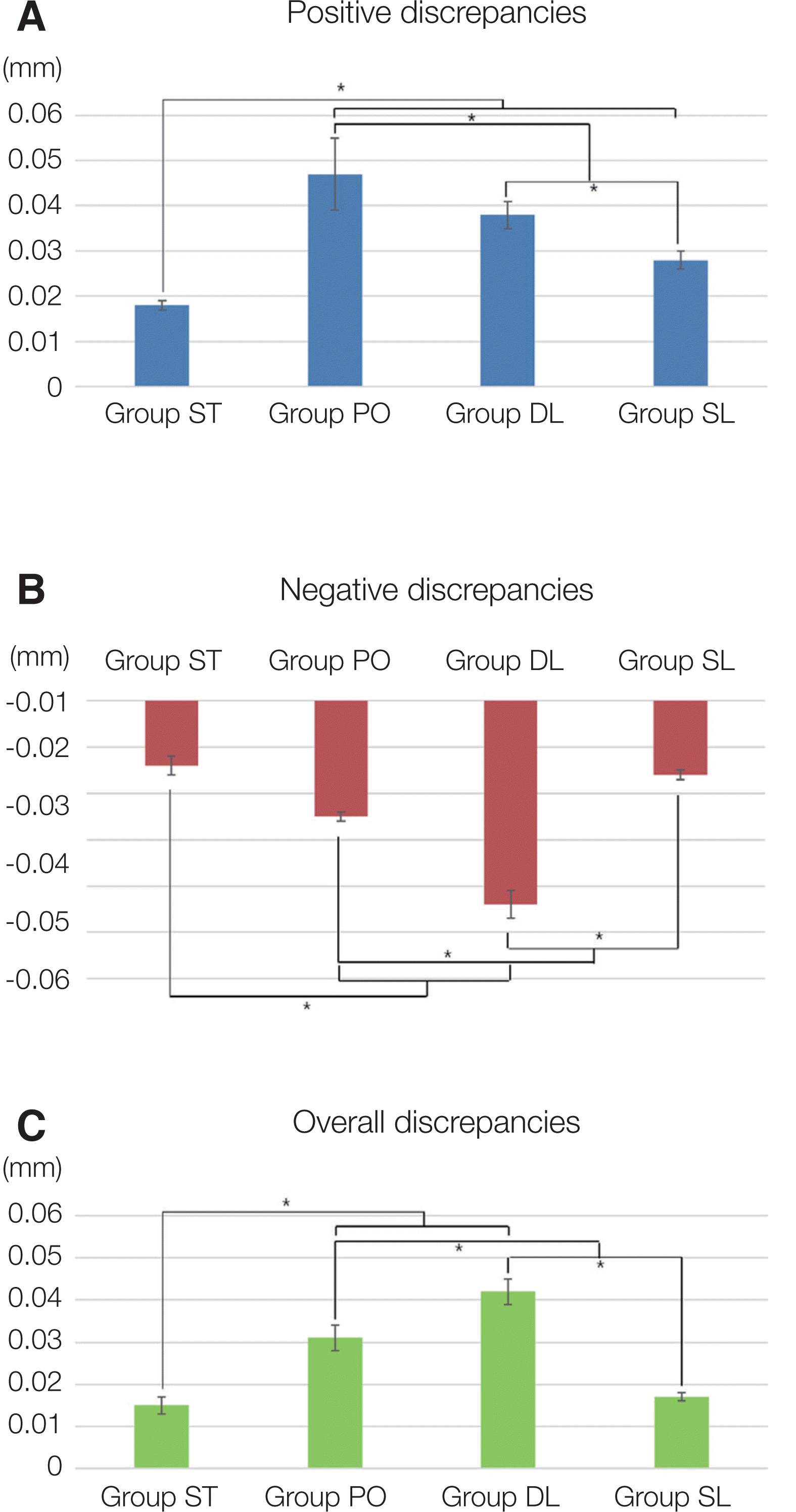
Measurements of standard die, conventional dies, and three different 3D printed dies were superimposed and the values of positive, negative, and overall discrepancies were exported. The values of overall discrepancies were obtained by the median of positive discrepancies and absolute value of negative discrepancies. The median values and standard deviations of the discrepancies for all the tested groups are presented in Table 2.
Group ST showed the lowest positive discrepancy. Group PO showed the highest discrepancy.
Group ST showed the lowest absolute value of negative discrepancy. Group DL showed the highest absolute value of negative discrepancy.
Group ST showed the lowest discrepancy and group DL had the highest overall discrepancy, meaning the largest volumetric change. Group SL showed the least values among the 3D printed die groups.
Fig. 2 showed color maps of all specimens and showed the discrepancies between standard die and experimental die (Fig. 2A. Type Ⅳ dental stone, Fig. 2B. Polyjet, Fig. 2C. DLP, Fig. 2D. SLA). Positive discrepancies, areas in the light-to-dark blue spectrum indicate larger portions than standard die. Green areas indicate volumetric change within the accepted limits (± 50 μm). Negative discrepancies, areas in the yellow-to-red spectrum indicate smaller portions than standard die.
Statistically significant differences were observed in the positive discrepancies between groups, ST and all the other groups, PO and DL, PO and SL, DL and SL (P < 0.05, Table 3, Fig. 3A).
Statistically significant differences were observed in the negative discrepancies between groups, ST and PO, ST and DL, PO and DL, PO and SL, DL and SL except for group ST and SL (P < 0.05). For negative discrepancy, no statistically significant variations were observed between group ST and SL (P < 0.05, Table 4, Fig. 3B).
Just as the case of negative discrepancy, statistically significant differences were observed in the overall discrepancies between groups, ST and PO, ST and DL, PO and DL, PO and SL, DL and SL except for group ST and SL (P < 0.05). For overall discrepancy, no statistically significant variations were observed between group ST and SL (P < 0.05, Table 5, Fig. 3C).
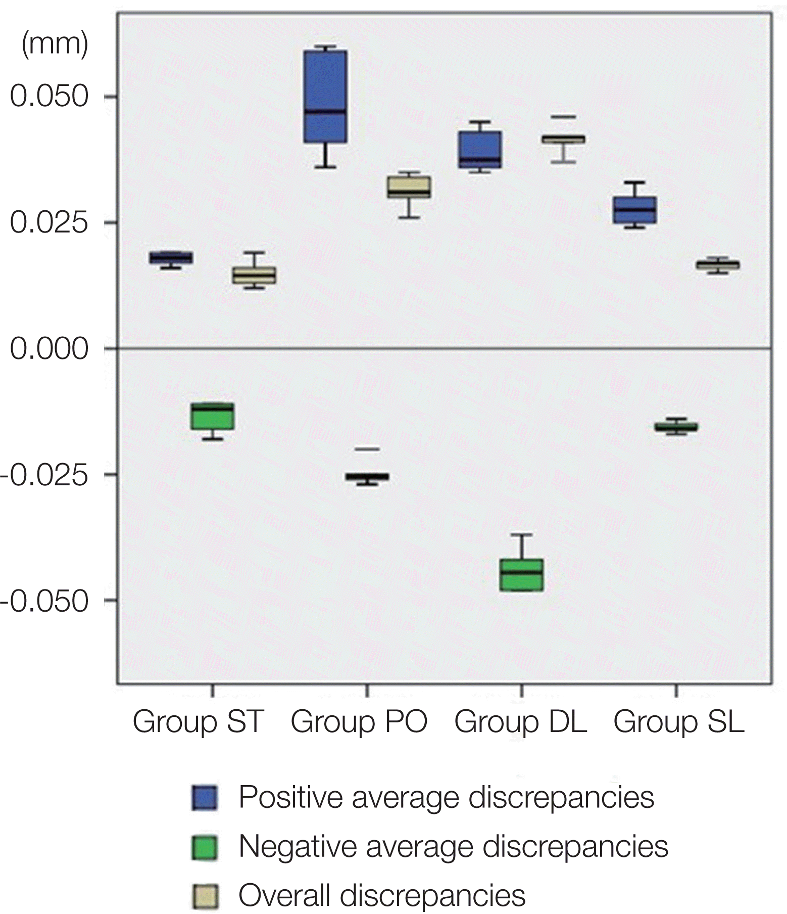
No statically significant variation in negative, overall change was detected between groups ST and SL (P < 0.05). Box and whisker plots of the obtained positive, negative and overall discrepancies were shown in Fig. 4.

Discussion

The null hypothesis of this study was that there is no difference in the dimensional accuracy of dental dies, fabricated by conventional method and various 3D printing systems. This hypothesis was rejected, since statistically significant difference emerged. In this study, SLA models showed greater accuracy than DLP, Polyjet models. These errors may be found at any of the some stages of the process, such as model shrinkage during building, post-curing.14,22 The minimal thickness of the layers can cause differences in final model production.14,22
Accurate replication of a prepared tooth surface is crucial for the dental die fabrication and final restoration of a tooth.23 The accuracy of die is influenced by impression method, the types of impression materials, and type of die material.23 The desirable requirements of dies include dimensional accuracy, reproduction of fine detail, surface hardness, ease and efficiency in fabrication, durability, and compatibility with impression materials.23,24 An ideal universal die material is yet to be produced.25
Type Ⅳ dental stone is the most commonly used material in dentistry for making dies used in the lostwax technique.18 ISO (International Standards Organization) Type Ⅳ dental stone is the predominant die material due to dimensional accuracy, ease of manipulation, low cost, and suitability for use with elastomeric impression materials.26,27 However, several studies have revealed that dental stone expands slightly while setting.18,27-30 The maximum range of setting expansion of die stone exhibits 0.1% as defined by the American Dental Association Specification No.25 for dental gypsum products (ANSI/ADA, 1987).27 This slight expansion is preferable to slight shrinkage of impression material.24 Also, the disadvantages of Type Ⅳ dental stone as dies are lack of hardness, abrasion resistance.18 Although Type Ⅳ dental stone has been successfully used for many years, numerous attempts have been made to develop die materials with improved properties.31
Epoxy and polyurethane resin die had acceptable mechanical properties.24 Whereas Type Ⅳ dental stone did not reproduce detail smaller than 20 μm and porous due to gypsum crystal size, epoxy or polyurethane resins are well reproduced smaller than 1 to 2 μm without porosity.32 Epoxy resins have shown slight shrinkage because of polymerization shrinkage.18,31,33 Combined with the impression material’s shrinkage, resins produced an undersized model.18,24,31 Although polymeric materials used in 3D printing are various like as powder, filament and sheet form, 3D printing machines in dentistry usually utilize the active polymerization of photo-sensitive resins.1 The photo-curing of liquid photopolymer resins as a methodology for 3D printing is attractive, because of higher resolution, more smooth surface, the ability to fast builds possible and print clear objects and good z axis strength due to chemical bonding between layers.1 So, 3D printing techniques in this study are UV or visible light-based approaches that polymerize photo-sensitive resins.1
There are two impression methods. One is conventional method by using elastomeric impression materials and the other is digital method by using intra-oral scanners.34 The accuracy of conventional impression methods is investigated by numerous studies, which evaluate volumetric change.35-37 The digital intraoral impression method was developed in the 1980’s.38-40 Several studies have evaluated to precision of digital impressions focusing on singleunit or FPD preparations.35,41-43 In these small parts of a dental arch, digital impression shows high precision and adequate for use instead of conventional impression methods.34 Digital impression method using the oral scanner and 3D printer is utilized to minimize errors compared to conventional fabrication technique.34 In the other study, the scan data was less accurate for irregular objects.44 The light beam from light scanner goes in straight lines so irregular surfaces or higher light reflection materials such as resin will not be scanned in detail.44 Discrepancies of 3D printed models in this study were influenced by these problems of scanning.
Recently, three-dimensional measurements of models are being increasingly used in dental area.14,34 The advantages of this measurement over accuracy are the high number of measuring points and the possibility of evaluating the local spots of deviation. 34 In this study, the volumetric changes were compared and analyzed using 3D surface contrast software.
Group ST showed the lowest dimensional discrepancy in all three discrepancies (P < 0.05). The discrepancy values were lower than the acceptable discrepancy of type Ⅳ dental stone, 0.050mm.45 This discrepancy was caused by setting expansion of stone.27
Among dies fabricated by three 3D printer systems, dimensional variations were shown statistically significant difference. Group PO was found smooth surface since Polyjet system did not require finishing, only a jet of pressurized water to remove supporting structures.5,16 In cases where the parts built are thin, small or delicate, the water jet can damage these parts.15 However, average positive discrepancy of group PO was the highest value among dies (0.047 mm). This value is lower than acceptable discrepancy of type Ⅳ dental stone (0.050 mm).45
Absolute value of negative discrepancy of group DL was the highest (-0.044 mm) and this led to relatively high overall discrepancy value (0.042 mm). Nevertheless, these values were lower than acceptable discrepancy of type Ⅳ dental stone (0.050 mm).45
Except conventional method, group SL is the lowest median positive (0.028 mm), negative (-0.016 mm), and overall discrepancy (0.017 mm) among different three 3D printer systems. Most values were statistically significant different to each other. However, no statistically significant differences were shown between group ST and group SL (P < 0.05). This statistic value implied similar volumetric change between two fabrication techniques. Overall discrepancy is calculated by medians of positive discrepancy and negative discrepancy. In this study, the lowest value of overall discrepancy means the lowest deviation range of expansion and shrinkage. Accuracy and reproducibility are influenced by different types of 3D printer system.22 In this study, SLA system is considered superior than any other types of 3D printer system.
The models fabricated by 3D printing will be influenced by the techniques applied, these techniques can cause differences in final model dimensions.46 Errors produced by 3D printer were made during data preparation and exchange.12 When incomplete data appeared to float, it fabricated floating contour artifacts.12 Also, due to insufficient support structure, structural sagging and distorting the final restoration occurred during the actual fabrication of the model.12 Postproduction resin shrinkage is depending on the dimensional plane and modeling material, and can alter the precision of models fabricated by 3D printer system.12
DLP models had similar low differences compared to plaster models, although these had a higher mean systematic difference in the clinical crown height measurement.14 Several studies show that Polyjet models provided greater dimensional precision with uniformly smooth surface than Fused deposition modeling (FDM), Three dimensional printing (3DP), and Selective laser sintering (SLS) models.16,19,45 However, in this study, DLP and Polyjet models showed higher discrepancies than SLA models. Few authors reported that SLA demonstrated more details than FDM, found sufficient to reconstruct the detail and accuracy of models.44,47 No studies reported the accuracy of dental dies between DLP, Polyjet and SLA.
In the color map of conventional dies, acceptable discrepancy of green areas appeared mostly. Also, green areas are prominent in the color map of 3D printed dies. However, in the color map of group PO, yellow spectrum of expansion showed in the occlusal third of axial plane and light-blue spectrum of shrinkage showed in the cervical third of axial plane. In the color map of group DL, yellow spectrum showed in the axial plane and light-blue spectrum showed in the occlusal plane. In the color map of group SL, yellow spectrum showed in the axial plane and it was the least tendency among the 3D printed models. Because 3D printing process is additive manufacturing, this process may adjust dimensions vertically, but does not correct horizontal changes.16
Only a few studies have reported the accuracy and reproducibility of dental dies fabricated by different 3D printing systems. When the span in the FPDs is longer, the accuracy of the final restorations is lower. In order to be precise, accuracy must be improved from the die stage. 3D printing has lots of advantages, however, it still has many challenges that remain to be overcome.13 In this study, acceptable discrepancy was defined by that of type Ⅳ dental stone (0.050 mm).45 Based on the breadth of results from this study, further research should be performed to increase a higher degree of accuracy. The materials used in 3D printing are limited to the materials required for each technique.13 Given the limited number of commercially available materials, it may be challenging to mechanical properties, pore size, control of degradation rate, and surface properties.13 Future studies to evaluate other 3D printing methods with different 3D printing machines and materials should be continued.

Conclusion

Within the limitations of this study, following conclusions could be drawn:
  1. Conventional dies showed smaller dimensional discrepancy compared to 3D printed dies.

  2. The differences between dies fabricated by conventional method and different 3D printing systems were statistically significant, but they were within clinically acceptable range. Dies fabricated by SLA system showed the least dimensional changes among three different 3D printing systems and showed similar accuracy to conventional methods.

This study confirms that 3D printed dies manufactured with SLA, DLP, Polyjet systems are clinically acceptable for die fabrication.

References

1. Stansbury JW, Idacavage MJ. 2016; 3D printing with polymers: Challenges among expanding options and opportunities. Dent Mater. 32:54–64. DOI: 10.1016/j.dental.2015.09.018. PMID: 26494268.
2. Sun J, Zhang FQ. 2012; The application of rapid prototyping in prosthodontics. J Prosthodont. 21:641–4. DOI: 10.1111/j.1532-849X.2012.00888.x. PMID: 22823471.
3. van Noort R. 2012; The future of dental devices is digital. Dent Mater. 28:3–12. DOI: 10.1016/j.dental.2011.10.014. PMID: 22119539.
4. Ian G, David R, Brent S. 2015. Additive manufacturing technologies: 3d printing, rapid prototyping, and direct digital manufacturing. 2nd ed. Springer;New York:
5. Faber J, Berto PM, Quaresma M. 2006; Rapid prototyping as a tool for diagnosis and treatment planning for maxillary canine impaction. Am J Orthod Dentofacial Orthop. 129:583–9. DOI: 10.1016/j.ajodo.2005.12.015. PMID: 16627189.
6. Marshall B. 1993. Automated fabrication: improving productivity in manufacturing. 1st ed. Prentice Hall;Englewood Cliffs:
7. Kruth JP, Leu MC, Nakagawa T. 1998; Progress in additive manufacturing and rapid prototyping. CIRP Annals. 47:525–40. DOI: 10.1016/S0007-8506(07)63240-5.
8. Chua CK, Leong KF. 1998. Rapid prototyping: principles and applications in manufacturing. 1st ed. Wiley;New York:
9. Pham DT, Gault RS. 1998; A comparison of rapid prototyping technologies. Int J Mach Tools Manufac. 38:1257–87. DOI: 10.1016/S0890-6955(97)00137-5.
10. Kwak KH, Park SH. 2013; Trend of the global 3D printing industry technology. JKSME. 53:58–60.
11. Hull CW. 1986. Apparatus for production of three-dimensional objects by stereolithography. US Patent 4575330.
12. Chang PS, Parker TH, Patrick CW Jr, Miller MJ. 2003; The accuracy of stereolithography in planning craniofacial bone replacement. J Craniofac Surg. 14:164–70. DOI: 10.1097/00001665-200303000-00006. PMID: 12621285.
13. Wu GH, Hsu SH. 2015; Review: Polymeric-Based 3D Printing for Tissue Engineering. J Med Biol Eng. 35:285–92. DOI: 10.1007/s40846-015-0038-3. PMID: 26167139. PMCID: PMC4491116.
14. Hazeveld A, Huddleston Slater JJ, Ren Y. 2014; Accuracy and reproducibility of dental replica models reconstructed by different rapid prototyping techniques. Am J Orthod Dentofacial Orthop. 145:108. DOI: 10.1016/j.ajodo.2013.05.011. PMID: 24373661.
15. Chua CK, Leong KF. 2014. 3D printing and additive manufacturing: principles and applications. 4th ed. World Scientific;Singapre: DOI: 10.1142/9008.
16. Ibrahim D, Broilo TL, Heitz C, de Oliveira MG, de Oliveira HW, Nobre SM, Dos Santos Filho JH, Silva DN. 2009; Dimensional error of selective laser sintering, three-dimensional printing and PolyJet models in the reproduction of mandibular anatomy. J Craniomaxillofac Surg. 37:167–73. DOI: 10.1016/j.jcms.2008.10.008. PMID: 19056288.
17. Nandini Y, Vinitha KB, Manvi S, Smitha M. 2013; Comparison of dimensional accuracy of four different die materials before and after disinfection of the impression: an in vitro study. J Contemp Dent Pract. 14:668–74. DOI: 10.5005/jp-journals-10024-1383. PMID: 24309347.
18. Bailey JH, Donovan TE, Preston JD. 1988; The dimensional accuracy of improved dental stone, silverplated, and epoxy resin die materials. J Prosthet Dent. 59:307–10. DOI: 10.1016/0022-3913(88)90178-3. PMID: 3279186.
19. Salmi M, Paloheimo KS, Tuomi J, Wolff J, Makitie A. 2013; Accuracy of medical models made by additive manufacturing (rapid manufacturing). J Craniomaxillofac Surg. 41:603–9. DOI: 10.1016/j.jcms.2012.11.041. PMID: 23333490.
20. Valderhaug J, Fløystrand F. 1984; Dimensional stability of elastomeric impression materials in custom-made and stock trays. J Prosthet Dent. 52:514–7. DOI: 10.1016/0022-3913(84)90336-6. PMID: 6389833.
21. Artopoulos A, Juszczyk AS, Rodriguez JM, Clark RK, Radford DR. 2013; Three-dimensional processing deformation of three denture base materials. J Prosthet Dent. 110:481–7. DOI: 10.1016/j.prosdent.2013.07.005. PMID: 24189112.
22. Murugesan K, Anandapandian PA, Sharma SK, Vasantha Kumar M. 2012; Comparative evaluation of dimension and surface detail accuracy of models produced by three different rapid prototype techniques. J Indian Prosthodont Soc. 12:16–20. DOI: 10.1007/s13191-011-0103-8. PMID: 23449946. PMCID: PMC3332309.
23. Bloem TJ, Czerniawski B, Luke J, Lang BR. 1991; Determination of the accuracy of three die systems. J Prosthet Dent. 65:758–62. DOI: 10.1016/S0022-3913(05)80007-1. PMID: 2072316.
24. Derrien G, Sturtz G. 1995; Comparison of transverse strength and dimensional variations between die stone, die epoxy resin, and die polyurethane resin. J Prosthet Dent. 74:569–74. DOI: 10.1016/S0022-3913(05)80307-5. PMID: 8778379.
25. Newman A, Williams JD. 1969; Die materials for inlay, crown and bridge work. Br Dent J. 127:415–20. PMID: 5260564.
26. Kenyon BJ, Hagge MS, Leknius C, Daniels WC, Weed ST. 2005; Dimensional accuracy of 7 die materials. J Prosthodont. 14:25–31. DOI: 10.1111/j.1532-849X.2005.00007.x. PMID: 15733132.
27. Millstein PL. 1992; Determining the accuracy of gypsum casts made from type IV dental stone. J Oral Rehabil. 19:239–43. DOI: 10.1111/j.1365-2842.1992.tb01098.x. PMID: 1500967.
28. Minneci C, Mello AM, Mossello E, Baldasseroni S, Macchi L, Cipolletti S, Marchionni N, Di Bari M. 2015; Comparative study of four physical performance measures as predictors of death, incident disability, and falls in unselected older persons: the insufficienza Cardiaca negli Anziani Residenti a Dicomano Study. J Am Geriatr Soc. 63:136–41. DOI: 10.1111/jgs.13195. PMID: 25597564.
29. Lee H. 1986; Use of the personal computer to design processing conditions for improving dental die accuracy. J Prosthet Dent. 55:141–5. DOI: 10.1016/0022-3913(86)90095-8. PMID: 3511230.
30. Brukl CE, McConnell RM, Norling BK, Collard SM. 1984; Influence of gauging water composition on dental stone expansion and setting time. J Prosthet Dent. 51:218–23. DOI: 10.1016/0022-3913(84)90265-8. PMID: 6583400.
31. Duke P, Moore BK, Haug SP, Andres CJ. 2000; Study of the physical properties of type IV gypsum, resincontaining, and epoxy die materials. J Prosthet Dent. 83:466–73. DOI: 10.1016/S0022-3913(00)70043-6. PMID: 10756298.
32. Derrien G, Le Menn G. 1995; Evaluation of detail reproduction for three die materials by using scanning electron microscopy and two-dimensional profilometry. J Prosthet Dent. 74:1–7. DOI: 10.1016/S0022-3913(05)80221-5. PMID: 7674178.
33. Nomura GT, Reisbick MH, Preston JD. 1980; An investigation of epoxy resin dies. J Prosthet Dent. 44:45–50. DOI: 10.1016/0022-3913(80)90045-1. PMID: 6929922.
34. Ender A, Mehl A. 2011; Full arch scans: conventional versus digital impressions - an in-vitro study. Int J Comput Dent. 14:11–21. PMID: 21657122.
35. Caputi S, Varvara G. 2008; Dimensional accuracy of resultant casts made by a monophase, one-step and two-step, and a novel two-step putty/light-body impression technique: an in vitro study. J Prosthet Dent. 99:274–81. DOI: 10.1016/S0022-3913(08)60061-X. PMID: 18395537.
36. Walker MP, Ries D, Borello B. 2008; Implant cast accuracy as a function of impression techniques and impression material viscosity. Int J Oral Maxillofac Implants. 23:669–74. PMID: 18807563.
37. Wöstmann B, Rehmann P, Balkenhol M. 2009; Accuracy of impressions obtained with dual-arch trays. Int J Prosthodont. 22:158–60. PMID: 19418862.
38. Beuer F, Schweiger J, Edelhoff D. 2008; Digital dentistry: an overview of recent developments for CAD/CAM generated restorations. Br Dent J. 204:505–11. DOI: 10.1038/sj.bdj.2008.350. PMID: 18469768.
39. Birnbaum NS, Aaronson HB. 2008; Dental impressions using 3D digital scanners: virtual becomes reality. Compend Contin Educ Dent. 29:494496498–505. PMID: 18935788.
40. Christensen GJ. 2009; Impressions are changing: deciding on conventional, digital or digital plus in-office milling. J Am Dent Assoc. 140:1301–4. DOI: 10.14219/jada.archive.2009.0054. PMID: 19797561.
41. Gordon GE, Johnson GH, Drennon DG. 1990; The effect of tray selection on the accuracy of elastomeric impression materials. J Prosthet Dent. 63:12–5. DOI: 10.1016/0022-3913(90)90257-D. PMID: 2404101.
42. Rudolph H, Luthardt RG, Walter MH. 2007; Computeraided analysis of the influence of digitizing and surfacing on the accuracy in dental CAD/CAM technology. Comput Biol Med. 37:579–87. DOI: 10.1016/j.compbiomed.2006.05.006. PMID: 16844107.
43. Ziegler M. 2009; Digital impression taking with reproducibly high precision. Int J Comput Dent. 12:159–63. PMID: 19413271.
44. Keating AP, Knox J, Bibb R, Zhurov AI. 2008; A comparison of plaster, digital and reconstructed study model accuracy. J Orthod. 35:191–201. discussion 175DOI: 10.1179/146531207225022626. PMID: 18809782.
45. International Organization for Standardization (1998). Dental gypsum products (ISO Standard No. 6873).
46. Lee KY, Cho JW, Chang NY, Chae JM, Kang KH, Kim SC, Cho JH. 2015; Accuracy of three-dimensional printing for manufacturing replica teeth. Korean J Orthod. 45:217–25. DOI: 10.4041/kjod.2015.45.5.217. PMID: 26445716. PMCID: PMC4593866.
47. Kasparova M, Grafova L, Dvorak P, Dostalova T, Prochazka A, Eliasova H, Prusa J, Kakawand S. 2013; Possibility of reconstruction of dental plaster casts from 3D digital study models. Biomed Eng Online. 12:49. DOI: 10.1186/1475-925X-12-49. PMID: 23721330. PMCID: PMC3686614.

Fig. 1
Specimens. (A) Type IV dental stone, (B) Polyjet, (C) Digital light processing, (D) Stereolithography.
jdras-36-4-242-f1.tif
Fig. 2
Color map of dental die model. (A) Conventional method, (B) Objet EDEN260V® (STRATASYS Ltd), (C) LC-3DPrint® (NextDent), (D) EQ-1® (CMET Inc.).
jdras-36-4-242-f2.tif
Fig. 3
(A) Positive discrepancies in dies, (B) Negative discrepancies in dies, (C) Overall discrepancies in dies.
* denotes the significant difference at the 0.05 level.
ST: Type IV dental stone, PO: Polyjet, DL: Digital light processing, SL: Stereolithography.
jdras-36-4-242-f3.tif
Fig. 4
Box and whisker plots, showing median value and interquartile range (mm) of volumetric change in the groups (blue: positive discrepancies, green: negative discrepancies, yellow: overall discrepancies).
jdras-36-4-242-f4.tif
Table 1
Classification of the experimental groups
Method (Type) No.
Group ST Conventional method Dental stone pouring 10
Group PO 3D printer Polyjet 10
Group DL 3D printer Digital Light Processing 10
Group SL 3D printer Stereolithography 10

ST: Type Ⅳ dental stone, PO: PolyJet, DL: Digital Light Processing, SL: Stereolithography.

Total of 10 specimens were fabricated in each group (Table 1, Fig. 1A: Type Ⅳ dental stone, Fig. 1B: Polyjet, Fig. 1C: DLP, Fig. 1D: SLA).

Table 2
The median value and standard deviation of positive, negative average and overall discrepancies (Unit: mm)
Group ST* Group PO Group DL Group SL§
Median S.D. Median S.D. Median S.D. Median S.D.
Positive average 0.018 0.001 0.047 0.008 0.038 0.003 0.028 0.002
Negative average -0.014 0.002 -0.025 0.001 -0.044 0.003 -0.016 0.001
Overall 0.015 0.002 0.031 0.003 0.042 0.003 0.017 0.001

* ST: Type Ⅳ dental stone, † PO: PolyJet, DL: Digital Light Processing, SL: Stereolithography.

Table 3
Statistical analysis of positive discrepancies in dies
Group ST* Group PO Group DL Group SL§
Group ST*
Group PO < .001a
Group DL < .001a .019
Group SL§ < .001a < .001a < .001a

* ST: Type Ⅳ dental stone, † PO: PolyJet, DL: Digital Light Processing, SL: Stereolithography.

“a” denotes the significant difference at the 0.05 level.

Table 4
Statistical analysis of negative discrepancies in dies
Group ST* Group PO Group DL Group SL§
Group ST*
Group PO < .001a
Group DL < .001a < .001a
Group SL§ .218 < .001a < .001a
Table 5
Statistical analysis of overall discrepancies in dies
Group ST* Group PO Group DL Group SL§
Group ST*
Group PO < .001a
Group DL < .001a < .001a
Group SL§ .043 < .001a < .001a

* ST: Type Ⅳ dental stone, † PO: PolyJet, DL: Digital Light Processing, SL: Stereolithography.

“a” denotes the significant difference at the 0.05 level.

TOOLS
Similar articles