

 PDF
PDF Citation
 Print
Print


Introduction
Polymethyl methacrylate (PMMA) resin became one of the most commonly used dental materials since it was introduced in the 1930s.1,2 Denture resins should have sufficient strength and toughness to ensure dimensional stability under various temperature conditions of intraoral environment for decades.1-3 Nonetheless, denture fracture has been reported continuously as the main cause of denture treatment failures.4 According to Zarb et al.1 68 percent of acrylic dentures were broken within a couple of years after their delivery. This result implies that denture fracture is one of the most common failures of denture treatment.5 Therefore many attempts have been made to enhance mechanical properties of denture base resins by mixing additives to change microstructure and by increasing the ratio of fluid/powder.6-9 Additionally, various producing methods have been introduced to simplify the polymerization process and to strengthen the physical properties of dentures.10,11
Denture bases are repeatedly subjected to bending force caused by bite force applied over the years resulting in fracture or crack of denture bases.12 Furthermore, the unevenly distributed stresses in denture occurred by the irregular absorption of alveolar ridges contribute to the denture fracture.13 Therefore, sufficient strength and toughness are needed to ensure that denture bases to endure the stress when they function in the oral environment. A high degree of flexural strength is required to prevent fracture of denture since the flexural strength represents the maximum bending stress of the material at the moment of fracture.14,15 Based on American Society for Testing and Materials (ASTM) D790 that complies with the standard of ISO 20795-1:2013 for the denture base polymer.16-18 3-point bend test which can evaluate strength and resistance of the material is recommended to estimate the flexural strength of denture base resins.16-18
With the development of Computer-Aided Design/Computer Aided-Manufacturing (CAD/CAM) technique, the complete denture manufacturing based on the CAD/CAM system is actively being performed.19,20 Many clinical researches have reported that they made dentures with CAD/CAM technique, which cuts off the resin blocks by milling or with 3D printing which reduces the polymerization contraction. These works are to overcome the problems of the conventional denture base resins that accompany the contraction and warp issues during the polymerization.21,22 The resin blocks for milling are industrially fabricated under high temperature and pressure. Accordingly, they are highly condensed and have less fine porosity, which leads to little polymerization contraction during the manufacturing process, ensuring less residual monomers.23 Due to these attributes, the companies that manufacture CAD/CAM denture base resins argue that CAD/CAM denture base resins have better mechanical properties than the conventional denture base resins.24 Therefore, the dentures with thinner thickness and enhanced density may be possible, making patients more satisfied.24 However, there are few researches about the mechanical properties of CAD/CAM denture base resins compared to the conventional denture base resins. Also, it is barely possible to find research that examines the flexural strength of denture base resins according to different thicknesses.
Therefore, the main purpose of this study is to compare flexural strength and flexural modulus of CAD/CAM denture base resins with conventional denture base resins based on different thicknesses. The null hypotheses were set as follows.
The flexural strength and flexural modulus of CAD/CAM denture base resins P-DO not have significant differences with those of conventional denture base resins.
As the thickness of denture base resins reduces, the flexural strength of denture bases resins decreases significantly.
Go to : 
Materials and Methods
This study used one type of conventional denture base resin, two types of 3D printing denture base resins and two types of CAD/CAM denture base resins for milling. The information regarding the five different types of denture base resins adopted in this study is described in the Table 1. All materials complied with the standards of ISO 20795-1:2013 (Dentistry-Base polymers - Part 1: Denture base polymers).18
Table 1
Compositions and manufacturers’ specifications of tested materials
![]()
Fabrication of specimens
The materials were divided into three groups with different thicknesses: 1.6 mm, 2.0 mm, 2.5 mm. The lengths between supports of each thicknesses were 25.6 mm (thickness 1.6 mm), 32.0 mm (thickness 2.0 mm), 40.0 mm (thickness 2.5 mm). The width of the specimen was 12.7 mm (1/2 inch) and the length of each edge of the specimen was 65.0 mm. In this study, 150 rectangle shaped specimens were used, 15 subgroups were set depending on the materials and thickness. The number of specimens of each subgroup was designed to be 10. Table 2 summarizes the abbreviations of each material and the subgroups by different thickness.
Table 2
Abbreviations of tested material groups
![]()
To fabricate specimens for the conventional denture base resins, pink base-plate wax was flasked and invested using ISO type 3 dental stone (Microstone, Whip Mix Co, Louisville, USA) according to the manufacturer’s instruction. Then the flasks were heated for 8 minutes and detached. The waxes were washed with clean water and the flaskes were cooled to the room temperature. When the denture base resin, Lucitone 199® (Dentsply Sirona, York, USA), reached the dough stage the resins were condensed by figure pressure. Then the flasks were closed using flask press (OL57, MANFREDI, Torino, Italy) applying the pressure up to 45 kgf/cm2. The flasks were fixed into a spring clamp and put into a polymerization unit (Hanau Curing Unit, Hanau Engineering Company Inc., Buffalo, USA). Then, the flasks were heated for 1 hour 30 minutes at 73°C and another 30 minutes at 100°C according to the manufacturer instructions. After the flasks were pulled out from the polymerization unit, the flasks were cooled to the room temperature for 30 minutes and immersed in 21°C water for 15 minutes. After unboxing the investment box, the cuboid shape specimens were cut by a slicing machine (Samsung Clover, Seoul, Korea) with a diamond disk and ground by a grinding machine (SPL-15 Grind X; OKAMOTO Co., Tokyo, Japan) to meet the final dimensions. Finally, the specimens were cleansed by an ultrasonic device (SD-120H, Mujigae Co., Seoul, Korea) and washed in distilled water.
The specimens for 3D printing denture base resins were produced by requesting standard tessellation language (STL) files from DENTCA™ CAD/CAM DENTURE (DENTCA - Denture Base II, DENTCA Inc., Torrance, USA) and DIO (DIOnavi - Denture; DIO IMPLANT CO., LTD, Busan, Korea). After fabrication, it was confirmed that the specimens meet the final dimensions.
Vipi Block Gum (VIPI Industria, São Paulo, Brazil) and IvoBase® CAD (Ivoclar Vivadent Inc., Schaan, Liechtenstein) were used as CAD/CAM denture base resin blocks for milling. The resin blocks were cut with the slicing machine (Samsung Clover) using the diamond disk and ground by the grinding machine (SPL-15 Grind X, OKAMOTO Co.) to achieve the final dimension. The specimens were cleaned by an ultrasonic device (SD-120H, Mujigae Co.) and washed in distilled water.
All specimens were evaluated to investigate any voids or irregularities. Based on the standard of ISO 20795-1:2013, the specimens were polished with the 500-grit, 1000-grit and 1500-grit abrasive papers (DAESUNG, Incheon, Korea) under water cooling. Then the specimens were measured using the digital caliper (IP65; Mitutoyo, Kawasaki, Japan) at 5 different parts within 0.01 mm error range. Before a 3-point bend test, all specimens were stored in distilled water at 37°C temperature for 50 hours.
3-point bend test and scanning electron microscope (SEM) analysis
The 3-point bend test was conducted based on the standard of ASTM D790.19 In this study, a universal testing machine (QM100TS Universal Testing Machine, Instron Ltd, Norwood, USA) was used. Specimens were laid on supports with a diameter of 5.0 mm and the middle part of specimens were pressed at a crosshead speed of 5 mm/min. The experiment was continued until the specimens fractured. The strength and the modulus of elasticity were measured using a software program MC_Tester Version 12.1.0 (Universal Testing Machine of Software, Instron Ltd). The maximum strength was recorded with Newton’s (N) measurement and the modulus of elasticity was calculated automatically by using the linear of the stress-strain curve in the software program. Additionally, the transformation of the specimen (mm) and the following stress (N) is measured. The flexural strength and flexural modulus were calculated by the following equations: Flexural strength (MPa) = 3PL/2bd2, Flexural modulus (MPa) = L3m/4bd3. In this equation, P = maximum strength (N), L = length of support fixtures (mm), b = width of specimen (12.7mm), d = thickness of specimen (1.6, 2.0, 2.5 mm), m = gradient of the initial linear of the stress-strain curve (N/mm2)
Two samples were randomly selected from each groups and executed a surface analysis for the fractured section of specimens by using a SEM (×500, × 1000 magnification) (ZEISS GeminiSEM 500, Carl Zeiss Co., Oberkochen, Germany).
Statistical analysis
The analysis was conducted by using SPSS (v23.0, IBM Co., Armonk, USA) software program. A normality test was implemented with a KolmogorovSmirnov test and One-way analysis of variance (ANOVA) was used to evaluate differences between groups. Tukey HSD test was used for post-hoc analysis (P = 0.05).
Go to :
Results
Table 3 describes the average values and standard deviations of the flexural strength of each group. Table 4 shows the average values and standard deviations of the flexural modulus of each group.
Table 3
Means and standard deviations of flexural strength in all groups
![]()
Table 4
Means and standard deviations of flexural modulus in all groups
![]()
Within all thicknesses, M-VP shows the highest average for flexural strength. The average flexural strength of P-DO is not significantly different from that of C-LC. P-DC and M-IV show lower average flexural strength compared to C-LC (P < 0.001). P-DO has higher flexural strength compared to the other 3D printing denture resin (P < 0.001), P-DC. The post-hoc analysis results are presented in Table 5 and Fig. 1.
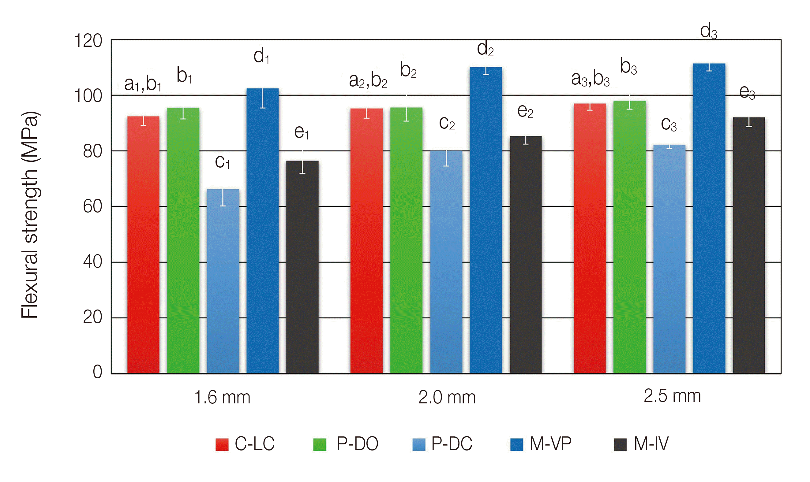 | Fig. 1Mean flexural strength of the different materials in the same thickness. Different single letters denote statistical difference. C-LC, Lucitone 199®; P-DO, DIOnavi - Denture; P-DC, DENTCA - Denture Base II; M-VP, Vipi Block Gum; M-IV, M-IVobase® CAD.
|
Table 5
Tukey HSD test of flexural strength between groups in 1.6 mm, 2.0 mm and 2.5 mm thickness
![]()
The average flexural moduli under the same thickness were significantly different among the materials except for the comparison between P-DC and M-IV at the thickness of 2.5 mm. M-VP showed the highest average flexural modulus. C-LC, P-DO, P-DC and M-IV follows the next accordingly. The post-hoc analysis results are summarized in Table 6 and Fig. 2. As the thickness increases, the average flexural strength is also increased in each group. However significant differences were not found all the time. Under the group of C-LC, the significant differences were only found under the thicknesses of 1.6 mm and 2.5 mm (P = .006). In the group of P-DO, there was no significant difference in the flexural strength depending on the changes in thickness. The P-DC and M-VP groups showed significant differences between 1.6 mm and 2.0 mm (P < 0.001, P = 0.003) as well as between 1.6 mm and 2.5 mm (P < 0.001). The group of M-IV showed significant differences across all thicknesses (P < 0.001). The post-hoc analysis results are described in Fig. 3.
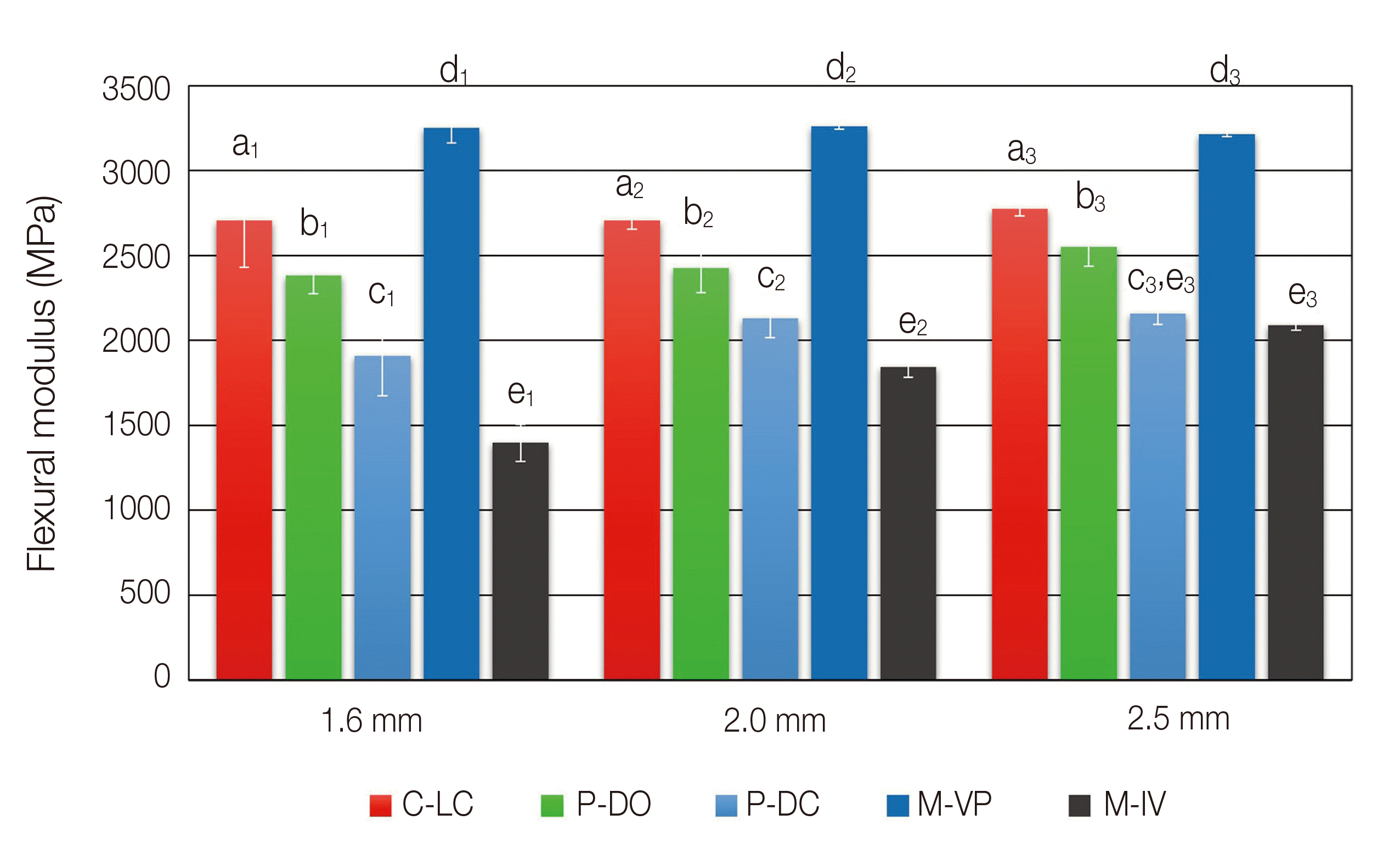 | Fig. 2Mean flexural modulus of the different materials in the same thickness. Different single letters denote statistical difference. C-LC, Lucitone 199®; P-DO, DIOnavi - Denture; P-DC, DENTCA - Denture Base II; M-VP, Vipi Block Gum; M-IV, M-IVobase® CAD.
|
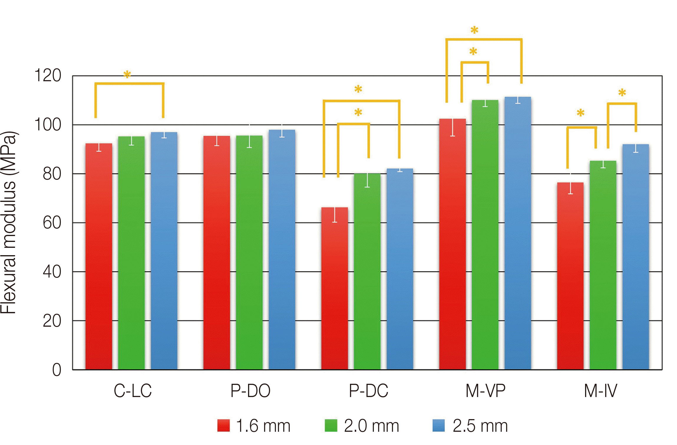 | Fig. 3Mean flexural strength of the different thickness in the same materials. * denotes significant difference at the level of 0.05. C-LC, Lucitone 199®; P-DO, DIOnavi - Denture; P-DC, DENTCA - Denture Base II; M-VP, Vipi Block Gum; M-IV, M-Ivobase® CAD.
|
Table 6
Tukey HSD test of flexural modulus between groups in 1.6 mm, 2.0 mm and 2.5 mm thickness
![]()
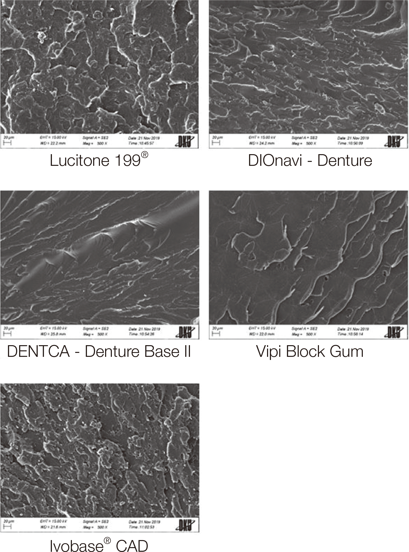
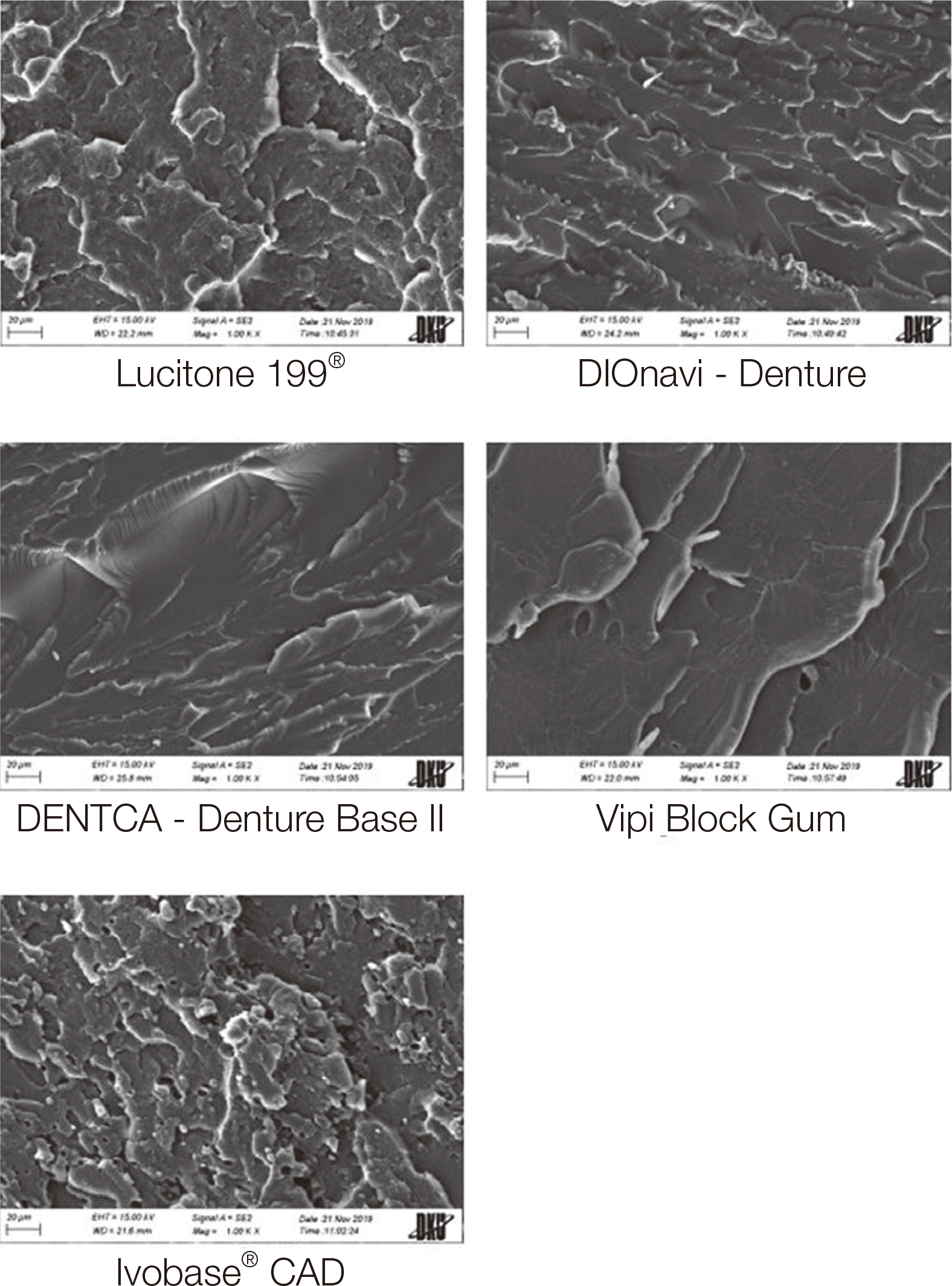
The images of SEM (Fig. 4 and 5) illustrate differences in the surface structure of each denture resin. M-VP had a relatively smooth and even surface. The surfaces of C-LC, P-DO, P-DC and M-IV were noticeably rough and showed various coarse mass forming stripes. Notably, the surface of M-IV showed a considerable amount of white spherical particles that are likely to contain various microspheres.
Go to :
Discussion
The size of specimens used in this study did not coincide with the standard size for a 3-point bend test suggested by ISO 20795-1:2013. When the size of the specimen changes, it is reasonable to follow the ASTM D790 international standard.17 The reason that the minimum thickness was set to be 1.6 mm (1/16 inch) is that if the thickness of the specimen becomes thinner than 1.6 mm (1/16 inch), then the length and width of the specimen should be changed accordingly. Thus, to keep the consistency in dimensions of the specimens with the other thickness groups, 1.6 mm was set to be the minimum thickness. As mentioned in ASTM D790, the most critical factor to reduce errors by conducting a 3-point bend test is the ratio between the length of the support beams and the thickness of the specimen, which is 16. Accordingly, 25.6 mm, 32.0 mm and 40.0 mm were set as the lengths of support beams. In addition to this, ASTM D790 defines that if the thickness of the specimen is thinner than 3.2 mm (1/8 inch) and thicker than 1.6 mm (1/16 inch), then the width of the specimen should be 12.7 mm (1/2 inch). Also, the lengths of the specimen’s edges should be either at least 10 percent of the whole length of support beams or 6.4 mm (1/4 inch) to prevent the specimen from slipping into the support beam. Therefore, the final dimensions were designed to be 65.0 mm × 12.7 mm × 1.6 mm / 2.0 mm / 2.5 mm.
According to the results from this study, the first null hypothesis was partially rejected. Furthermore, the second null hypothesis was also partly rejected. M-VP showed a significant flexural strength difference compared to the conventional denture base resins. However, the other PMMA block, M-IV had significantly lower flexural strength than the conventional denture base resins. Most of the previous studies reported that PMMA denture base resins for milling have significantly higher flexural strength than conventional denture base resins.25-27 The study from Aguirre et al.25 used Lucitone 199® (Dentsply Sirona Inc.), as conventional denture base resins and Vertex PMMA (Avadent Original, Global Dental Science, Scottsdale, USA) as PMMA resins for milling. The average flexural strength of conventional denture base resins was 116.6 ± 3.1 (MPa) and CAD/CAM denture base resins’ strength was 146.6 ± 6.6 (MPa). They described that CAD/CAM denture base resins have significantly higher flexural strength than conventional denture base resins. Furthermore, Al-Dwairi et al.26 used Meliodent (Heraeus Kulzer, Hanau, Germany) as conventional denture base resins and derived the average flexural strength as 93.33 ± 8.64 (MPa). Avadent PMMA pucks (Avadent Digital Dental Solutions, Scottsdale, USA) and Tizian Blank PMMA (Schütz Dental, Rosbach, Germany) were taken for CAD/CAM denture base resins. The average flexural strength of materials was 123.11 ± 9.47 (MPa) and 130.67 ± 10.48 (MPa) each. They concluded that CAD/CAM denture base resins have significantly higher flexural strength than conventional denture base resins.
According to Murakami et al.11 CAD/CAM denture base resins for milling are polymerized, especially with high pressure and temperature. This process reduces the residual monomers and the internal space and increases the average number of molecules of PMMA polymer, which leads to improved mechanical properties.11,24,28 The high flexural strength of CAD/CAM PMMA denture base resins for milling proved in this study and the other previous researches seems to have a strong connection with the conversion rate and void level characterized by its manufacturing process. Further, the components and the forms of PMMA chain are expected to be relevant with the flexural strength of CAD/CAM PMMA resins.
However, Pacquet et al.29 reported the opposite result. They used Probase Hot (Ivoclar Vivadent Inc.) for conventional denture base resins and Ivobase® CAD (Ivoclar Vivadent Inc.) for CAD/CAM denture base resins for milling. The average flexural strengths were 97.31 ± 4.96 (MPa) and 87.98 ± 7.37 (MPa) each. They concluded that Ivobase® CAD has significantly lower flexural strength than the conventional denture base resins.
The differences of flexural strength among several types of CAD/CAM PMMA resin blocks were reported in the previous studies.24,28,30 These studies claim that the differences would be associated with different density or porosity caused by different manufacturing processes. Also, the difference in the components of CAD/CAM PMMA resin blocks would lead to the flexural strength difference. Aguirre et al.25 note that denture base resins classified as “Highimpact” resins include rubbery comonomers such as butyl acrylate that cause dispersion of rubber inclusion. M-IV are classified as a “High-impact” resins according to the manufacturer, “High-impact” resins can enhance its impact strength by its advanced elasticity, on the other hand, can impact negatively on the flexural strength. After analyzing the fractured patterns of specimens from the 3-point bend test, the specimens in the group of M-IV showed more transformations than other resins. Ductility and fractured patterns of M-IV seem to be related to its relatively low flexural strength.
Since there are almost no researches that investigate the flexural strength of 3D printing denture base resins, it is hard to compare with previous studies. In this study, P-DO did not show any significant flexural strength difference with the conventional denture base resins. However, P-DC had significantly lower flexural strength than the conventional denture base resins. These differences could be explained by the components of each material. P-DC contains diurethane dimethylacrylate (DUDMA) as monomers. According to the Safety Data Sheet (SDS) provided by the manufacturer, there is approximately 30 - 50 percent of DUDMA in P-DC. Based on Gajewski et al.31 urethane dimethacrylate (UDMA) has low viscosity and high elasticity. UDMA is synthesized by the reactions from hydroxyalkyl methacrylates and diisocyanates.32 UDMA obtains elasticity and increases its conversion rate by hexaethylene diurethane.33,34 In the 3-point bend test of this study, it was observed that more transformations occur in P-DC before fracture. As mentioned before, the denture base resins with strong elasticity can have relatively low flexural strength.
This study also evaluated the changes in flexural strength depending on the thickness of denture base resins. Some materials showed a significant decrease in flexural strength as the thickness reduced. However, the average flexural strength of all groups was higher than 65 MPa, which is the minimum flexural strength suggested by ISO 20795-1:2013. If the thickness of denture base decreases, denture wearers would be more satisfied with reduced weight and increased retention. Also, when the interarch distance is restricted, dentures with lower thickness would be more useful.35
A limitation of this study is that the research regarding cyclic loading and fatigue resistance of a material against heat circulation are not covered. In addition to this, the specimens did not reflect the real shape of dentures. Also the actual dentures in the oral environment function more complicatedly, undergoing several types of strength in several directions. Furthermore, additional studies on the other mechanical properties such as impact strength, fracture toughness should be done.
Go to :
Conclusion
The following conclusions can be drawn within the limitations of this study.
The flexural strength of different CAD/CAM denture base resins used in this study varied according to the composition and properties of each material.
The flexural strength of CAD/CAM denture base resins was higher than the standard suggested by ISO 20795-1:2013 at a thickness of 1.6 mm or more though the thickness decreased.
For clinical use of dentures with lower thickness, further researches should be done regarding other properties at lower thickness of denture base resins.
Go to :
 XML Download