

 PDF
PDF Citation
 Print
Print


Introdcution
A common prosthodontic treatment of an edentulous maxilla includes a conventional complete denture (CD) or an implant-supported overdenture (IOD).1-4 These treatment options are useful when the patient has excessive ridge resorption and unusual maxillomandibular relationship. However, these prostheses need to be removed from the mouth for recovery of the tissue and maintenance of oral health.5-7 Some patients may be compliant to these requirements because of discomfort with no teeth in their mouth, especially maxillary anterior teeth.8,9
The patient’s comfort and oral function can be restored by means of an implant-supported fixed complete denture (ISFCD).1,10 However, the ISFCD has limitation to patients who present with deficient bone quantity on posterior region and systemic condition excluding placement of multiple implants.1,4 Alternatively, the combination of fixed and removable prosthesis can be considered. The anterior segment can be restored with an implant-supported fixed dental prosthesis (ISFDP) and the posterior segment with of a conventional removable partial denture (RPD).8,9,11-13 However, there is a concern with regard to potential stress accumulation on the implants supporting the ISFDP and being engaged by an RPD.14-17 Yet, no long-term study is available with this type of treatment modality.
This case report describes 10-yr clinical outcome of a patient whose edentulous maxilla was restored by means of an ISFDP in the anterior segment and a distal-extension RPD in the posterior segment. The ISFDP was designed to splint 4 endosseous implants as one unit and engaged by the RPD. The edentulous mandible was restored by means of an implant-supported overdenture.
Go to : 
Case Report
A 68-yr-old edentulous woman visited and expressed dissatisfaction with her existing conventional CDs. She wanted an ISFDP for her maxillary anterior teeth. Her edentulous mandible was severely atrophic. Her medical history was unremarkable. In 2008, the patient’s edentulous maxilla was restored by means of an ISFDP for the anterior segment and a distal-extension RPD for the posterior segment. Her edentulous mandible was restored by means of a mandibular IOD.13
A total of 8 endosseous regular-neck dental implants (Standard Implant, Straumann, Basel, Switzerland) were placed: 4 in the maxilla and 4 in the mandible. The maxillary implants were placed for supporting the ISFDP in anterior to the maxillary sinus. The custom abutment was fabricated with precious metal (Dental Casting Gold Alloy, Heesung catalysis, Cheongju, Korea). The metal framework of ISFDP was fabricated with casting using a nickel-chrome-cobalt alloy (T-3 C&B Alloy, CMP Industries, New York, USA). The artificial denture teeth (Endura Anterio A3, Shofu Dental Corp, Kyoto, Japan) were arranged and the gingiva was veneered with self-cure acrylic resin (Vertex SC, Vertex Dental, Zeist, The Netherlands). The ISFDP was designed to demonstrate screw access holes on the occlusal surface and was luted intraorally to abutments. Although the prosthesis was cemented to the abutments, the ISFDP was retained with screw to the fixture.
The RPD was designed to engage the ISFDP by means of direct and indirect retainers. Occlusal rests were placed directly over supra-structure of implants, and 19-gauge base metal alloy wrought wire retentive clasp arms were contoured to engage 0.01-inch mesiofacial undercuts. A modified palatal strap was chosen as the major connector to obtain additional support and stability from the palate. The implant fixtures of lower arch were connected by an implant connecting bar (Dental Casting Gold Alloy, Heesung catalysis). An mandibular IOD was constructed to engage the bar by means of attachment elements: Hader bar segment (Hader-EDS Bar System, Attachments International, California, USA) between right and left lateral incisor and right and left first premolar and extra-coronal resilient attachments (ERA Attachment, Sterngold Dental, Attleboro, USA) at each end of bar. The teeth were arranged to centralize the masticatory forces with lingualized occlusal scheme. The anterior guidance was controlled to reduce lever action exerted on ISFDP (Fig. 1).
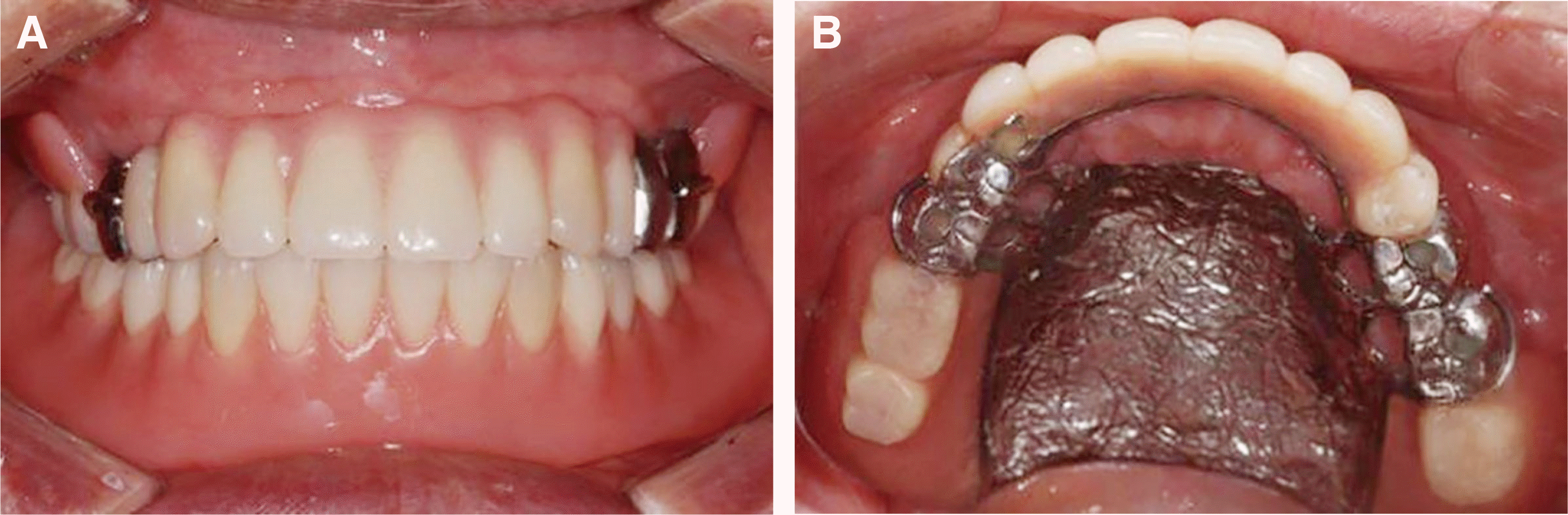 | Fig. 1(A) Maxillary implant-supported fixed partial denture and removable partial denture opposing mandibular implant overdenture placed, (B) Four occlusal rests were placed on premolar and molar. Ninety-gauge wrought wire retentive clasps were engaged 0.01-inch undercut. Major connector was modified palatal strap.
|
The patient was recalled every 3 - 6 months after placement of the maxillary and mandibular prostheses. At each follow-up appointment, the patient was provided oral hygiene instructions with oral prophylactic procedures. The removable prostheses were cleaned in an ultrasonic cleaner. Clinical examination and radiographic survey were followed to evaluate stability of the ISFDP and health of the peri-implant mucosa. At 1-yr follow-up, the patient complained of decreased retention of the maxillary RPD. The wire clasps were adjusted using a plier, to engage the undercuts and demonstrate a passivity at rest.
The patient was compliant to instructions and did not demonstrate any signs or symptoms of implant failure for 30-mo follow-up period except for breakage of resin veneer layered on the right maxillary canine (Fig. 2). The ISFDP was removed from the mouth with the abutments for repair. The abutments were unscrewed from the implant fixtures through the screw access holes built in the ISFDP. The healing abutments were connected to the implants to prevent collapse of the peri-implant mucosa. The ISFDP was repaired with an acrylic resin (Vertex SC, Vertex Dental) and screwed to the fixtures up to 35 Ncm using a torque rachet (Straumann). The screw access holes were filled with silicone material (EZ seal, Megagen, Daegu, Korea) and composite resin (Filtek Z350 XT, 3M ESPE, Minnesota, USA). During the course of 4-yr follow-up, there were no negative signs or symptoms related to the prosthodontic treatment, except for dislodgement of one of resins placed in the screw access holes.
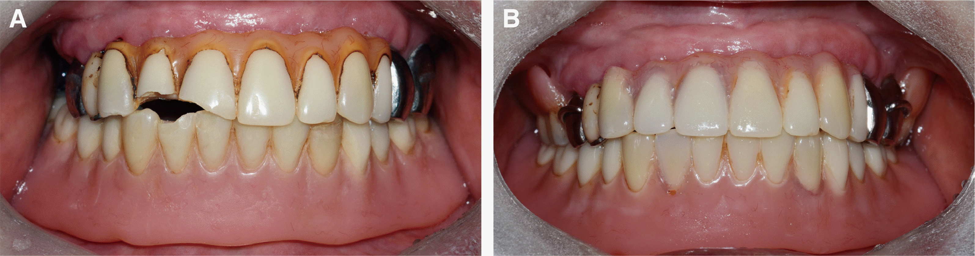
Eight and half years later, the ISFDP sustained breakage of resin veneers layered on the right central and lateral incisors, and the right lateral incisor of the mandibular IOD broke at the incisal level (Fig. 3A). The patient indicated she had lost her balance and fell on floor, inflicting facial injuries and breakage of artificial teeth. The prostheses were otherwise stable with no signs of screw loosening or breakage. The ISFDP was retrieved from the mouth and was returned to the mouth after the broken teeth were repaired with an acrylic resin (Vertex SC, Vertex Dental) (Fig. 3B).
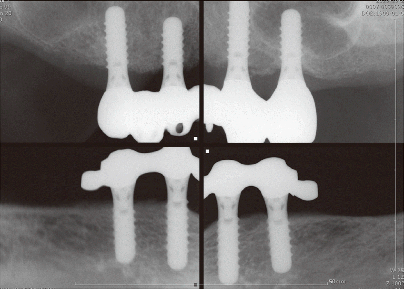
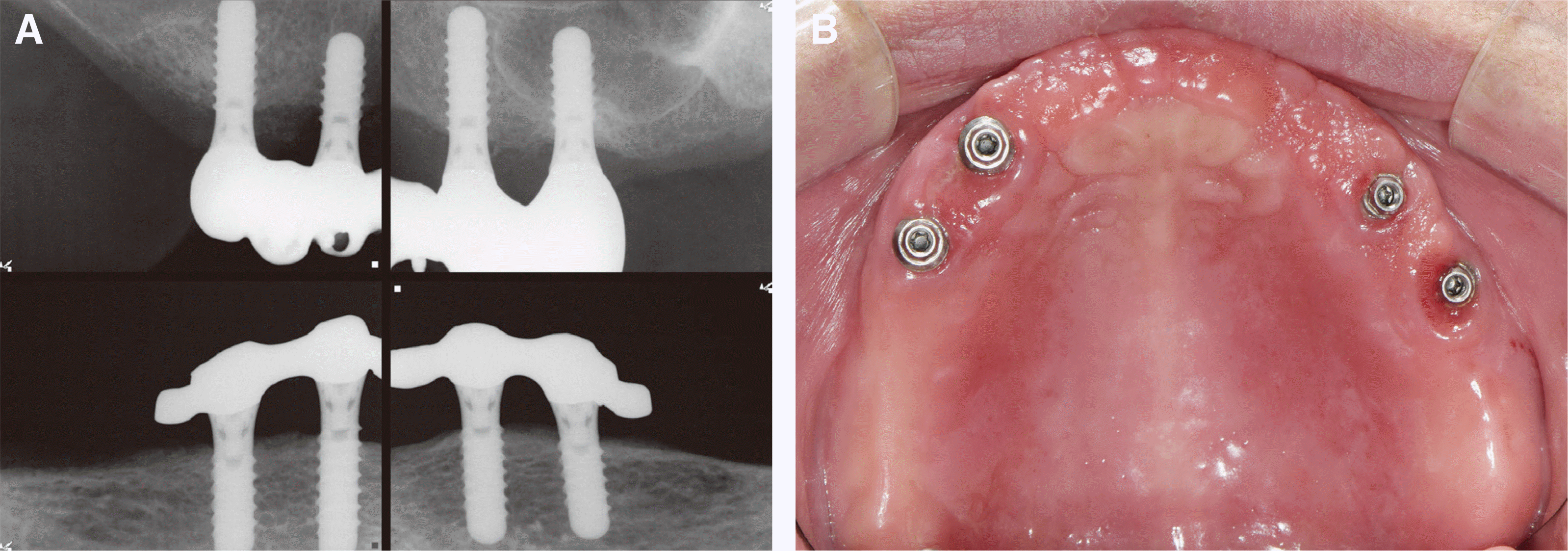
During 10-yr follow-up period, the implants had kept stability with minimal loss of crestal bone (Fig. 4A). The peri-implant mucosa was healthy with maintaining attached gingival cuff, although a mild inflammatory reaction was noted around the maxillary left implants (Fig. 4B). Throughout the follow-up period, the IOD kept. No negative event occurred with the mandibular IOD other than replacement of nylon retention inserts of the attachment element. The patient was regularly followed and instructed to use interproximal brush and oral irrigator to keep oral hygiene, especially underneath the implant connecting bar and ISFDP. At 10-yr follow-up appointment, the ISFDP showed a discoloration and wear of acrylic resin.
Go to :
Discussion
The anterior ISFDP has rest seats, guide planes, and undercuts to receive a distal-extension base RPD.11-13 Upon occlusal loading, the maxillary ISFDP may receive high forces driven from the RPD.15 These forces can be significant to cause biological and/or mechanical complications of the implant because of an leverage action from clasp assembly of the RPD.4,15-17
To reduce the risk of complications, the ISFDP was designed to splint the maxillary implants, and the RPD embraced a stress releasing concept with using mesial rests and 19-gauge wrought wire clasp arms.4,13 The RPD framework was designed to engage broad surface of the palate and tuberosity and fitted through the process of physiologic adjustment.13,14 The occlusal rest was designed to seat directly over implant support and adjusted to control the rotational movement of the RPD.14
The patient was followed regularly to evaluate oral hygiene and assess stability of the prostheses.5-7 The peri-implant mucosa appeared unremarkable with minimal inflammatory reaction. The importance of oral hygiene was emphasized at each visit to keep stability of implant support. Over the course, the ISFDP was retrieved twice to repair broken veneers of acrylic resin. The RPD kept functional stability without a need of lining of the base, although the wire clap arms were adjusted as needed to enhance the retention.
The ISFDP was luted to the abutments in the mouth to ensure a passive fit.16,17 This prosthesis was also retrievable because of the screw connection of the abutments to the fixtures.11-13 The abutments were individually screwed to each implant, and then the implants were splinted across the arch with metal framework of ISFDP. The ISFDP was designed to demonstrate screw access holes on the occlusal surface. The majority of excess luting agent was extruded through the holes. This effect of venting was advantageous in minimizing the residual cement around the margin of the ISFDP and avoiding potential development of peri-implantitis associated with cement remnants.4,18 The substructure of the ISFDP was cast in a base metal alloy to reduce the cost, although the abutments were cast in a high noble alloy.11-13 No adverse reaction was noted with the combination of noble alloy abutments and base metal alloy prosthesis.
The antagonist may play an important role for prosthodontic success.2-4 The edentulous mandible was restored with an IOD. No mechanical complications were noted both in the maxillary and mandibular prostheses. There may be a cushioning effect through the resiliency of IOD attachments and mucosa of the posterior edentulous ridge of the mandible.2,13,19
Implant overdenture (IOD), compared to ISFDP in patients with edentulism who have severe alveolar bone resorption but do not want extensive surgery, can be an alternative treatment option that can reduce costs while meeting the patient’s aesthetic and functional needs.20 Also, it is favorable to maintain oral hygiene. However, when the denture is removed, the patient can be dissatisfied with the absence of anterior teeth.
For aesthetic reasons, the patient wanted the teeth to remain even after the prosthesis was removed. As in this case report, the method of restoring the anterior teeth with the ISFDP and posterior teeth with the posterior-extended RPD can maintain the aesthetics of the patient and provide psychological satisfaction because the anterior teeth remain even after the denture is removed.
The patient expressed high satisfaction with the combination of fixed and removable prosthodontic treatments, especially with the capacity of retaining her maxillary anterior teeth at night. No major issue was noted with these prostheses over the course of 10-yr follow-up. The maintenance was minimal for repair of acrylic veneer and replacement of resin teeth and nylon retention inserts of attachment elements. However, patient selection and compliance can be crucial for success of this type of treatment and avoid consequences associated with parafunctional activities.21
Go to :
Conclusion
A patient with complete edentulism can be treated with the combination of fixed and removable prostheses. Maxilla was restored with an ISFDP in the anterior region and a distal-extension RPD in the posterior region. Mandible was restored with an IOD. The ISFDP was engaged by means of the RPD and opposed the IOD. During 10-yr follow-up period, no severe mechanical and biological complications occurred except the discoloration of resin veneers and artificial teeth. The implants were maintained stable state. The patient expressed high satisfaction with the capacity of retaining the maxillary anterior teeth in her mouth at night.
Go to :
 XML Download