

 PDF
PDF Citation
 Print
Print


Introduction
There are many clinical and laboratory steps in the manufacture of implant prosthesis.
1
To obtain the accurate fit of the prosthesis, the errors in each step should be minimized.
2
Therefore, inaccurate oral records may reduce the accuracy of the prosthesis, which may lead to failure of treatment.
3
The accuracy of definitive cast for the manufacture of implant prosthesis is affected by several factors such as impression method, impression material, type of stone material, and pouring technique.
3-5
Inaccurate impressions can lead to inadequate restorations and biologic or mechanical complications, so clinicians should always strive to achieve good fit.
The method of implant impression is traditionally divided to the open-tray method using pick-up impression coping and the closed-tray method using transfer impression coping. Much research has been done comparing the accuracy of the two impression methods. Although there is no significant difference in the number of implants below 3, the open-tray method is more accurate in most studies with more than 4 implants.
3,6-9
And implant placement angle may affect the accuracy of the impression.
10
Traditional impression taking and model fabrication process may cause errors due to shrinkage, uneven thickness, separation, distortion, and swelling.
11
Recent years, digital intraoral impression method has been introduced with advances in dental CAD/CAM systems, and it is possible to reduce the error that may occur during the laboratory process by a simple manufacturing process.
12,13
The digital impression taking of implant requires a transfer post, mainly a scan body is used for it.
14
However, both the traditional impression and the digital impression method using a scan body require removal of the healing abutment, and it has been reported that there is a possibility of damage to the soft tissue around the implant when removing or reconnect the healing abutment.
15
Therefore, reducing the frequency of healing abutment removal will help maintain health of surrounding soft tissue and minimize the patient’s discomfort.
To simplify implant impression techniques, a new implant restorative system using CAD/CAM technology was introduced called Encode restorative system. There is a digital identification code on the encoded healing abutment which informs the location of the implant, so that it is possible to make a digital impression directly without removing the healing abutment.
16-18
This can prevent mucosal injury that may occur when the abutment is removed or reconnect, and can reduce patient discomfort, impression time and cost.
19
There are not many studies about encoded healing abutment, and a study has shown that the shorter diameter and length of the scan body, the higher the error rate.
20
Therefore, it is necessary to study the accuracy of the impression method using the encoded healing abutment. In this study, the purpose was to compare the accuracy of three different implant placement angles with the encoded healing abutment, scan body, and pick-up impression coping impression method.
Go to : 
Materials and Methods
Fabrication of master model
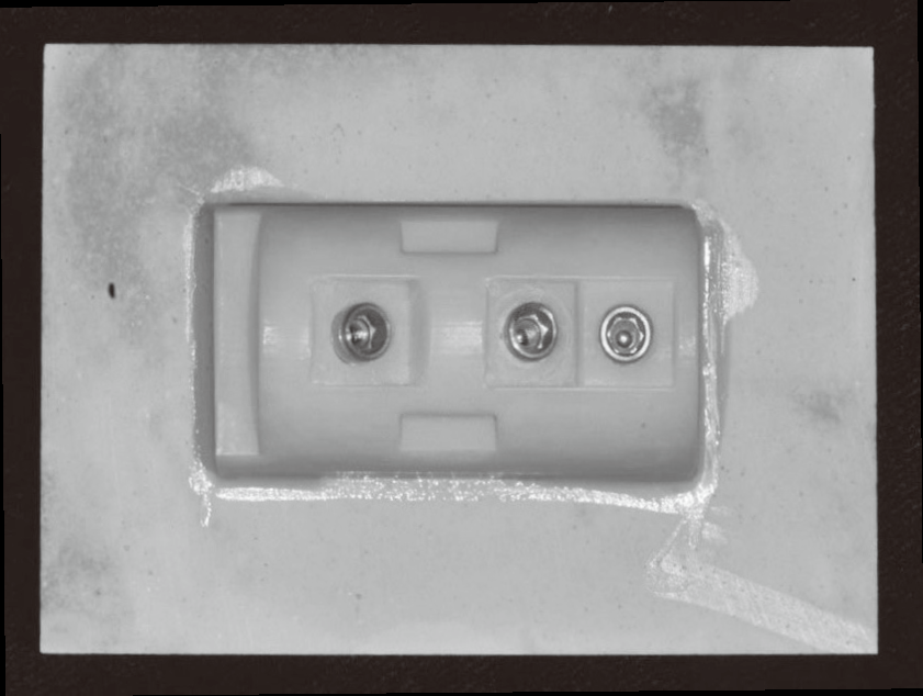
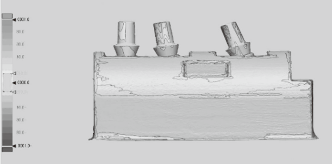
40 mm long edentulous ridge block was designed with 3D modeling software (Autodesk 123D design, Autodesk Inc., San Rafael, USA). A cylindrical recipient spaces of Ø4.5 × 10.0 mm were formed for placement of the implant analog at the first premolar (PM1), second premolar (PM2), and second molar (M2) region. PM1 was formed to be parallel to tooth axis, PM2 was formed to 10° mesial inclination and M2 to be 20° mesial inclination. Then, a vertical box was formed to the distal side of the block so that it could serve as a stop when taking impression using individual tray. The design files were saved as STL files and printed out using 3D printer (Objet EDEN260V®, Stratasys Ltd., Eden Prairie, USA) with acrylic printing material (Objet Verodent MED670, Stratasys Ltd.).
Ø4.3 × 10.0 mm implant analogues (ISLA500, Neobiotech, Seoul, Korea) were placed into each recipient space, and fixed with resin cement (Super Bond C & B, Sun Medical Co. Ltd., Moriyama, Japan) (Fig. 1).
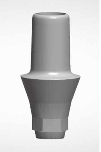
The abutment to be secured into the master model was designed with 5.0 mm diameter, 3.0 mm cuff and 5.0 mm height using CAD software (Dental system™, 3shape, Copenhagen, Denmark). The abutment was milled using a CAM machine (Plus Mill BX4, Dental Plus, Seoul, Korea) and then, abutment was sandblasted to improve the accuracy of scanning. Fig. 2 shows the abutment image on CAD.
Fabrication of reference model
The abutment was tightened with 15 Ncm to the implant of master model. To increase the accuracy of the scanner, a 3D scanner (Freedom HD, DOF Inc., Seoul, Korea) was used after applying scan powder (EASY SCAN, ALPHADENT Co. Ltd., Goyang, Korea). Then the reference model data was saved to STL file.
Fabrication of the test models
The classification of test group according to impression method is as follows. Fig. 3 shows copings that used to make impression at each group.
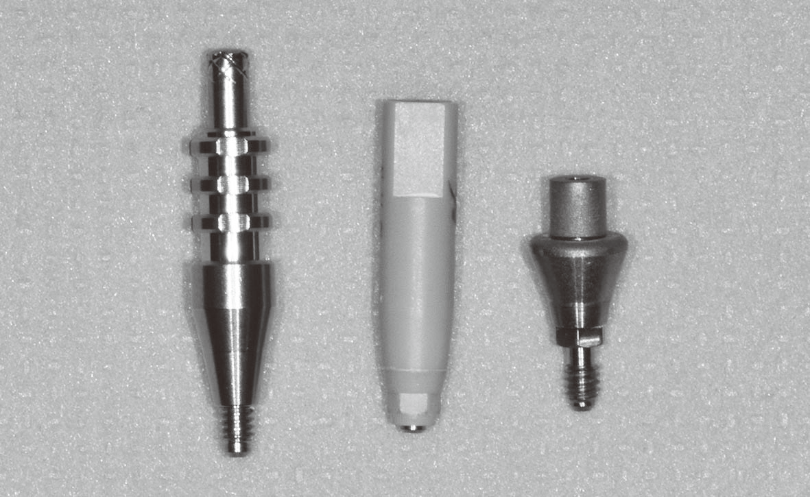
Group P : Pick-up impression coping (open-tray) - 15 files
Group S : Scan body digital impression - 15 files
Group E : Encoded healing abutment digital impression - 15 files
Pick-up impression model
The individual trays were made with two base plate waxes to provide uniform thickness of impression material. The open-tray type individual tray was made with tray resin (Quicky, NISSIN Dental products Inc., Kyoto, Japan) so as to have a stop on the stop part of the master model and the reference box. The pick-up impression copings (Ø4.5, ISIPS411, Neobiotech) were tightened into implant with a constant torque of 15 Ncm and then impression was taken with polyvinyl siloxane (PVS) impression material (Honigum, DMG, Hamburg, Germany). After impression material was set and removed form master model, the implant analogues were secured with 5 Ncm to the pick-up impression copings in the individual tray. This process was repeated 15 times and a new pick-up impression coping and individual tray were used. Models were fabricated with the type IV dental stone (Fuji rock EP, GC Corp., Tokyo, Japan), and carefully trimmed to minimize discrepancies from the master model.
After the stone models by pick-up impression taking were completed, the abutments (Ø 5.0, cuff 3.0, height 5.0 mm) were secured to the implants with 15 Ncm. Then, the 3D scanner (Freedom HD, DOF Inc.) was used for scanning and save as STL file.
Scan body impression model
The Ø4.0 scan body (ISPSBH40NB, Neobiotech) was tightened with torque of 15 Ncm on the implant of the master model, and a digital impression was made 15 times using intraoral scanner (TRIOS, 3Shape). Acquired impression files were imported into the CAD software (Dental system™, 3shape) and the position of the implants were obtained by superimposing the library data provided by the manufacturer of the scan body. Then, digital abutments were placed in each implant position and the 3D image data was converted using CAD software (Exocad®, Exocad GmbH, Darmstadt, Germany) and save as STL files.
Encoded healing abutment impression model
The Ø5.3 encoded healing abutment (ISEHA503S, Neobiotech) were tightened with 15 Ncm torque to each implants of the master model. To increase the accuracy of the impression, a scan powder was applied and impression was made 15 times using an intraoral scanner (Fig. 4). Acquired impression files were imported into the CAD software and the positions of the implants were obtained by superimposing the library data provided by the manufacturer of the scan body. After that, 3D images data was converted to a STL file in the same way as scan body impression.
Data analysis
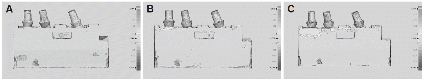
Each STL file was imported into 3D analysis software (Geomagic® Control X, 3D systems Inc., USA) and the unnecessary parts were removed for accurate overlap. Then, the reference model was set as the reference object, each test group model was set as the measurement object, and the overlapping was performed using the best fit alignment which is the most frequently used overlapping method for accuracy analysis.21 The color coded map was used for the analysis of three dimensional error and the acceptable error was set to 15 µm and the maximum error range was set to ± 100 µm.
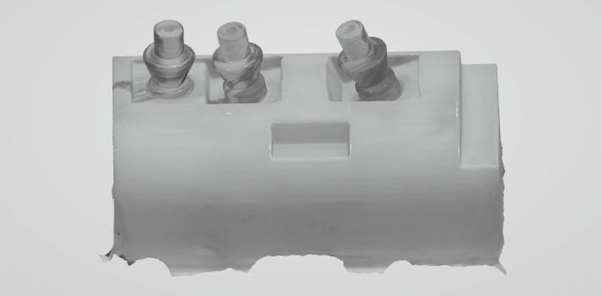
The root mean square (RMS) value was calculated to compare the discrepancies between reference and experimental abutments. In order to find the difference according to the impression method, RMS values were measured in three abutments in group P, S and E (Fig. 5), and each group was divided into three subgroups (Table 1). The RMS values were compared in the abutments of PM1 (0°) for subgroup a0, PM2 (10°) for subgroup a1, and M2 (20°) for subgroup a2.
 | Fig. 53D comparison between reference model and test models illustrated in color-coded map of all abutments.
|
Table 1
Classification of all groups in this study
![]()
Statistical analysis
The mean and standard deviation of RMS values were measured for each group and statistical analysis was performed using SPSS v23.0 (IBM SPSS Inc., Chicago, USA). Kolmogorov-Smirnov analysis and Shapiro-Wilk analysis were performed for the regularity test. One-way ANOVA test was performed and the Dunnett T3 test was performed for post-analysis. The Jonckheere-Terpstra test was used to examine the correlation between the angulation of the implant and the error (P = 0.05).
Go to :
Results
Table 2 shows the mean and standard deviation of RMS values for each group. The RMS values were highest in group E (38.29 ± 4.12 μm), followed by group S (35.27 ± 2.56 μm) and group P (25.56 ± 2.53 μm). And Fig. 6 shows a typical features of color coded map in each group. In the color coded map, green color means that the experimental model is within 15 μm of the reference model, red color is positive, which is outside the reference model, and blue color is negative, which is inside the reference model.
The normality test of the RMS values revealed normal distribution in all the groups and all subgroups. One-way ANOVA test was performed to find out the difference between impression methods, and result of the test showed statistically significant (P < 0.05, Table 3, 4), therefore Dunnett T3 test was performed for post-hoc analysis. As a result, the RMS values of the group P were smaller than those of the group E and S (P < 0.05), and there was no significant difference between the group E and the group S (Table 5).
Table 3
Results of one-way ANOVA for RMS values between Group P, S and E
| Sum of Squares | df | Mean Square | F | P | ||
|---|---|---|---|---|---|---|
| Group P, S, E | Between Groups | .001 | .001 | .001 | 39.600 | .001 * |
| Within Groups | .001 | 42 | .000 | |||
| Total | .002 | 44 | ||||
![]()
To determine whether the implant placement angle affects accuracy of impression, one-way ANOVA and Dunnett T3 test were performed on the subgroups. RMS values of a0 and a2 in group P were significantly smaller than those of group S and E (P < 0.05), and there was no significant difference in RMS values between a0 and a2 of group S and E (Table 6, 8). RMS values of a1 were the lowest in group P, and significantly higher in group S and group E (P < 0.05, Table 7).
The result of Jonckheere-Terpstra test for the correlation of the RMS values according to the angulation of the implant showed a correlation in group E (P < 0.05), which RMS value was 33.83 ± 2.88 μm (Ea0), 39.69 ± 4.31 μm (Ea1) and 37.62 ± 4.69 μm (Ea2). No significant correlation was shown in group P and group S (Table 9).
Table 9
Results of Jonckheere-Terpstra test of RMS value for Group P, S and E
| Group P | Group S | Group E | |
|---|---|---|---|
| Number of Levels in Angle | 3 | 3 | 3 |
| N | 45 | 45 | 45 |
| Observed J-T Statistic | 420.0 | 425.0 | 433.0 |
| Mean J-T Statistic | 227.5 | 337.5 | 337.5 |
| Std. Deviation of J-T Statistic | 48.022 | 48.011 | 48.006 |
| Std. J-T Statistic | 1.718 | 1.822 | 1.989 |
| Asymp. Sig. (2-tailed) | .086 | 0.068 | .047 * |
![]()
Go to :
Discussion
The purpose of this study is to compare the accuracy of the impression method using the encoded healing abutment with the traditional open-tray method and the conventional digital impression scan body method and to determine whether the angulation of implant affects the accuracy of the impression. In 2006, Grossmann et al.16 introduced an impression method using an encoded healing abutment, but there are not many studies about the accuracy of this method.22-25 In addition, they mainly analyzed the linear deformation or angular deformation using the center point of the implant platform or the upper spherical structure, which may have limitations in confirming the error that may occur when fabricating the prosthesis in clinic, and other errors may be generated accordingly since the coordinates are manually determined.22-24
In this study to compare the accuracy with situation simulating more clinical, the abutment was designed and milled with CAD/CAM system and RMS of three-dimensional error value of the abutment part was measured and compared. As a result, the differences of accuracy were found according to the impression methods and the null hypothesis that there is no significant difference according to the impression was rejected. The impression method using the encoded healing abutment showed a higher RMS value than the pick-up impression coping method (P < 0.05) regardless of the angle of implant and conventional pick-up impression coping method showed the highest accuracy. Other previous studies reported the encoded impression group showed larger error than the pick-up impression group, and in this study the error of encoded healing group was 33 - 50 μm and that of pick-up impression group was 18 - 32 μm. Eliasson and Ortorp showed differences of 79.5 μm and 31.2 μm, Simon et al. showed 65 - 107 μm and 13 - 20 μm, Howell et al. showed 35 - 242 μm, 2.4 - 161.9 μm, and Abdullah et al. showed 48 - 228 μm and 14 - 25 μm in the difference of encoded impression and pick-up impression, respectively.22-25 The error values of this study showed similar to those of Eliasson and Ortorp and Simon et al. On the other hand, the results of the study by Howell et al. and Abdullah et al. showed a larger error than those of this study since they measured at the position of the occlusal plane.22-25 This study is conducted in three-dimensional and more similar to the clinical situation.
In group E, there was a positive correlation between the implant angulation and error. Encoded healing abutments are more likely to show inaccuracies with inclinations because of their smaller impression surface. Although previous studies reported that increase in angulation did not increase the error in the encoded healing group,23,25 another study reported the encoded healing abutments at 15° angulation showed larger error than 0° implant.24 And there is a possibility that the collar portion of the encoded healing abutment may be embedded in the gingiva due to the tilt of the implant.25 Therefore, it is considered that collar area should be located at least 1 mm above the gingiva during impression when using an encoded healing abutment in inclined implant. In this study, group S and P were not correlated with the error according to the implant angulation (Table 7), and similar results were obtained in other recent studies.26-28 Moura et al. compared accuracy of traditional impression method and scan body method with a 15° implant angulation and there was no significant difference.26 However, Assunaco et al. reported that the error was increased with the inclination of the implant, and a larger error was shown with the angulation of 25° implant.11,29 Therefore, according to the results of present study, it is difficult to conclude that the pick-up impression method and the scan body impression method are free of implant angulation, and further studies on the implants with larger angulation seem to be needed.
The ‘best fit algorithm’ is a method currently used to superimpose the STL datasets and analyze the accuracy.21 As a result of the best fit alignment between the reference model and the test model, positive or negative values are produced, which is difficult to represent the actual deviation value by canceling each other. These inaccuracies could be avoided by using root-mean-square (RMS) calculations to analyze 3D deviations.30 Previous studies used ‘least squares method’ or ‘zero method’ at best alignment and set scan body as the overlap reference.31-33 However, it might be difficult to measure the actual deviation value with the scan body which is the target of the alignment. In color coded map of this study, most areas in group P were green color range which means less error, and red or blue color were more observed in the group S and E. In group S and E, blue color was observed in the mesial lower part of the abutment and red color was observed in the distal upper part of the abutment at PM2 or M2, which could be interpreted as impression model is less inclined than the master model. Additional attention should be paid to the impression of an inclined implant when using an intraoral scanner.
Although a highly reproducible coordinate measuring machine (CMM) can be used for accuracy analysis (2 μm), results of CMM are generated as solid data and the data loss occurs causing additional errors during transforming into STL data for best-fit alignment.22,23 Furthermore, CMM is quite expensive, and its measurement processing time is long. Measuring a specimen with a complicated inner surface is difficult, because the CMM probe requires contact with the surface of the specimen. On the other hand, the 3D scanner used in this study has many advantages such as reproducibility of 15 μm, low cost and short time.
In group S and E, an intraoral scanner was used. Although the intraoral scanner has a disadvantage of that larger errors may appear depending on the operator, the intraoral scanner is used in the group S and E in order to simulate the actual clinical situation.33
In the reference model and group P, milled abutment is used and error occurs during milling process (10 - 20 μm).
34,35
Therefore it could be considered that the group S and group E have more RMS values than the group P due to the error of the milled abutment used in the reference model. In recent research, the scan body is as accurate as pick-up impression coping.
36
Recently, many clinical cases using an encoded healing abutment have been reported.
16,19,37-40
Representative advantages of an encoded healing abutment are simple impression procedure, economical and no need of removing the healing abutment until the final restoration. Additionally, it may be useful when the scan body or impression coping could not be screwed by the inclined adjacent tooth. In the case of an impression using the conventional scan body, the shape of the gingival form of the healing abutment and the scan body are different each other, resulting in errors and damage to the gingiva and pain.
37,40
However, because the encoded healing abutment has both gingival height and diameter information, it is possible to fabricate the abutment which is same with the upper gingival shape of the implant. Therefore, the use of an encoded healing abutment is likely to increase if further studies are performed and the disadvantages are supplemented.
Go to :
Conclusions
In this study, the accuracies of three types of impression methods (encoded healing abutment, scan body, pick-up coping) were compared at various implant angles and the following conclusions were drawn.
1. The pick-up impression coping method was significantly more accurate than the encoded healing abutment and scan body impression method (P < 0.05).
2. The accuracy of encoded healing abutment and scan body impression method was not significantly different.
3. When using the encoded healing abutment impression method, the accuracy of impression significantly decreased as the angulation of implant was increased. Scan body and pick-up impression coping method, however, did not show a statistically significant correlation.
According to the results of this study, the use of the encoded healing abutment as a scan body can be clinically used. However, it should be used with caution when implants are placed with an angle.
Go to :
 XML Download