

 PDF
PDF Citation
 Print
Print


Introduction
Osseointegration has been reported to be a dynamic process in which the alveolar bone come in direct contact with the placed implant surface.1 This phenomenon has also been demonstrated in various preclinical models2,3 and histologic reports in a few humans.4-6 However, the status of osseointegration does not accurately specify the amount of ideal contact ratio between the implant surface and the bone, thus various methods have been proposed to evaluate the degree of osseointegration.7,8
Donath and Breuner proposed the Sage-Schliff (sawing and grinding) technique producing undecalcified specimen to investigate the interface between the implant surface and surrounding alveolar bone 9. This technique has provided a fundamental method to evaluate bone-implant contact (BIC), and as a result, the degree of osseointegration can be assessed histologically.
For successful implant treatment, it is essential to obtain substantial osseointegration and secondary stability of the implant over long-term period. Although BIC does not represent the overall osseointegration status in clinical situation, however, it is generally accepted that higher BIC is recommended for long-term success of dental implant.
As mentioned above, it is important to understand the bone-to-implant interaction in patients showing successful treatment outcome of implant. However, only histological analysis can properly assess biochemical host response and the value of BIC, and the researchers had difficulty to retrieve the successfully osseointegrated implants from the patients due to ethical reasons. Therefore, there are few, if any, studies which evaluated histologic BIC of successfully osseointegrated implants in humans and these specimens can only be obtained under limited conditions requiring explantation such as implants placed in an inappropriate position, causing pain or sensory disturbance, or showing fracture of screw or fixture.10 Thus, performing a histologic analysis of these implants is a unique opportunity to assess osseointegration through BIC measurements and to identify the biomechanical interaction between the implant and bone.
The current study is a clinical histological case report of retrieved implant which was fractured after 3-year functional load.
Go to : 
Case Report
This study was approved by the Institutional Review Board (IRB) at Dankook University (IRB No. DKUDH IRB 2019-06-002).
Clinical Observation
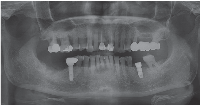
A female patient, 60-years old, came to department of periodontology, Dankook University dental hospital with missing tooth on #36 and wanted treatment using dental implant. The tooth had been extracted because of periapical lesion a year ago. After careful clinical and radiographical examination, a ø 4.5 × 8.5 mm bone-level implant (Luna®, Shinhung, Seoul, Korea) was placed according to one-stage protocol (Fig. 1). The implant-supported prosthesis was placed four months later.
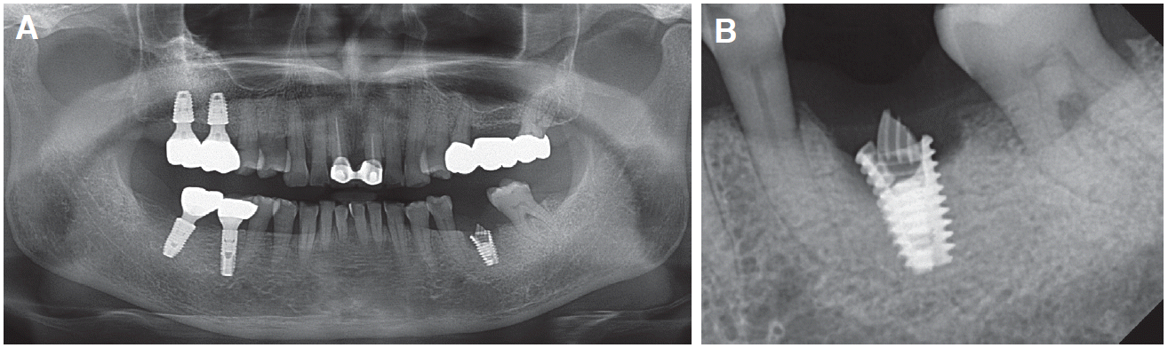
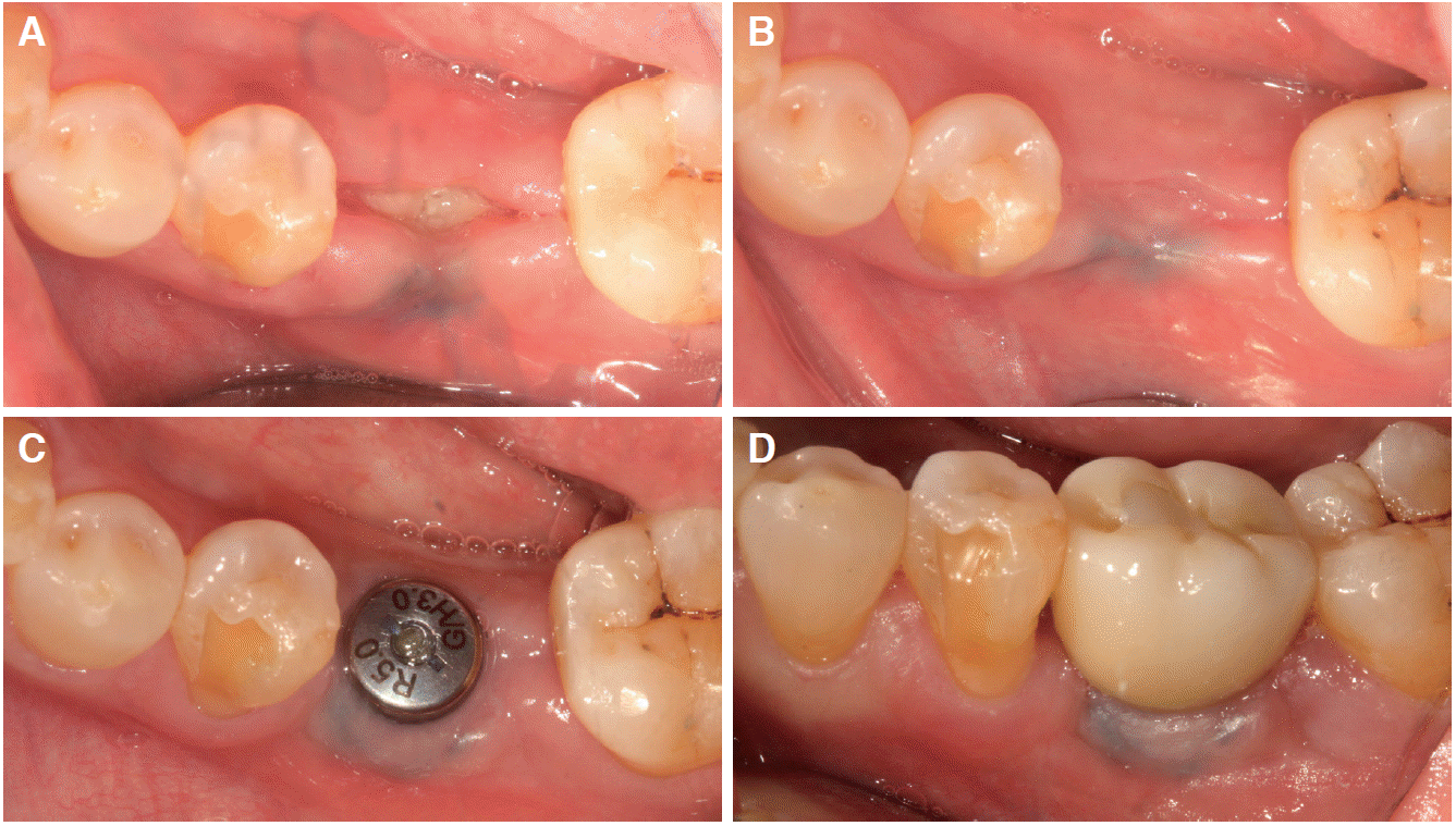
During three years after prosthesis delivery, no signs or symptoms were shown in annual follow-up (Fig. 2). However, after three years of function, the patient returned to the clinic with the complaint of loose implant (Fig. 3). From the clinical and radiographic examination, the marginal bone loss was observed with probing depth of 4 - 5 mm, and it was suspected that the fixture collar and abutment screw had been fractured. Due to the failure to remove the fractured screw with the removal kit, less invasive implant retrieval was not feasible. As a result, the explantation of the implant using trephine bur followed by alveolar ridge preservation were planned. After the explantation using 5-mm diameter trephine bur (Trephine kit, GenOss, Seoul, Korea), the socket was thoroughly debrided and open healing alveolar ridge preservation was performed using 250 mg of deproteinized bovine bone mineral with 10% porcine collagen (Bio-Oss Collagen, Geistlich, Wolhusen, Switzerland) and resorbable collagen membrane (Collagen graft, GenOss, Seoul, Korea) (Fig. 4). Five-month after the alveolar ridge preservation, a new ø 5.0 × 8.5 mm bone-level implant (Luna®, Shinhung, Seoul, Korea) was placed by one-stage protocol without any further ridge augmentation procedure (Fig. 5, 6).
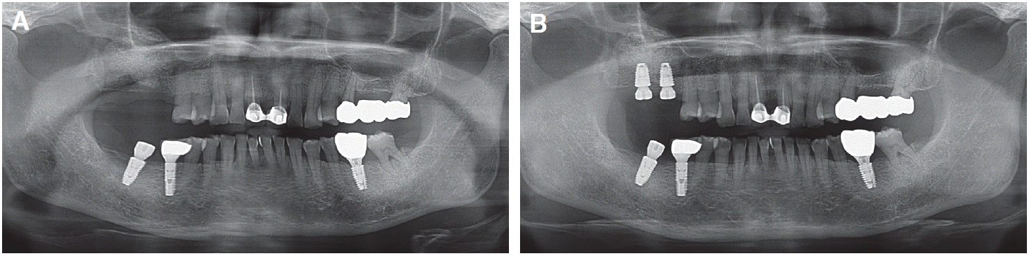
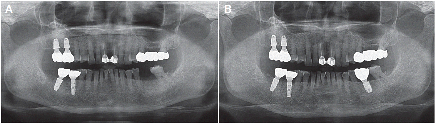
 | Fig. 3Radiographs when returned to the clinic with the complaint of loose implant 3.5 years after implant placement. (A) Paroramic view, (B) Periapical view.
|
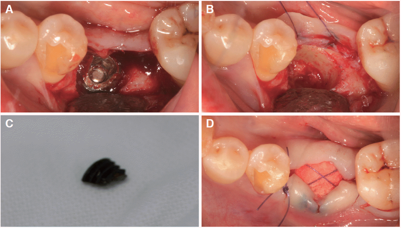 | Fig. 4Clinical photographs illustrating surgical procedure. (A) After flap reflection. Granulation tissue around fractured implant fixture can be observed, (B) After explantation using trephine bur, (C) Removed fragment of implant fixture, (D) After alveolar ridge preservation with deproteinized bovine bone mineral with 10% porcine collagen (Bio-Oss Collagen, Geislitch, Wolhusen, Switzerland) and resorbable collagen membrane (Collagen graft, GenOss, Seoul, Korea).
|
Histologic processing
The specimen including implant fixture was fixed in neutral-buffered formalin for 2 weeks and dehydreated in ascending concentrations of ethanol. The dehydrated specimen was embedded in light-curing one-component composite resin and sectioned from the center of sample using a diamond saw (EXAKT 300 CP, Apparatebau, Norderstedt, Germany). The section was reduced to a final thickness of 40 μm by microgrinding with a grinding device (KULZER EXAKT 400 CS, Apparatebau, Norderstedt, Germany). The specimen was stained with the Goldner trichrome stain.
Histologic and histomorphometric analysis
The stained slide was scanned on a digital slide scanning device (Pannoramic 250 Flash III, 3D Histech, Budapest, Hungary), and the image was visualized using viewing software (Caseviewer ver. 2.0, 3D Histech, Budapest, Hungary). For determining percentage of BIC, the regions of BIC along the implant perimeter were divided from the total perimeter in region of interest. The BIC was measured in two ways: including whole implant surface and including only sub-crestal area below the fracture line with a ×100 magnification
Go to :
Results
Clinically, the implant fracture extended beyond fourth thread in buccal aspect (Fig. 4), similar to the abutment fracture line although it seemed to involve only the first thread on the lingual side (Fig. 7). Based on this level, inflammatory cell infiltration was observed above and peri-implant bone below. The outer area of this bone between the fourth and fifth thread showed a different response to the staining compared to the other bone areas.
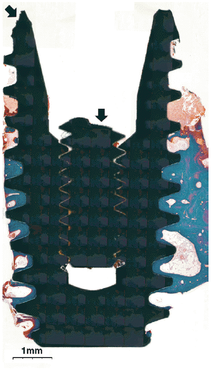
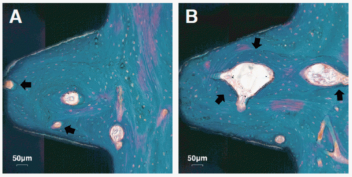
Below the fifth thread, close bone-implant contact was observed along irregular implant surface without the intervention of fibrous tissues (Fig. 8A). Upper two inter-thread spaces were filled with dense mature bone (Fig. 8B) and lower two inter-thread spaces were surrounded by band-shaped bone and marrow space (Fig. 8C). Fig. 9 provides magnified views of aforementioned the dense mature bone. Primary osteons had irregularly deposited lamellae and misaligned bone lacunae. Osteocytes were observed in interstitial lamellar bone and around bone-implant interface as well as in the primary osteons. Newly-formed bone could be distinguished by staining characteristic darker than previously-formed one. Irregular cement lines were observed at the boundary of the osteons, the interstitial lamellar bone, or the woven bone. Neither inflammatory cells nor epithelial cells were observed on the bone-implant interface or in the bone.
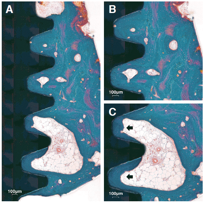 | Fig. 8Medium magnification views of bone tissue in the sectioned specimen. (A) The implant threads are well surrounded by compact mature bone (×40). (B) Close contact is continued between the implant surface and the compact bone (×50). (C) Implant threads are surrounded by band-shaped bone with marrow space (Black arrows, ×50).
|
The overall BIC along the whole implant surface was 53.1%, and the BIC when measured only in subcrestal area was 70.9%.
Go to :
Discussion
It is important to note that the histologic analysis of explanted implants in humans is not only capable of measuring BIC, but also of examining the response at the interface between the implant and bone.11 Therefore, the current study provides valuable information to observe the natural bone response to implant, especially under functional load.
Much effort has been devoted to surface modification of implant to increase both the quality and quantity of osseointegration. Studies found that the micro-roughness and porosity of implant surface facilitates not only angiogenesis but also cell migration process, activity, and function,12 so that BIC and the degree of mechanical engagement between bone and implant has been improved.2,13 Various methods were attempted to obtain such micro-roughness. In the case of surface blasted with aluminum oxide (Al2O3), there was a significant increase in the degree of bone attachment under the functional load, and in the acid-etched surface can further increase BIC.2 Therefore, most of the recent-marketed implant brands have sandblasted, large-grit, acid-etched surface to improve the BIC, and the histologic analysis of this case was also consistent with other studies using implant with above-mentioned surface.
In the present study, histologic analysis revealed that a dense bone was engaged between implant threads and newly formed bone. The BIC is influenced by various factors such as age, sex, habit, and systemic diseases affecting bone metabolism of the patient. There is lack of evidence regarding critical percentage of BIC for achieving osseointegration needed to successfully stabilize implants. The BIC in the above sample was 70.9%, which is consistent with the high BIC reported in other studies. Brunel et al reported 74% of BIC with hydroxyapatitie coated implant under 14 months loading,5 and Hayakawa et al also reported a similar results with sandblasted, acid etched implant.14 Although there was a fracture at the implant body, the remaining surface showed equally high BIC along the surface. At this moment, it is not clear why the implant fixture has broken, however, the remaining fixture appeared well-integrated. Further studies are needed to determine how much BIC is clinically necessary for successful maintenance of implant.
Various methods have been introduced to remove successfully osseointegrated implants including counter-torque ratchet, piezo tips, high-speed burs, forceps, and trephine bur. The bone removal techniques like using piezo tips, high-speed burs, and trephine bur have limitation of being traumatic and of damaging the explantation socket. Therefore, if possible, less invasive techniques like counter-torque ratchet technique and reverse screw technique are recommended for implant removal. However, if these techniques failed to remove the fracture screw, the bone removal techniques should be considered. In the present study, the abutment screw fracture occured below the shoulder of the implants, and the authors failed to engage with the removal kit. Instead, the authors chose to use trephine bur for explantation. As previously stated, the explantation procedure using trephine bur has some risk like overheating. It should be carefully planned and performed under copious irrigation with cooled saline.
A number of studies showed that unfavorable forces during mastication may result in mechanical complications of the implant (i.e. screw fracture, screw loosening, fixture fracture).15 When parafunctional load is applied to the implants due to oral habits such as bruxism, a bending force and a torque are generated the implant and the superstructure. In addition, the risks from mechanical load seem to be the highest in the molar region with one or two implant, especially the implants under loading not parallel to major axis of implant from cantilever or heavy occlusal force per se such as bruxism.16 As a result, fracture of fixture and/or screw frequently occurs in implants for single tooth replacement.17 In addition, the design of implant system,18 holes in hollow-cylinder implant,19 and reduced implant diameter20 are also responsible for fracture of the implant. In order to prevent these mechanical complications of the implant, clinicians should consider proper diameter of fixture, adjustment of oral habit, and design and size of implant prosthesis for edentulous site.
Go to :
Conclusion
In the present study, the implant appeared well surrounded by compact mature bone, and high percentage of BIC were observed. Also, the implant surface showed excellent osteoconductivity, as seen almost all threads were filled with compact bone without inflammatory cell infiltration, epithelial cell downgrowth. Within the limitation of the present case, a successful osseointegration have been achieved after a long loading period, showing a positive interaction between bone and implant.
Go to :
 XML Download