

 PDF
PDF Citation
 Print
Print


Introduction
Peri-implantitis is an infectious disease caused by bacteria from dental biofilms.1 A history of periodontitis, cigarette smoking, poor oral hygiene, and lack of periodic supportive periodontal therapy are considered risk factors for peri-implantitis.2-4 The composition of biofilm microorganisms around the implant is similar to that observed in periodontitis, which may increase the risk of peri-implantitis in patients with active periodontal disease.5 Dental biofilm formation begins with the pellicle from saliva that covers the surface of the tooth within a few minutes of mechanical cleansing. Early colonizers including Streptococcus and Actinomyces spp. directly adhere to the pellicle.6,7 As inflammation progresses, some gram-negative bacteria present in the biofilm, including Fusobacterium nucleatum, play an important role by linking several periodontal pathogens, such as Porphyromonas gingivalis, Treponema denticola, and Tannerella forsythia, to form the “red complex”.8,9 A strong association has been observed between the “red complex” and occurrence of peri-implantitis.10 Thus, early intervention of peri-implant mucositis and cleansing of early bacterial colonization play important roles in the control of peri-implant disease.
Periodontal disease begins with plaque deposition in the gingival crevice; thus, peri-implant disease progression is associated with plaque deposition on the implant abutment surface and surrounding crevice of the peri-implant area. Therefore, the properties and surface modification of the implant abutment material may affect peri-implant conditions. Zirconium oxide (zirconia) has been recently used as an implant abutment for aesthetic purposes, as its color is similar to that of natural teeth.11,12 In addition, zirconia has a high-loading capacity, and excellent corrosion resistance and biocompatibility, indicative of its suitability as an implant abutment material.13,14 Nascimento et al.15 revealed no significant difference between the bacterial species attached to zirconia and titanium disks but observed a significant difference in the degree of colonization; titanium disks presented higher counts of bacteria than zirconia disks. Another study observed no significant differences between the species and the amount of bacteria present on zirconia and titanium surfaces.16 These results indicate that zirconia, like titanium, is vulnerable to periodontal bacteria in the mouth, and that surface cleaning of zirconia abutments may be significant in the prevention of peri-implant disease.
Treatment studies of peri-implant disease have focused on the cleansing of the implant and abutment material surfaces. Limited accessibility around the implant and abutment may complicate the removal of the bacterial load with mere mechanical debridement.17 Also, mechanical devices are known to damage the surfaces of implants and abutments. Although local and systemic antibiotics have been used to manage this problem, these bacteria have shown resistance to antibiotics. Several methods have been proposed for cleaning the surface of implants, but none of these methods have demonstrated superiority over other methods.18
Photodynamic therapy (PDT) is a newly proposed method for treating periodontitis and peri-implant diseases that applies a combination of light, photosensitizers, and oxygen.19 Irradiation with light of a specific wavelength results in the transition of the photosensitizer from a low energy state to a singlet state. Clinical plaque disclosing agent erythrosine is a potential photosensitizer for the PDT of oral plaque biofilms.20 This process produces reactive oxygen species such as free radicals and singlet oxygen, which are extremely toxic to bacteria.21 Bassetti et al.22 compared the effects of PDT and local drug delivery on the treatment of peri-implant disease and found that PDT could essentially replace local drug delivery. Recently, adjunctive PDT with mechanical debridement in the management of peri-implantitis has been suggested to improve periodontal conditions.23,24 We recently evaluated the antibacterial effect of a newly devised toothbrush with light-emitting diodes (LEDs) on Porphyromonas gingivalis attached to sandblasted and acid-etched (SLA) titanium surfaces. The antibacterial effect of the LED toothbrush with erythrosine was better than that of a commercial PDT kit.25
Researchers have used an in vitro periodontitis-associated dental biofilm model for evaluating the antimicrobial effects of various treatment options for peri-implant disease because in vivo periodontitis-associated biofilm can be altered by host problems and many ethical considerations.26 Frankline et al.27 suggested that an in vitro dental biofilm model using a Center for Disease Control and Prevention (CDC) biofilm reactor (dynamic method) can create an environment similar to the saliva and gingival crevicular fluid in the oral cavity. However, few studies have reported the efficacy of an LED toothbrush on dental biofilm attached to a zirconia surface by CDC biofilm reactor. Therefore, the present study aimed to evaluate the antimicrobial effects of the LED toothbrush on periodontitis-associated dental biofilm attached to a zirconia surface prepared by static and dynamic methods.
Go to : 
Materials and methods
Zirconia disk preparation
Zirconia disks (HASS Corporation, Gangneung, Korea) measuring 12 mm in diameter and 2.5 mm in thickness were manufactured. One side of the zirconia disk was covered with a putty-type hydrophilic vinyl polysiloxane material (Eli-dent Group S.P.A., Fiorentino, Italy) so that bacteria attach to only one side of the disks. The disks were soaked in 70% ethanol for 60 s and sterilized in an autoclave. The disks were then placed in the wells of a 24-well polystyrene cell culture plate (SPL Life Sciences Co., Ltd., Pocheon, Korea) with 2 mL of artificial saliva (Kolmar Korea Co., Ltd., Sejong, Korea), and the plate was incubated at 37°C with gentle shaking for 4 h to form acquired pellicles.
Bacterial strain and culture conditions
The two strains of periodontitis-associated bacteria used in this study were Streptococcus gordonii DL1 and F. nucleatum ATCC 23726. All bacteria were grown in trypticase soy broth (Becton, Dickinson and Company, Sparks, USA) supplemented with 1 mg/mL yeast extract (Becton, Dickinson and Company, (Becton, Dickinson and Company, Sparks, USA), 5 μg/mL hemin, and 1 μg/mL menadione. S. gordonii and F. nucleatum were incubated in an anaerobic chamber (90% N2, 5% CO2, and 5% H2; Sheldon Manufacturing Inc., Cornelius, USA) at 37°C.
Static method
For biofilm formation using the static method, 25 μL of S. gordonii bacterial suspension (1 × 10⁹ CFU/mL) in 2 mL trypticase soy broth containing 5 μg/mL of hemin and 1 μg/mL of menadione was inoculated in 24-well cell culture plates (Corning Inc., New York, USA) in the presence of the zirconia disks. The plates were incubated for 24 h at 37°C in an anaerobic environment. After 24 h, the bacterial suspension was removed. In addition, 25 μL of F. nucleatum bacterial suspension (1 × 10⁹ CFU/mL) in 2 mL trypticase soy broth supplemented with 5 μg/mL hemin, and 1 μg/mL menadione was inoculated in the same 24-well cell culture plates and incubated for 5 days at 37°C to induce the formation of a multi-species biofilm. Fresh medium was added for 3 days.
Dynamic method
For biofilm formation using the dynamic method, disks were mounted on polypropylene coupon holders and placed in a CDC biofilm reactor (BioSurface Technologies Corporation, Bozeman, USA). The reactor was filled with 350 mL of S. gordonii suspension at a concentration similar to that used in the static method described above. After 24 h, the S. gordonii suspension was removed and 350 mL of F. nucleatum suspension was added at concentration similar to that used in the static method described above. The culture was incubated for 5 days at 37°C to induce the formation of a multi-species biofilm. Fresh medium was added for 3 days.
Instrumentation and quantitative evaluation of bacterial removal
After 5 days of incubation, the zirconia disks with multi-species biofilms formed using the dynamic and static methods were divided into five groups, comprising eight disks each (Table 1): negative control; commercial photodynamic therapy (PDT; Periowave system, Ondine Biomedical Inc., Vancouver, Canada) as positive control; brushing with toothbrush (B; Smart E-care, AinA Co., Ltd., Daegu, South Korea) only; brushing with an LED light (BL): and brushing with an LED light and erythrosine (BLE). The LED light comprised one red LED (630 nm, 44 mW), two blue LEDs (465 nm, 64 mW), and one white LED (550 nm, 64 mW). The disks from the PDT group were placed in methylene blue (1 mL, 100 μg/mL) for 60 s, followed by irradiation (670 nm, 160 mW) with the diode laser for 60 s. The disks from the B group were only brushed for 60 s. A brush attached electric toothbrush was applied by a constant speed and direction for desirable experimental results. The disks from the BL group were brushed with an LED light for 60 s. The disks from the BLE group were placed in erythrosine (1 mL, 22 μM) for 60 s before being brushed with an LED light for 60 s. After treatment, each disk was placed in a test tube and vortexed with 3 mL phosphate-buffered saline (PBS) and glass beads (0.15 mm diameter, DAIHAN Scientific, Wonju, Korea) for 60 s to detach the bacteria. The solution containing detached bacteria was spread directly onto trypticase soy agar plates containing 1 mg/mL yeast extract, 1 μg/mL menadione, 5 μg/mL hemin, 5% sheep blood (Hanil-Komed Co., Ltd., Seongnam, Korea), and 1.5% Bacto agar (Becton, Dickinson and Company) using a spiral plate system (IUL, Barcelona, Spain). The plates were incubated under anaerobic conditions for 96 h at 37°C. An automatic counter (IUL) was used to determine the number of colony-forming units (CFUs). The percentage of surviving bacteria was determined by counting the CFUs after incubation by dividing the number of CFUs on the treatment group disks with the number on the control group disks.
Scanning electron microscopy
Scanning electron microscopy (SEM) was used to visualize the changes in bacterial cell walls and observe the number of attached cells. The disks with attached bacteria were fixed in 2.5% glutaraldehyde for 2 h at room temperature. The fixed samples were washed 3× with PBS for 10 min each and dehydrated for 30 min in graded ethanol solutions (30%, 50%, 70%, 90%, and 100%). After critical point drying, samples were mounted on a stub, coated with gold, and observed with SEM. The surface of the disk was observed using variable pressure field emission SEM (SUPRA55VP, Carl Zeiss, Oberkochen, Germany).
Statistical analysis
The data were analyzed with a statistical program (SPSSTM 22.0, IBM Inc., Armonk, USA). Paired t-test and one-way analysis of variance with the Duncan correction were applied to assess differences in application. The level of significance was set at P < 0.05.
Go to :
Results
Table 2 presents the mean values of log CFU/mL and proportion of surviving bacteria according to static and dynamic culture methods. The mean values of log CFU/mL of biofilms in the dynamic method group were lower than those in the static group, but no significant difference was observed in the level of cell growth between the two methods. Regardless of culture method, the proportion of F. nucleatum was higher than that of S. gordonii, but not statistically significant. Fig. 1 shows SEM images of bacteria attached to the zirconia surface according to static and dynamic culture methods. No significant difference was observed in the SEM images between the two methods.
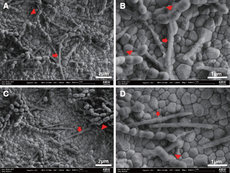 | Fig. 1Scanning electron microscopy images of attachment on zirconia surface according to culture method. (A, C Biofilm in well plate (static method), (B, D) Biofilm in CDC biofilm reactor (dynamic method). The arrowheads indicate S. gordonii and the arrows indicate F. nucleatum. CDC: Center for Disease Control and Prevention
|
Table 3 shows the mean counts of viable bacteria and percentages of bacterial reduction according to static and dynamic culture methods and treatments. Regardless of culture method, the control group showed significantly higher mean counts of viable bacteria than all of the experimental groups (P < 0.05). Among the static method groups, 93.3% and 97.8% of bacteria were removed in the BL and BLE groups, respectively. Among the dynamic method groups, the BL and BLE groups showed bacterial reductions of 93.5% and 97.1%, respectively, compared with the control groups. There were statistically significant differences among the treated groups, with the BLE group demonstrating the greatest antimicrobial effects for both culture methods.
Table 3
Counts of viable bacteria and percentages of bacterial reduction on the zirconia surface in different treatment groups
![]()
Fig. 2 shows SEM images of S. gordonii and F. nucleatum attached to the zirconia surface by CDC biofilm reactor. In all SEM images, F. nucleatum and S. gordonii adhered to the zirconia surface of the disk, and the number of total bacteria decreased significantly in the BL and BLE groups compared to the other groups (Fig. 2A - 2E). The control and B groups showed intact bacterial morphology, however, damage to the bacterial cell wall was observed in the PDT, BL, and BLE groups (Fig. 2F - 2J). Especially, complete destruction of the cell wall and smaller amounts of bacterial attachment were observed in the BLE group.
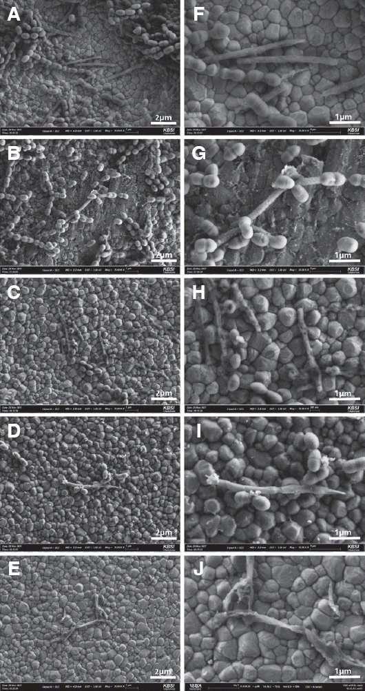 | Fig. 2Scanning electron microscopy images of S. gordonii and F. nucleatum attached to zirconia surface by CDC biofilm reactor. (A, F) control group; (B, G) brush alone group; (C, H) photodynamic therapy group; (D, I) brushing with a LED light group; (E, J) brushing with a LED light and erythrosine group.
CDC, Center for Disease Control and Prevention; LED, light-emitting diodes.
|
Go to :
Discussion
This study was designed to evaluate the antimicrobial effects of LED toothbrushes on periodontitis-associated dental biofilm attached to zirconia surfaces by CDC biofilm reactor in vitro. We used S. gordonii and F. nucleatum to form a biofilm model by CDC biofilm reactor and 24-well plate. The CDC biofilm reactor was designed to create an environment similar to the saliva and gingival crevicular fluid in the oral cavity in vitro.27 We recently compared in vitro oral biofilms made by static and dynamic methods with F. nucleatum and P. gingivalis and discovered that the dynamic method (CDC biofilm reactor) formed looser biofilms containing fewer bacteria than the static method (well plate).28 However, both methods are useful for mimicking periodontitis-associated oral biofilms. In this study, the mean log CFU/mL of biofilms in the dynamic method group were lower than those in the static group, but no significant difference was observed in the level of cell growth between the two methods. Our results concur with the previous study, even though different periodontal bacteria were used in the two studies. F. nucleatum is the most abundant gram-negative anaerobic bacteria in biofilms from healthy gingiva and increases with periodontal disease progression. This study evaluated the antimicrobial effect of an LED toothbrush on the initial biofilm that could be formed on a zirconia abutment and crown, using S. gordonii and F. nucleatum to reproduce the conditions observed in peri-implant mucositis, an early peri-implant disease.
The incomplete removal of plaque around an implant may result in bacterial settlement, leading to peri-implant mucositis. The consequences give rise to peri-implantitis, leading to supportive marginal bone loss. Thus, cleaning the initial bacterial deposits plays an important role in the prevention of peri-implant disease. Several methods have been proposed for the initial treatment of peri-implant disease in clinical settings.29 A few conventional methods, including adjunctive antiseptic rinse, powered toothbrush, and irrigation, allow patients to control plaque; however, their efficacies are unproven,30 leading to the development of new instruments such as the LED toothbrush. The LED toothbrush may have dual action for dental biofilm; brushing is essential for oral hygiene to break the biofilm and LED light may be as effective as photodynamic therapy (PDT). Park et al.31 applied brushing with dentifrice to resorbable blasting media titanium disks incubated with P. gingivalis. The authors described significantly reduced bacterial adherence, but some bacteria remained in pits on the titanium. Schwartz et al.32 applied PDT to implants with peri-implant diseases and confirmed clinical efficacy within a short time. In the current study, regardless of culture method, the BL and BLE groups showed more than 90% bacterial reduction compared with the control groups, with the BLE group showing the greatest antimicrobial effects. The SEM images of the BL and BLE groups revealed significantly decreased numbers of total bacteria (Fig. 2A - 2E), and the PDT, BL, and BLE groups revealed damaged bacterial cell walls (Fig. 2F - 2J). Especially, the BLE group demonstrated complete destruction of the cell wall and smaller amounts of bacterial attachment; this effect was stronger in the presence of the photosensitizer erythrosine. SEM examination revealed morphological alterations of the bacteria in the BL and BLE groups, which seemed to be due to the antibacterial effects of the 2 blue LEDs (465 nm, 64 mW) and the commercial PDT kit. Song et al.33 compared the antimicrobial effects of blue light on periodontal pathogens in planktonic and biofilm conditions and noted that exposure to blue light for periodontal bacteria in the biofilm state is less effective than exposure in planktonic conditions. Habiboallah et al.34 studied the photodynamic sterilizing effect of visible light in the presence of erythrosine, a photosensitizer for gram-negative bacteria, and found that visible blue LED light (440 - 480 nm, 570 mW) in conjugation with erythrosine significantly reduced bacterial viability. Thus, PDT alone may have limitations in the removal of bacteria and requires additional mechanical methods and photosensitizers. The wavelength of blue light used in this experiment failed to match the maximum absorption wavelength of erythrosine (525 nm), but contained the maximum absorption wavelength of erythrosine in the range of white light (500 - 550 nm, 22.7 mW), which exhibits an antimicrobial effect on Streptococcus spp.35 The results of present study are in accordance with those previously reported.
A limitation of this study is that only two different bacterial strains were used for biofilm formation. Roder et al.36 suggested the possibility of including as many strains or natural environmental samples as possible for the development of biofilms with characteristics similar to those under natural conditions. Hence, further studies should consider the cultivation of several species of bacteria for biofilm formation.
Go to :
Conclusion
Within the limitations of this study, the use of a blue LED toothbrush with erythrosine was shown to be more effective than conventional PDT for the removal of bacteria attached to zirconia surfaces. This method induced cell wall destruction of S. gordonii and F. nucleatum, the bacterial strains associated with early biofilm formation and early peri-implant disease. No significant difference was observed in biofilm formation between dynamic and static methods; both methods showed good reproducibility with biofilm formation on the zirconia disk surface. As tooth brushing is conventionally performed one to three times a day, an LED toothbrush would be useful patient-administered equipment for cleaning zirconia abutments exposed in the oral cavity. In addition, clinicians could apply LED with erythrosine after mechanical cleaning for the effective treatment of peri-implant mucositis.
Go to :
 XML Download