

 PDF
PDF Citation
 Print
Print


Introduction
To manage decayed lesions, conventional concept of cavity design for restoration not only focuses on dental caries prevention, but also focuses on retention and resistance of the restoration. This means that the cavity must be deep enough with design features of parallel walls and flat floors. In addition, all unsupported enamel structure should be removed. A traditional approach to control caries inevitably leads to an excessive tooth reduction.1
The concept of minimal interventional dentistry has evolved due to improved understanding of caries processes and the development of adhesion restorative materials.2 With the paradigm shift from retentive restorations to conservative restorations, less invasive cavity preparation is increasingly emphasized.3-6 From a clinical point of view, it is questionable whether fragile enamel walls without supporting dentin should be removed or preserved. It has been assumed that bonded composite will strengthen the tooth when the enamel has lost its dentin support. However, such assumption is based on clinical evaluations or clinical cases reports.7-11 Such biomechanical assumption that the composite can strengthen the teeth has not been fully verified.
Several studies have reported the effect of cavity design on fracture resistance of teeth and restoration by occlusal loading.12-14 Most works on fracture mechanism in restored teeth are related to in vivo or in vitro experimental analyses.15-18 Modern computer aided design and finite element analysis (CAD-FEA) methodologies play an essential role in biomedical investigations of clinical situations in various dental fields.19-26
When practiced in living subjects, some dental research studies are expensive and ethically doubtful. Conversely, using virtual models and simulations can improve investigation performance, reduce the cost of in vitro and in vivo experiments, and improve profitability.27
The aim of this study was to compare stress distribution and maximum von Mises stress generated in intracoronal restorations and tooth structures of mandibular molars using three-dimensional FEA method. The following independent variables were investigated: (1) type of cavity design (conventional versus minimal); and (2) type of restorative materials (composite resin versus gold alloy).
Go to : 
Materials and Methods
1. Three-dimensional solid model generation
Mandibular molar tooth was scanned using a 3-D scanner (Freedom HD, DOF Inc, Seoul, Korea). Obtained surface contours and meshes were then imported into SolidWorks 2015 software (Dassault Systems Solid-Works Corp, Waltham, USA). Threedimensional solid model of intact mandibular molar was generated using a “SCANto3D” add-in module (Fig. 1A).
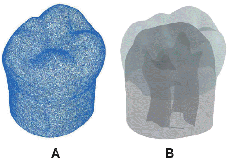
Interfacial surface between pulp chamber and dentin and interfacial surface between dentin and enamel were made by lofting technique of the CAD program according to the anatomy of natural tooth (eHuman 3-D Tooth Atlas 7.6, eHuman Inc, Fremont, USA). Once enamel, dentin, and pulp 3-D volumes were generated, Boolean operations were used to ensure congruence between related interfacial surfaces. For instance, dentin volume was created by subtracting pulp cavity volume. Enamel 3-D volume was then obtained by subtracting dentin volume (Fig. 1B). All solid models were derived from the three-dimensional solid model of the intact mandibular molar.27
2. Cavity preparation design
Based on the three-dimensional CAD model of un-restored mandibular molar tooth, inlays with conventional cavity model and composite filling with minimal invasive preparation model were made. Four 3-D experimental models were designed and created:(1) O cavity with conventional design (OR-C, OG-C models); (2) MO cavity with conventional design (MR-C, MG-C models); (3) O cavity with minimal design (OR-M model); and (4) D cavity with minimal design (MR-M model). Shape and dimensions of intracoronal restorations were taken from the literature.28
All inlays with conventional design cavities had pulpal and axial walls with at least 0.6 mm dentin thickness over the pulp while gingival walls of proximal boxes were located 0.4 mm above cementoenamel junction. The narrowest portion of the preparation was 1.0 mm faciolingually, which was located between buccal and lingual cusp tips. The cavity extended the full length of the occlusal groove, including mesial and distal pits with their radiating grooves. The pulpal wall was flat horizontally. The occlusocervical thickness of inlay was between 0.7 mm and 2.6 mm in O cavity. In MO inlay cavity, proximal boxes were extended proximally from the occlusal cavity. The shape of the proximal box was on straight lines or planes with thickness of at least 0.8 mm. Minimally invasive models (OR-M, DR-M) preserved the unsupported enamel and removed the minimum tooth structure in spherical form, which was limited to the area of dental caries lesion (Fig. 2). Volumes of restorations were 24.92 mm3 in conventional O cavity and 46.35 mm3 in conventional MO cavity. Those of OR-M model and MR-M model were 4.13 mm3 and 9.73 mm3, respectively, in minimally invasive cavity (Table 1).
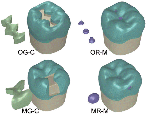 | Fig. 2Two conventional inlay models of O cavity (CGC), MO cavity (MG-C), and two minimal invasive designs for occlusal caries (OR-M) and proximal caries (MR-M) models were made.
|
Table 1
Experimental models and cavity volume for each restoration
![]()
3. Finite element analysis
In minimal cavity design groups, restorative material was composite resin. In conventional cavity design groups, two types of restorative materials were tested: (1) gold alloy (E = 95.6 GPa, υ = 0.35),20 and (2) composite resin (E = 9.5 GPa, υ = 0.24).16 Material properties of dentin (E = 18.6 GPa, υ = 0.32)21 and enamel (E = 84.1 GPa, υ = 0.3)20 were assigned. All materials were assumed to have linear, elastic, and isotropic properties. The bonding interface between dentin and composite or enamel was considered to be perfect in this experiment. Three-dimensional solid models were meshed with tetrahedral elements. The number of elements and nodes varied according to models (59,009 - 73,064 elements and 89,352 - 108,739 nodes). Fixed zero-displacement in three spatial dimensions (X, Y, and Z) was assigned to nodes at the bottom surface of the tooth, preventing rigid body displacement for all models. To simulate biting force, a total amount of 200 N load was applied vertically on the tooth at 10 occlusal contact points (5 buccal cusp points, 3 central fossa points, and each point on both marginal ridges) (Fig. 3). A static finite element analysis (FEA) was performed to predict the stress distribution generated by occlusal loading.
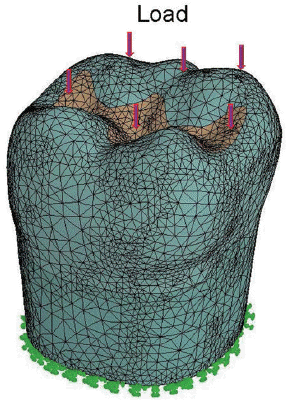 | Fig. 3A total amount of 200 N axial load was applied at 10 occlusal points: 5 points in buccal cusp area and 5 points in marginal ridges and central fossa area (model OG-C). Solid model with restoration, enamel, dentin, and pulp chamber of the mandibular molar was meshed with tetrahedral elements. Bottom of the model was fixed in all directions as a boundary condition.
|
Go to :
Results
In order to analyze stress distribution and location, all structures created were isolated from the rest of the model. For each group, peak stresses on restorative materials and abutment teeth were evaluated separately.
1. Stress distribution in restorations
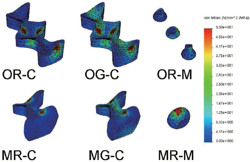
Maximum values of von Mises stress within restorations with minimal cavity design generated were significantly lower (OR-M model: 26.8 MPa; MR-M model: 72.7 MPa) compared to those with conventional cavity design (OR-C model: 397.2 MPa; OG-C model: 341.9 MPa; MR-C model: 362.5 MPa; MG-C model: 352.6 MPa).
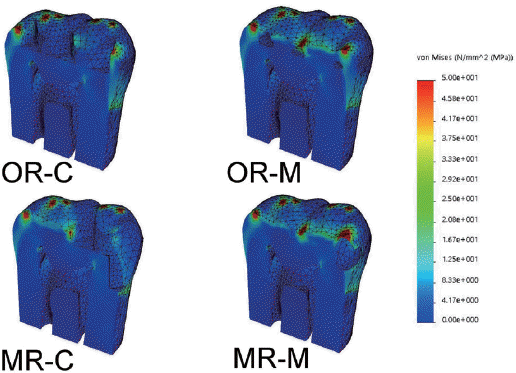
Regarding the effect of cavity design, minimal invasive designs (OR-M, MR-M) generated 5 to 10 times smaller maximum von Mises stresses than those with conventional inlay designs (Fig. 4). Regarding the effect of dental material, composite resin (OR-C, MR-C) exhibited slightly higher maximum von Mises stresses than gold alloy (OG-C, MG-C) in restorations. Gold inlay (OG-C) showed more favorable and well distributed stresses in the restoration than composite resin inlay (OR-C) (Fig. 5, Fig. 6). Overall, the order of stress intensity in restorations with conventional inlay/minimal cavity filling designs was as follows: OR-C > MR-C > MG-C > OG-C > MR-M > OR-M.
 | Fig. 4Minimal invasive cavity designs (OR-M, MR-M) produced very small maximum von Mises stress magnitude compared to conventional inlay designs in the restoration. There were no significant differences in maximum stress magnitudes within the abutment tooth among models.
|
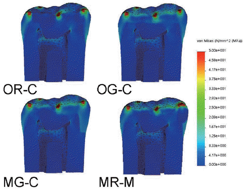
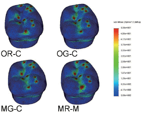
The differences in stress magnitudes over the adjacent enamel along cavosurface margins of composite restorations (OR-C, OR-M, MR-M) were distinguished. In terms of stress location, high concentrations of von Mises stress on surfaces of restorations were found near the occlusal contact areas where biting forces were applied (Fig. 7).
2. Stress distribution in abutment teeth
In tooth structure, magnitudes of maximum von Mises stresses in models with conventional design were between 372.8 MPa and 412.9 MPa, while those in models with minimal cavity designs were between 361.1 MPa and 384.4 MPa. The gold O cavity inlay (OG-C) produced the highest von Mises stress (412.0 MPa) while the composite minimal O cavity design (OR-M) generated the lowest von Mises stress (361.1 MPa) (Fig. 4). When comparing a l l experimental models, ma x imum stress values generated in the abutment teeth were close to one another (i.e., between conventional/minimal invasive cavity designs and tested restorative materials).
In terms of stress distribution patterns in enamel and dentin, similar results were observed for all experimental models. High stress concentrations were found at the enamel surface near buccal cusp tips, central fossa, and marginal ridges where axial occlusal forces were applied (Fig. 6, Fig. 7, and Fig. 8).
Go to :
Discussion
Preservation of sound tooth structures is the primary goal of restorative dentistry. Even if removal of additional dental tissue is necessary, protecting the remaining tooth structure from undesirable mechanical responses should be considered. Tooth preparation designs proposed for posterior inlay restorations have been based upon recommendations made by GV Black for cast metal and amalgam, resulting in considerable tooth structure removal with opposing walls that are parallel.28 The preparation design for an indirect restoration must satisfy a balance between preserving the tooth structure and maximizing the strength of the restoration. However, there is a problem within the concept of the original GV Black classification since it identifies the position of lesion and prescribes cavity design regardless of the size and extent of carious lesion. Recently, the Academy of Operative Dentistry European Section has considered adhesively bonded resin composites for use in direct minimal intervention approaches to restore posterior teeth, emphasizing the importance of the practice of evidence-based minimal intervention dentistry to extend the longevity of restorations.3
Masticatory loads in the posterior region are much higher than those in the anterior region. Stress concentrations can manifest themselves in various forms of clinical failures such as tooth fracture and fracture of restorative body. The main purpose of this research was to evaluate the maximum stress values and stress distribution in intracoronal restorations and the tooth after occlusal loading to identify failure possibility under various types of cavity designs and materials.
St-Georges et al.14 have reported fracture resistance of prepared teeth restored with bonded inlay for MOD preparations can weaken the teeth by approximately 59%. Under compressive load testing, composite and ceramic inlay restorations do not restore the original strength of the teeth. Removal of marginal ridges, increase in the depth and width of inlay cavity, and increased preparation in the proximal box formation are main reasons for the decrease in resistance. The current minimally invasive dentistry advocates conservative principles of cavity preparation. Small isolated lesions should be treated individually, not interconnected, as common practice for conventional inlay preparations. Furthermore, the preparation should not be extended beyond dimensions of the caries lesion so that the enamel unsupported by dentin is preserved.
The traditional approach to control caries inevitably leads to an excessive tooth reduction. FEA results from Wayne et al.26 have revealed that larger restoration volume proportion will result in higher dentinenamel stresses under static loading. This result suggests that minimal invasive cavity can produce stresses that are more favorable biomechanically. In our study, tooth structure prepared with conventional inlay designs removed five times greater volume than that with minimal invasive cavity designs (Table 1). Within restorations, minimal invasive models generated smaller peak stress while similar magnitudes of stresses were produced within the abutment tooth. Low magnitudes of von Mises stresses observed in our experiment models with minimal invasive cavity contradicted GV Black’s classical principles of cavity preparation from the biomechanical point of view. Our findings could serve as a basis for preserving as many intact tooth structures as possible (Fig. 4, Fig. 5, Fig. 6, and Fig. 7). A strengthening effect of the enamel without dentin support in minimally invasive technique could be expected in clinical situation of bonded composite restoration.
Guven et al.22 have analyzed the influence of inlay cavity design by FEA and reported that cavities with rounded corners showed less stress than those with rectangular corners due to improved stress distribution capabilities of rounded corners. The model of conventional inlay has a box-shaped cavity with sharp margins. This might have increased the maximum stress of the model in our study.
Currently, composite resin as well as metal alloy and dental ceramic represent logical options for restorations in posterior teeth. The restorative material is a factor that can affect the biomechanics during occlusal loading. Gold restorative material tends to concentrate more stress inside the inlay, resulting in lower cusp deflection than the resin.15 In our study, gold inlay (OG-C) showed well distributed smaller peak stresses in the restoration than the composite resin inlay (OR-C). Interestingly, in contrast to cavity design of inlays/filling, only a small difference was observed among maximum stresses by different restorative materials in each experimental group (Fig. 4). Thus, using proper cavity design may be more important than using a particular restorative material.
The fracture resistance of teeth restored with inlay/filling is very complex. It is impossible to include all variables encountered in the oral environment in a computer simulation.19 Although von Mises stress concentration cannot predict failure patterns in a computer simulation, higher stress concentrations are related to fracture of restorations and failure of teeth restored with inlays or filling. In oral cavity during function, teeth are loaded with complex and variable forces.
Low magnitudes stresses observed in our experiment models of minimal invasive cavity suggest preserving as many intact tooth structures as possible from a mechanical point of view. Several limitations and weaknesses of computer simulation need to be addressed in the future.
Go to :
Conclusion
Finite element analysis was performed to investigate the effect of different cavity preparation designs with various restorative materials on mandibular molar after exposure to a masticatory force. Within the limitations of this study, the following conclusions were drawn:
Models with minimal invasive designs (OR-M, MR-M) generated 5 to 10 times smaller maximum von Mises stress within restorations than those with conventional inlay designs when occlusal load was applied.
Peak stress was generated at the occlusal contact area around marginal ridges or central fossa in all models. Gold inlay (OG-C) showed well distributed and smaller stresses in the restoration than composite resin inlay (OR-C).
Lower magnitudes of von Mises stresses observed in models with minimal invasive cavity design suggest that bonded composite can strengthen the tooth when enamel has lost its dentin support.
Go to :
ORCID
Sunmi Yang https://orcid.org/0000-0002-9802-0282
Seon-mi Kim https://orcid.org/0000-0001-5103-767X
Namki Choi https://orcid.org/0000-0003-4830-8568
Jae-hwan Kim https://orcid.org/0000-0001-8088-6216
Sung-Pyo Yang https://orcid.org/0000-0003-4928-1838
Hongso Yang https://orcid.org/0000-0002-9138-4817
Go to :
 XML Download