

 PDF
PDF Citation
 Print
Print


Introduction
Dental implants for functional recovery of missing dentition present high success rates if certain factors are considered. In particular, initial implant stability plays an important role for successful clinical outcome. Implant stability can be described at two different levels: primary and secondary.1 Primary stability, defined as the mechanical engagement immediately following implant installation, is an important factor for the success of the dental implant. Primary stability is associated with the quality and quantity of the local bone, implant type, and surgical technique used. Secondary stability provides biological stability via bone formation and remodeling.2-4
Implant surfaces have been modified by various means in order to improve biocompatibility and accelerate osseointegration. Among the various methods, sandblasting and acid etching can be used to obtain excellent physical properties and may contribute to favorable bone response in biological environments. Implant design is also an important factor in achieving a successful primary stability.5-7 Tapered implants enhance primary stability by inducing cortical bone compression in the weak bony area and dispersing the force to the surrounding bone, and are therefore beneficial in immediate implant placement.8
Currently, various diagnostic methods have been suggested to measure implant stability. Resonance frequency analysis (RFA) is a noninvasive and objective diagnostic method that measures implant stability using vibration, and is the principle of structural analysis. The first study using RFA as a method to measure stability was performed in 1996.9 Integration Diagnostics AB (Savedalen, Sweden) launched the Osstell® system in 2000, which has been adopted universally. The Osstell Mentor® system has an electronic tuning fork that automatically converts kHz to an Implant Stability Quotient (ISQ) value. The device is portable, and emits signals with a force of 5 - 10 Ncm that are repeated by a transducer connected directly to the implant. The resonance frequency is then calculated from the response signal.
RFA can reveal minor differences in implant stability and is generally performed to assess the efficacy of modification of implant surface, implant configuration, and thread design, which leads to improved and accelerated osseointegration. An RFA value between 1 and 100 is obtained (degree of stability ranging from lowest [1] to highest [100]). Recently, various factors influencing RFA assessments have been determined. The quality of the bone in which the implant was installed was the most significant factor; further, the implant length affected ISQ values to a certain degree over time.10 In addition, the lateral or occlusal positioning of the probe in relation to the peg did not affect ISQ values, while mesiodistal vs. buccolingual directional assessments appeared to be a point of slight variance. Therefore, two-directional measurement may reveal more precise information than one-directional readings.
The aim of this study was to monitor the stability of two-stage submerged, sandblasted, large-grit, acid-etched (SLA) implants with tapered straight body design by applying RFA.
Materials and Methods
Ethical approval for this study was obtained from the Ethics Committee of Kyungpook National University Hospital (2015-08-022).
Study population
Twenty-six partially edentulous patients (13 women and 13 men, mean age: 54.5 years) participated in this study. The bone quality of the implant placement is described in Table 1.
Table 1
Bone quality of implant placement
| Anterior | Pre-molar | Molar | |
|---|---|---|---|
| Type I | 0 | 0 | 0 |
| Type II | 0 | 1 | 13 |
| Type III | 0 | 6 | 23 |
| Type IV | 0 | 0 | 1 |
| Total | 0 | 7 | 37 |
| 44 |
Control of periodontal disease, when necessary, was achieved by motivation and oral hygiene instruction. If required, initial periodontal therapy consisted of root scaling and planing and periodontal surgery. Patient inclusion criteria were as follows: 1) unremarkable medical history, no known allergies, and no metabolic bone diseases; 2) no heavy smoking (< 10 cigarettes/day); and 3) adequate quantity and quality of native bone in order to achieve primary implant stability. Patient exclusion criteria were as follows: 1) severe bruxism or clenching habits; 2) untreated periodontitis or periapical pathology; and 3) heavy smoking (> 10 cigarettes/day).
Implants
A total of 44 implants (OneQ-SL implants, Dentis, Daegu, Korea) were placed according to standard procedure. The implants were double thread screw, tapered (upper) and straight (middle)-shaped titanium implants with four different diameters (3.7, 4.2, 4.7, and 5.2 mm) and lengths ranging from 8 to 12 mm. All implants had an SLA surface.
Surgery
A drilling sequence and surgical procedure was applied, aiming for appropriate insertion torque (IT) and excellent initial stability. This was achieved using the following sequence: 1.8 mm guide drill, 2.0 mm twist drill, and 3.0 mm twist drill, for all sites. In cases where a wide implant was selected, the site was further prepared with 3.8 and 4.3 mm twist drills. Counter-sinking was performed to submerge the implant head in all bone qualities (Type 1, 2, 3, and 4) using the index described by Lekholm & Zarb (1985). Implants were inserted at low speed (30 rpm). Mucoperiosteal flaps were sutured with non-resorbable nylon. Systemic antibiotics (Amocla 3 × 375 mg/day, Gunil, Seoul, Korea) and analgesics (Etodin 3 × 200 mg/day, Myungmoon, Daegu, Korea) were prescribed for seven days. Sutures were removed on day 14 post-surgery.
Measurement parameters
Clinical monitoring was performed at 1, 4, 8, and 12 weeks post-surgery. Implant stability changes were evaluated over time by determination of total ISQ values at implant insertion (first OP) and healing abutment connection (second OP, 12 weeks), and the correlations between RFA and IT, bone quality, and jawbone were ascertained.
Implant survival
Clinical examination of the installed implants was performed at the second OP and two weeks after prosthesis delivery. Mobile implants were not regarded as failed implants and were not removed.
Resonance frequency analysis (RFA)
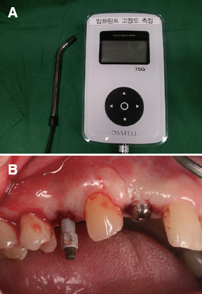
RFA was performed at implant insertion (n = 44) and healing abutment connection (n = 44) by applying the Osstell™ Mentor® (Integration Diagnostics AB, Göteborg, Sweden) (Fig. 1A). In each measurement, the implant fixture mounter and healing abutment were removed in order to access single implants. A transducer (Smartpeg™, Integration Diagnostics AB, Göteborg, Sweden) was connected to the implant (Fig. 1B).
Fig 1
(A) Resonance frequency analyzer (Osstell Mentor®), (B) Wireless transducer (Smartpeg™, Integration Diagnostics AB, Göteborg, Sweden) connected to the implant.
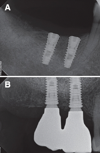
Measurement using the frequency response analyzer was performed four times from two different directions (e.g. from buccal and palatal directions). A radiographic examination was performed before and after surgery to obtain periapical radiographs and orthopantomograms (Fig. 2).
Results
Clinical observations
A total of 44 implants were inserted in 26 patients. The post-operative healing was uneventful. The survival rate of the inserted implants was 100% at healing abutment connection. No marginal bone loss or inflammation was observed.
Implant stability changes over time (total ISQ values)
The mean ISQ value of all the implants inserted was 69.4 ± 10.2 at the time of implant insertion and 81.4 ± 6.9 at healing abutment connection (Fig. 3).
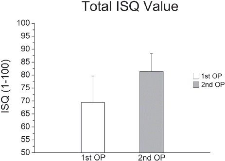
A significant difference was found between the first and second OP (P < 0.05).
Mean ISQ value for different variables
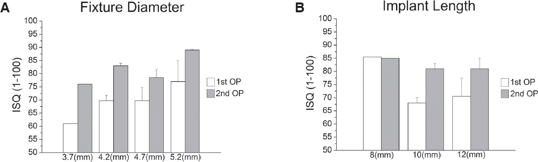
The correlations between RFA and IT, bone quality, and jawbone at implant insertion and healing abutment connection were assessed. Significant positive correlations were found between RFA and bone quality (P < 0.05; Fig. 4A) and between RFA and jawbone (P < 0.05; Fig. 4B). No significant correlations were found between RFA and IT or RFA and insertion area (P > 0.05; Fig. 4C, 4D). No significant differences were found between RFA and fixture diameter or RFA and implant length (Fig. 5A, 5B).
Fig. 4
Mean ISQ values for different variables I (immediately after implant, first OP). (A) Bone quality, (B) Jawbone, (C) IT, (D) Insertion area. Significant positive correlations were found between RFA and bone quality and between RFA and jawbone (*P < 0.05). No significant correlations were found between RFA, IT, and insertion area (P > 0.05).
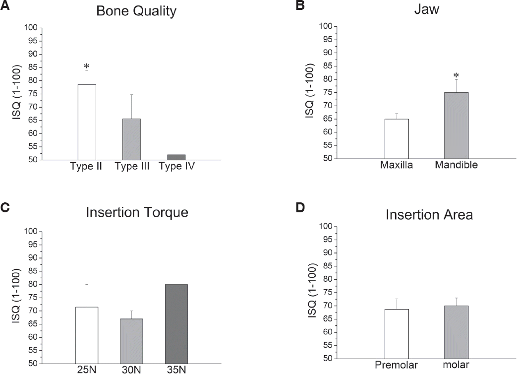
Fig. 5
Mean ISQ values for different variables II. (A) Fixture diameter, (B) Implant length. No significant correlations were found between RFA and fixture diameter or between RFA and implant length (P > 0.05).
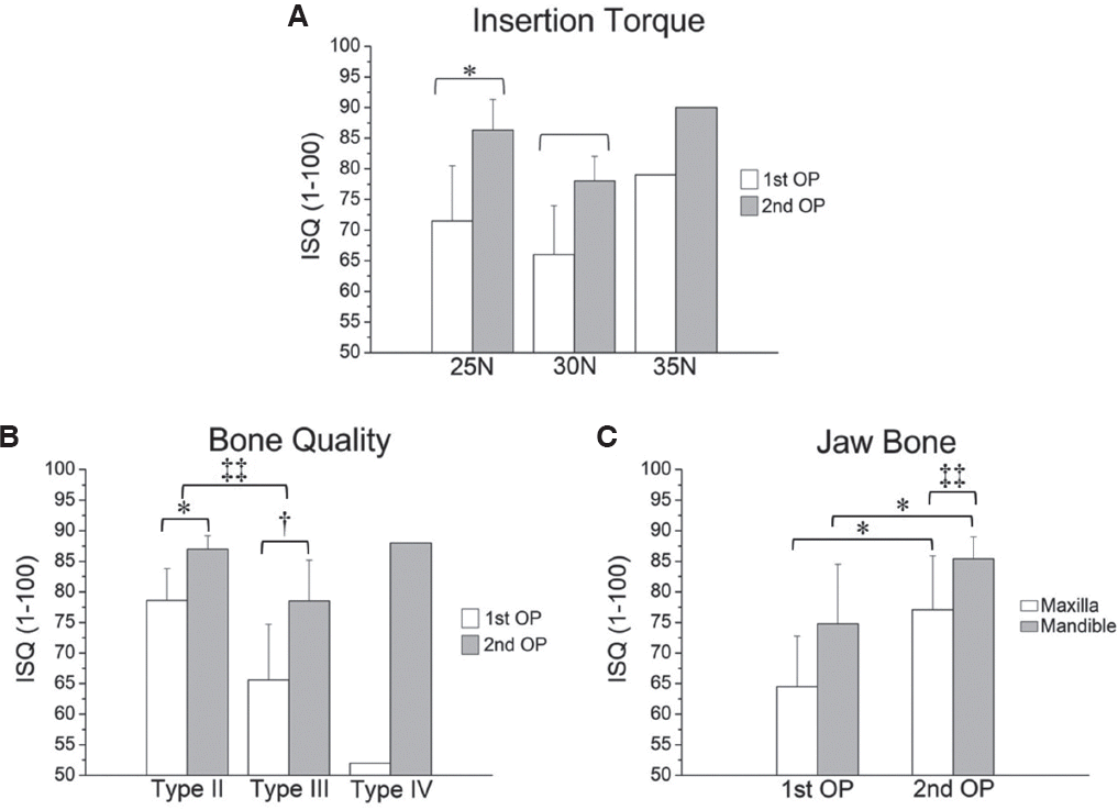
ISQ values measured at three ITs (25, 30, and 35 Ncm) were higher at the second OP (25 Ncm: 83.4 ± 5.3; 30 Ncm: 79.5 ± 7.3) than at the first OP (25 Ncm: 71.5 ± 11.3; 30 Ncm: 67.3 ± 8.7) (Fig. 6A). ISQ values of type II bone were higher than those of type III at the first OP (type II: 78.6 ± 5.2; type III: 65.6 ± 9.1) and second OP (type II: 87.0 ± 2.2; type III: 78.5 ± 6.7; P < 0.05) (Fig. 6B). Additionally, ISQ values of the mandible were higher than those of the maxilla at the first OP (mandible 74.8 ± 9.7; maxilla: 64.5 ± 8.3) and second OP (mandible: 85.4 ± 3.6; maxilla: 77.1 ± 8.8) (Fig. 6C).
Fig. 6
Implant stability changes over time. (A) IT, * correlation between the mean ISQ values of the first and second OP at an IT of 25 Ncm; † correlation between mean ISQ values of the first and second OP at an IT of 30 Ncm. (B) Bone quality, * correlation between the mean ISQ values of the first and second OP on type II bone; † correlation between the mean ISQ values of the first and second OP on type III bone; ‡‡ Significantly different ISQ values between type II and type III bone at the second OP. (C) Jawbone, * correlation between the mean ISQ values of the first and second OP in the maxilla (P < 0.05); † correlation between the mean ISQ values of the first and second OP in the mandible (P < 0.05); ‡‡ correlation between the mean ISQ values of the maxilla and mandible at the second OP (P < 0.05).
Discussion
Östman et al.11 have suggested the inclusion criterion of ISQ values for immediate loading of implant. They reported low failure rates when using ISQ 60 as an inclusion criterion for immediate loading implants in maxillae and posterior mandibles, with values above ISQ 65 indicative of immediate loading.11 In this study, it was shown that sandblasted and acid-etched implants with tapered straight body design achieved good primary stability at implant insertion, yielding ISQ values indicative of a favorable response to immediate loading (mean 69.4 ± 10.2). This suggests that sandblasted and acid-etched implants with tapered straight body design have a suitable implant thread design, body configuration, and surgical protocol to achieve primary stability for immediate loading.
For secondary stability (i.e. biocompatibility) of sandblasted and acid-etched implants with tapered straight body design, the RFA measurement was performed at healing abutment connection. The mean ISQ value of all implants was increased by 17% at implant insertion (81.4 ± 6.9). These ISQ values were higher than those reported in previous studies using the same clinical protocol.10,12-15 This indicates that sandblasted and acid-etched implants with tapered straight body design present an optimized SLA surface for improved osseointegration and are more osteoblast-compatible than the previous titanium surface modifications. Moreover, we can conclude that good initial stability of implants contributes to secondary stability and bone remodeling.
Several studies have suggested that IT values at 25 - 45 Ncm could avoid adverse micro-movement (threshold level between 50 and 100 μm) under loading, thus allowing the osseointegration healing process to commence.16,17 It is clear that a high IT provides the clinician a degree of certainty regarding the initial stability of the implant. However, an excessively high IT could increase overpressure to the surrounding bone and lead to micro-fracture of the bone, with resorption in the cortical area and a delayed healing process. In this study, the mean IT values of sandblasted and acid-etched implants with tapered straight body design were 28 Ncm. Therefore, the surgical protocol for implant insertion and the implant design were suitable for primary stability. There was no significant difference in ISQ values between 25 Ncm and 30 Ncm (P > 0.05).
Lai et al.18 reported that primary stability is affected by bone quality. Their study on 104 ITI SLA implants demonstrated significantly higher ISQ values for implants in type I bone than in type IV bone. In addition, Turkyilmaz et al.19 found a strong correlation between bone density and ISQ values. The highest ISQ value (36.1 kHz) in this study was for an implant inserted into type I bone; in contrast, an ISQ value of 9.9 kHz was for an implant in type IV bone by Huang et al.20 In this study, a significant correlation between bone quality and ISQ value at the time of implant insertion was observed for type II bone and type III bone (P < 0.05). There was also a significant difference between bone quality and ISQ value at the time of the second OP (P < 0.05). In this study, an ISQ value of 52 was obtained at the time of implant insertion into type IV bone, and an ISQ value of 88 was obtained at healing abutment connection. These results suggest that the stability of implants inserted in weak bones can be improved over time. This is consistent with a report by Friberg et al.21
Bischof et al.22 stated that implant length, position, and diameter did not affect primary stability. Regarding implant diameter and length and RFA, Horwitz et al.23 reported no correlation between fixation length and RFA; however, they suggested that larger implant diameters were associated with higher ISQ values. The current study is consistent with their results.
Several studies9,24,25 reported that the use of longer and wider implants increased primary stability due to the increased bone-to-implant contact surface area. However, our findings did not corroborate this observation.
Bone quality and implant stability are lower in the maxilla than in the mandible. According to several reports,23,26,27 primary stability is higher in the mandible than in the maxilla, a result also observed in the present study (P < 0.05).28 In addition, Nedir et al.29 reported that the value of ISQ was < 60 in the maxillary implants and > 60 in the mandibular implants. Regarding implant position (premolar and molar), we found no differences from the data published by Balleri et al.27
 XML Download