

 PDF
PDF Citation
 Print
Print


Introduction
The residual periodontal pocket of > 5 mm after the completion of active periodontal therapy is associated with an increased risk of disease progression.1-3 In addition, intrabony defects have been shown to worsen the long-term prognosis of the teeth.4 To treat intrabony defects, several approaches, including scaling root planing (SRP), open flap debridement, and resective and regenerative surgery, have been employed for several decades.5,6
Several bone graft materials, bioactive materials and growth factors can be adopted for regenerative surgery.7
The use of an enamel matrix derivative (EMD), an extract of the enamel proteins including amelogenins of various molecular weights, would generate fewer post-operative complications.8 Systematic reviews of several clinical trials have shown that some of these materials, when used in conjunction with surgical approaches designed to facilitate maximal preservation of soft and hard tissues, may indeed result in superior clinical outcomes.9 Xenografts can augment the effects of EMD in reducing the probing pocket depth (PPD), improving the clinical attachment level (CAL), and promoting defect filling compared to the EMD alone or open flap debridement in the treatment of intrabony periodontal defects.10 Despite the advantages of regenerative therapy, the achievement of primary soft tissue closure and revascularization are difficult, particularly in the interdental area. After periodontal surgery, a soft tissue crater is common in the interdental area, which is problematic for patients and periodontists. For better vascularization, several incision or surgical techniques [i.e. simplified papilla preservation flap (SPPF)11 etc.] have been proposed, but interdental soft tissue depression is still problem.
A periosteal graft in intrabony defects has the potential to stimulate bone formation and reduce the PPD when used as a graft material and barrier membrane.12,13 Moreover, the periosteal graft can augment a soft tissue volume in the field of periodontal surgery. This article reports 3 cases of the additional use of a periosteal membrane with a xenograft and EMD in interdental intrabony defects.
Case description
Case 1
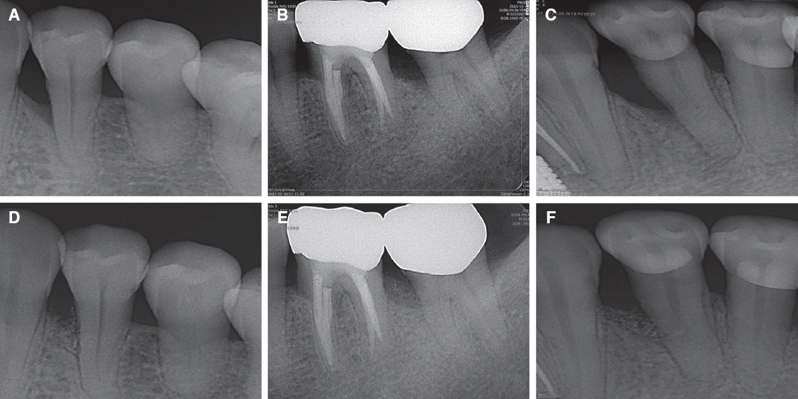
A 46-year-old woman with the chief complaint of dull pain in the mandibular left first premolar (#34) visited the Department of Periodontology, Pusan National University Dental Hospital. The patient had no remarkable systemic disease affecting the dental condition. A clinical examination revealed 7 mm PPD and 2 mm gingival recession on the mesiobuccal aspect on #34 (Table 1) and no tooth mobility. The gingival depression was noticeable at the mesiobuccal site of #34 and the gingival biotype was the thin type (Fig. 1A). In the periapical radiograph, an intrabony defect was detected on the mesial side of #34 (Fig. 2A).
Table 1
Comparison of the clinical parameters of 3 cases (mm)
Fig. 1
Regenerative surgical sequence with additional use of autogenous periosteal membrane graft combined with xenograft and Emdogain®. (A) Baseline PPD 7 mm and recession 2 mm. (B) After flap reflection, 2-wall intrabony defect with 6 mm depth. (C) Autogenous periosteal membrane graft combined with xenograft and Emdogain®. (D) Primary closure of the overlying flap.
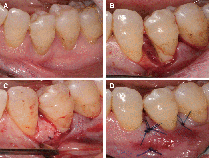
Fig. 2
Periapical radiograph before surgery (A, B, C) and 6 months after surgery (D, E, F). Note the resolution of intrabony defect after surgery (D, E, F).
Regenerative surgical therapy was considered after the initial periodontal treatment, including oral hygiene instruction and SRP. On the other hand, after regenerative surgery, the depression of the interdental papilla would be inevitable because the gingival biotype was thin and the location of the intrabony defect was interproximal. The patient was concerned about the wide embrasure and unaesthetic appearance. Therefore, several alternatives for regenerative surgery were considered and a decision was made to adopt an additional periosteal graft with a bovine bone graft and EMD (Emdogain®, Straumann, Basel, Switzerland).
All the surgical procedures were performed by the same periodontist (HK). After local anesthesia, access to the defect was achieved using a sulcular incision, SPPF and limited mesiodistal extension of the buccal and lingual flap of the one tooth neighboring the defect, and no vertical releasing incisions were made (Fig. 1B). After minimal flap elevation, a 2-wall intrabony defect was confirmed and the defect size was 3 (width) × 6 (depth) mm (Fig. 1B). The root surface and intrabony defect were debrided thoroughly and the root surface was conditioned using 24 % ethylenediaminetetraacetic acid (PrefGel, Straumann) for 2 minutes. After rinsing with saline, EMD was applied to the root surface and the defect was filled with a combination of EMD + deproteinized bovine bone mineral (DBBM, Bio-Oss®, Geistlich, Wolhusen, Switzerland).
A 3 × 14 mm sized and 1.0 mm thick autogenous periosteal graft was harvested from the palatal area. Briefly, after application of local anesthesia, a splitthickness flap “trap door”, which consisted of 1 horizontal and 2 vertical incisions, was elevated to access the donor graft tissue. After elevating the split-thickness flap, 4 incisions (mesial, distal, coronal, and apical) were made to the bone surface. Connective tissue with periosteum was obtained for use as a biologic barrier membrane. The graft was positioned over the combination of EMD + DBBM, stuck under the buccal and lingual flap and sutured with the overlying flap. Finally, the overlying flap was repositioned to cover as much of the periosteal graft as possible (Fig. 1C, 1D).
Amoxicillin 500 mg and acetaminophen 500 mg were administered three times a day for 3 days. The patient was instructed to avoid mechanical cleaning in the surgical area and to rinse with 0.12% chlorhexidine (Hexamedine®, Bukwang pharm, Seoul, Korea) twice a day for 1 month. The sutures were removed 14 days after surgery and supportive postoperative care was carried out monthly after surgery for 6 months. At 6 months after surgery, clinical and radiographic examinations were carried out.
The healing was uneventful and the volume and morphology of the interdental soft tissue was improved at 6 months after surgery (Fig. 3D). The PPD was reduced and the clinical attachment level was gained (Table 1). In addition, the graft material appeared to be well mixed with the surrounding bone (Fig. 2D).
Case 2
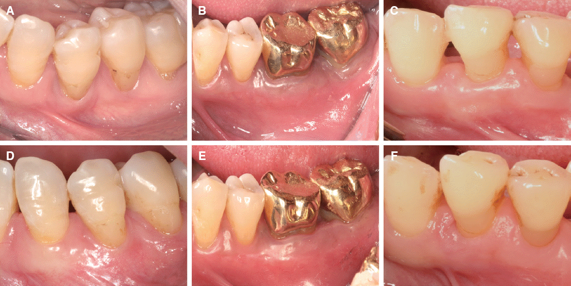
A 53-year-old man with a non-contributory medical history presented at the Department of Periodontology, Pusan National University Dental Hospital, with the chief complaint of dull pain of the mandibular left second molar (#37). He smoked approximately 10 cigarettes per day. The PPD was 7 mm (mesiolingual) and 5 mm (mesiobuccal) and a 1 - 2 mm gingival recession was detected at the mesial side of #37 (Fig. 3B). A radiographic examination revealed an intrabony defect with a moderate depth at the mesial side of #37 (Fig. 2B). An additional periosteal graft with a bovine bone graft and EMD were selected because the clinical situation and defect morphology were similar to those of case 1. After flap elevation and thorough debridement, a 2-wall intrabony defect (depth: 5 mm, width: 5 mm) was detected. An autogenous periosteal membrane (size: 5 × 14 × 1.0 mm) was obtained from the left palate and EMD + DBBM + periosteal membrane were applied using the same protocol of case 1.
Case 3
A 60-year-old woman presented to the clinic complaining of dull pain in the mandibular left first premolar (#34). The clinical and radiographic examinations revealed an intrabony defect and a PPD of 5 mm (mesiolingual), 4 mm (mesiobuccal) and a 2 mm gingival recession at the mesial side on #34 (Fig. 2C, 3C, Table 1). The same regenerative surgical protocol was selected because of the similar situation in the preceding 2 cases. A 3-wall intrabony defect (depth: 8 mm, width: 5 mm) and local factors were observed after flap elevation.
Discussion
Several problems, such as gingival recession and soft tissue crater are inevitable after periodontal surgery, particularly in regenerative surgery. Moreover, if the target site is the interproximal area, it would be more severe than in the buccal or lingual surface because the interdental papilla and connective tissue in the col area are devoid of adequate blood supply and healing capacity. To overcome these problems, several approaches (minimally invasive surgical technique,6 SPPF,11 single flap approach14 etc.) have been introduced. Despite the careful minimally invasive surgeries, in which a simplified papilla preservation flap was used, interproximal wound dehiscence with membrane exposure occurred in the majority of the regenerative surgical treatment.15 Regenerative surgery of interproximal intrabony defect is very difficult to perform, particularly in esthetics sensitive areas.
Moreover, meta-analysis of the effects of membrane exposure or wound dehiscence on the clinical outcome, showed that the sites with an exposed membrane had a negative effect on the regeneration such as the gain of the clinical attachment level.16 Bioactive materials and growth factors are introduced to boost the effects of regenerative surgery and although the actual clinical advantages are still uncertain, the EMD would generate fewer post-operative complications and better soft tissue healing.8 The soft tissue healing was more favorable and speedy when an EMD was added to the regenerative periodontal surgery from the author’s clinical experiences.
Previous studies17,18 on the use of autogenous periosteal graft as barriers in intrabony defects reported greater approximately 1mm CAL gain over sites treated by open flap debridement procedure alone. A periosteal graft in intrabony defects has the potential to stimulate bone formation and reduce the PPD when used as a graft material and barrier membrane.12,13 In these cases, EMD and autogenous periosteal membrane were adopted to enhance the outcome of regenerative surgery and soft tissue volume. The healing of 3 cases was uneventful and the morbidity and discomfort of the patients was minimal. Six months after surgery, the PPD had decreased by 2.83 mm, 1.17 mm, and 1.83 mm, respectively. In addition, the depth of the intrabony defect decreased significantly according to the radiography and the clinical attachment level gain were 3.16 mm, 1.50 mm, and 1.00 mm, respectively. After this regenerative surgical protocol, not only the gingival margin didn’t depressed severely, but also the soft tissue volume augmented aesthetically. These findings are in agreement with the results of a previous study.19
Although the trend of surgical treatment shifts to a minimally invasive surgery nowadays, the additional use of an autogenous periosteal membrane and EMD has a beneficial effect on regenerative surgery in the interdental area. In particular, in esthetic sensitive areas and the thin gingival biotype, these treatment options would be helpful for overcoming the unaesthetic problems after regeneration periodontal surgery.
 XML Download