

 PDF
PDF Citation
 Print
Print


Introduction
The main cause of inflammatory periodontal disease is functions of conditionally pathogenic microflora of oral cavity. Reduction of pathogenic bacteria and plaque is needed for periodontal treatment because the susceptible host for periodontal disease is difficult to be changed. Therefore, pathogenic bacteria and plaque must be decreased to achieve successful periodontal treatment.
It has been convincingly demonstrated that long-term stability of the clinical benefits obtained by periodontal therapy can only be achieved if a cause-related treatment is followed by effective periodontal maintenance care.1 Within maintenance care, it has also been demonstrated that self-performed plaque control is crucial to attain the best long-term result after the periodontal therapy.2 The daily use of a toothbrush is the most dependable way of achieving oral health benefits for all patients. For efficient plaque removal, toothbrushes vary in size and design as well as in length, hardness, and arrangement of the bristles.3
The use of low-level visible or near-infrared (IR) light from lasers for phototherapy, which is called low-level laser therapy (LLLT), has been the mainstream of phototherapy since the invention of lasers.4 But LLLT has also some limitations for personal use because of the complicated clinical setting, restrictions in wavelengths, and a limited area for light application. Besides, the narrow focal beam of lasers can be harmful for the eye and damage the tissue in our body.
Light-emitting-diode (LED) arrays were suggested for phototherapy as an effective alternative to LLLT. It was reported that the effect of light from LEDs would be same as that from lasers for therapeutic results.5 The main advantage of LEDs over lasers is that LED systems allow a larger area to be treated in a short time, with a large bandwidth (at several wavelengths), while typical laser systems irradiate only small spots.6
LEDs were applied by National Aeronautics and Space Administration (NASA) as a light source for growing plants in space stations by providing light energy for photosynthesis. NASA also found that the red part of the visible spectrum was useful for photosynthesis. Moreover, the red and IR light could also be absorbed by the human cells, modifying cellular functions, promoting cell survival, increasing the regeneration of tissue, wound healing, as well as curing inflammation and achieving pain relief.7-9
There are many previous studies about laser and LED, but the study which reported the effect of toothbrush with LED has not been reported.
The aim of the present study was to compare clinical antiplaque and antigingivitis effect between LED electronic toothbrush and electronic toothbrush without LED for gingivitis and mild periodontitis patients.
Go to : 
Materials and Methods
1. Patient selection
This study was performed at the department of periodontology, School of dental medicine, Dankook University, Korea. The study was reviewed and approved by the Institutional Review Board (IRB) of Dankook University in 2012 (IRB No. H-1203/003/ 001). To be included in this study, patients had to: have a minimum of 20 sound, natural teeth; have a negative history of antibiotic use during the preceding 2 weeks; have a mean Turesky-Quigley-Hein plaque index (PI) of at least 1.5; have a mean Löe- Silness gingival index (GI) of at least 1.0. Subjects with severe periodontal disease, orthodontic appliance, removable partial denture, intraoral lesion, or systemic disease that might directly affect periodontal condition were excluded. The participant flow for the study is shown in Fig. 1, according to the Consolidated Standards of Reporting Trials (CONSORT) group statement. A total of 30 patients participated in this study and were randomly assigned in 1:1 ratio to the experimental groups and control groups, respectively. All subjects signed an informed consent after nature of the study was fully explained to them. Inclusion and exclusion criteria were shown in Table 1.
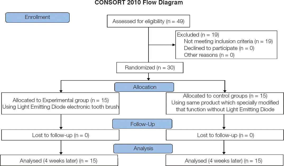 | Fig. 1Consolidated Standards of Reporting Trials (CONSORT) flow diagram for individual randomized, controlled trials of effect of Light Emitting Diode tooth brush.
|
Table 1
Inclusion and Exclusion criteria
![]()
2. Experimental material
Experimental group used LED electronic tooth brush (PROMEDIC-2010A, PROCARE Co. Ltd., Cheonan, Korea) which has red and white LED within its head, and control group used same product which specially modified that function without LED. Disclosing solution (GUM Red cote, Sunstar Butler, Chicago, USA) was used for plaque index evaluation. During experimental period, patients didn’t used any other self-performed plaque control device, had professional maintenance and only use toothpaste which didn’t have advantageous effect on gingival condition. (Closeup menthol fresh, Unilever, Ho Chi Minh, Vietnam)
3. Clinical parameter
Patient were evaluated PI and GI. Plaque accumulation was evaluated with PI. This index emphasizes the difference in plaque accumulation in the gingival third of the tooth and tends to overscore the incisal half of the crown, at the expense of the gingival margin. The scoring system is as follows:
0 = No plaque;
1 = Separate flecks of plaque at the cervical margin of the tooth;
2 = A thin continuous band of plaque (up to one mm) at the cervical margin of the tooth;
3 = A band of plaque wider than one mm but covering less than one-third of the crown of the tooth;
4 = Plaque covering at least one-third but less than two-thirds of the crown of the tooth;
5 = Plaque covering two-thirds or more of the crown of the tooth.
Gingivitis was evaluated GI. The gingival index was designed to estimate different degrees of inflammation in marginal gingiva. Severity of inflammatory change is scored from zero to three, as follows:
0 = absence of inflammation;
1 = mild inflammation; slight change in color and little change in texture;
2 = moderate inflammation; moderate glazing, redness, edema, and hypertrophy; bleeding on pressure;
3 = severe inflammation; marked redness and hypertrophy; tendency to spontaneous bleeding; ulceration.
4. Experimental design
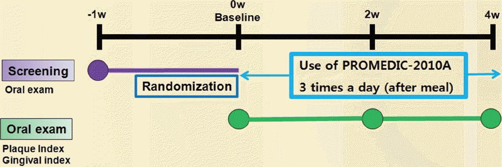
Clinical parameters (GI, PI) were measured at the baseline, 2 weeks and 4weeks later. During experimental period, patients used LED tooth brush and given toothpaste. Patients were requested not to change their lifestyle and toothbrush method. Experimental protocol is described in Fig. 2 and Table 2.
5. Statistical analysis
To compare the difference of control group and test group as time goes by, Wilcoxon signed rank test was performed. And to compare of clinical parameter among baseline, 2 weeks and 4 week later in each group, Mann-Whitney test was performed. These analyses were performed by SPSS software (release 18.0, SPSS Inc. Chicago, USA). A P value of P < 0.05 was considered statistically significant.
Go to :
Results
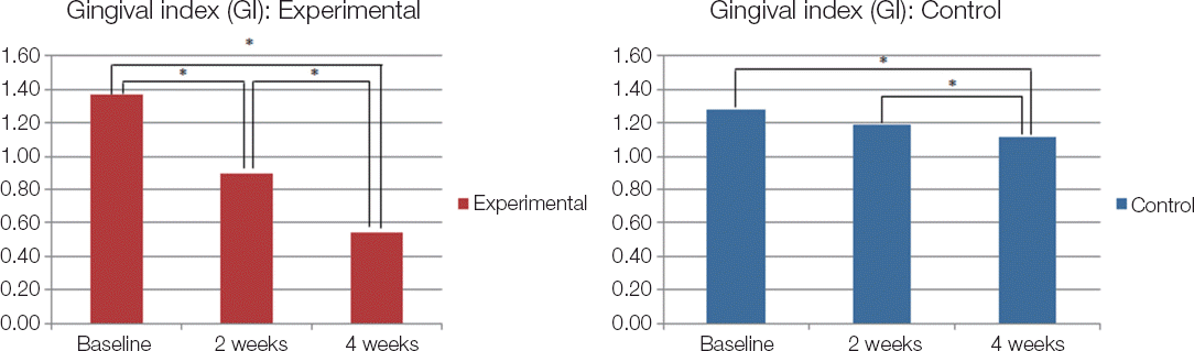
1. Change of Gingival index (GI)
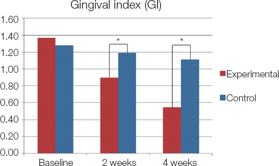
The mean GI in experimental group was 1.37 and in control group was 1.28 at baseline. Experimental group showed statistically significant differences at all the time. Control group didn’t show significant differences between baseline and 2 weeks, but others showed statistically significance (P < 0.05, Fig. 3). Lower GI values detected at 2 weeks and 4 weeks later in experimental group than control group (P < 0.05, Fig. 4).
2. Change of Plaque index (PI)
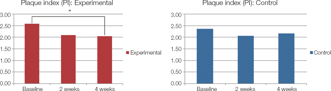
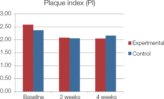
The mean PI in experimental group was 2.59 and in control group was 2.37 at baseline. Experimental group showed statistically significant differences between baseline and 4 weeks later. Control group didn’t show significant differences at all the time (P > 0.05, Fig. 5). Lower PI values detected at 4 weeks later in experimental group than control group, but not statistically significant (P > 0.05, Fig. 6).
Go to :
Discussion
Among the self-performed plaque control methods, daily use of a toothbrush is the most dependable way of achieving oral health benefits. Löe et al. reported that the time necessary to develop gingivitis varied from ten to twenty-one days, and institution of oral hygiene resulted in healthy gingival conditions and re-establishment of the original bacterial flora.10
In present study, patients were requested not to change their lifestyle and toothbrush method. And also they didn’t used any other self-performed plaque control device and had professional maintenance during experimental period. The reason is to eliminate the possibility of the change of GI or PI by scaling and oral hygiene institution and to evaluate only effect of LED.
In recent years, several reports on the benefits of using LEDs operating at several wavelengths, both in vitro and in vivo, for both normal and pathologic conditions have been published. Lee et al. reported that blue and red LED phototherapy is an effective, safe, pain-free, and easy-to-perform treatment for mild to moderately severe acne vulgaris.11 Leticia et al. reported that using infra-red LED to patient who treated with chemotherapy was a safe and effective method for preventing oral mucositis.12 Whelan et al. reported that the NASA LED effects the expression of genes involved in wound healing and possibly pain modulation thus enhancing the healing process. 13 De Sousa reported that The use of green and red LED light is effective in increasing fibroblastic proliferation.14
The toothbrush using in present study had red LED and white LED. As described before, the red LED penetrates deep into the skin and can be absorbed by the human cells. The red radiation from LEDs has been reported effective for wound healing, pain relief, skin rejuvenations, and curing inflammation. 15-18 The white LED was combination of blue, red, and green light sources. These reports were related with our research, using toothbrush with LED decrease gingival inflammation.
In present study, GI decreased statistically significance after 2 weeks and 4 weeks in both groups. The results effected by vibration of electronic toothbrush. However, compare of GI change between experimental and control group, lower GI values detected at 2weeks and 4 weeks later in experimental group than control group. The result supported that LED effected to decreasing GI.
Compare of PI change between experimental and control group didn’t show statistically significance. It seems like that LED didn’t effect to reducing plaque accumulation. However, change of PI between at baseline and at 4 weeks showed statistically significance and decreasing consistently. It was able to infer a conclusion that using toothbrush with LED for a long time can show significant change of PI.
There are a few limitations in the present study. The sample size is quite small to show the statistical power, and the short period of research. Further comparative studies with larger sample size should be conducted besides the longer period of research, so as to increase the scientific and statistical power.
Go to :
Conclusion
Based on these results and within the limits of this study, the electronic toothbrush with LED could reducing gingivitis in a short period and infer that decreasing plaque accumulation in a long period. However, longer research period and larger sample size will be needed to draw more definite conclusions on the reliability of toothbrush with LED.
Go to :
 XML Download