Journal List > J Dent Rehabil Appl Sci > v.33(2) > 1159967

고정성 보철물의 적합도에 대한 실험적 평가방법

초록

임상적으로 고정성보철물의 적합도는 성공적인 수복을 위해 필수적인 요소이다. 보철물의 적합도는 크게 변연적합도와 내면적합도로 분류되며, 이를 평가하기 위한 방법으로는 현미경 변연 측정법, 절단면 측정법, 실리콘 복제법, 3차원 스캔데이터 중첩법, 무게 측정법, 마이크로 CT 측정법 등 다양한 방법들이 소개되고 있다. 이에 본 연구에서는 기존의 문헌들을 바탕으로 각 방법들의 장, 단점을 비교분석함으로써 디지털환경에서 보다 편리하고 정확한 측정법을 제시하고자 한다.

Abstract

Clinically, the fit of fixed prosthesis is an essential element for successful restoration. The fit of prosthesis is largely classified into marginal fit and internal fit, and various methods to assess these have been introduced including microscopic margin measurement, cross-sectional measurement, silicone replica technique, 3-dimensional scanning data superposition, weight technique and micro CT scanning. Thus, this study is aimed at proposing a more convenient and accurate measurement method of fits in a digital environment by comparatively analyzing the advantages and disadvantages of each known method based on existing literature.

서론

치과환자들은 치아우식, 치주질환, 교정상담 등 여러가지 이유로 방문한다. 국내 한 치과대학병원의 환자 분석에서 교정의 이유로 내원한 환자 다음으로 치아우식을 이유로 내원한 환자의 빈도가 높았다.1 심한 치아우식으로 인한 근관치료 또는 발치, 치주질환으로 인한 치아상실 등의 치료 중 하나로 보철 진료를 하게 된다. 대다수 환자들이 궁금해 하는 것은 보철물의 예후이고, 그 보철물의 예후를 결정하는 중요한 요소는 내면과 변연의 적합도라 할 수 있다.
보철물의 시적을 평가하는 방법에는 여러 가지가 있다. 내면 및 변연적합도 검사, 인접면 검사, 교합 검사 등이 있고, 그 중 변연적합도는 보철물의 성공여부를 결정하는 중요한 요인 중 하나이다.2 변연적합도가 낮은 경우에는 미세 누출 증가, 지각 과민증 발생, 치태 침착 증가로 인하여 치아우식증이나 치주염이 발생하게 된다. 결과적으로 골 소실 등으로 인해 치아 상실 가능성을 증가시킬 수 있다. 그리고 보철물 적합성 향상을 위해서는 적절한 합착재 공간을 부여한 내면 적합도 또한 중요하다.3-5 Holmes 등은 1989년도 논문에서 변연적합도 평가를 Fig. 1과 같이 설명하였다.6
Fig. 1
Casting misfit terminology.
JDRAS_33_063_fig_1.tif
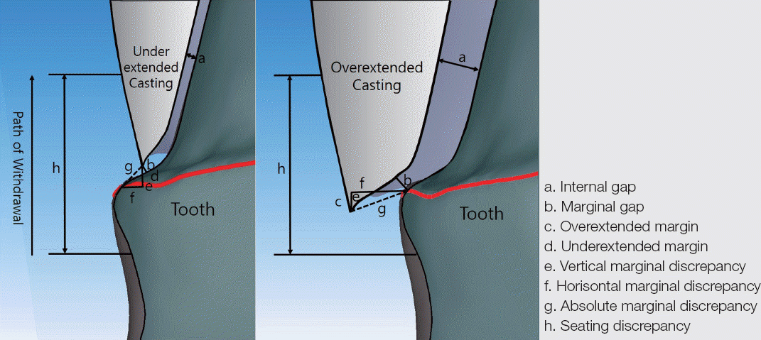
내면간격(internal gap)은 보철물 내면에서 지대치 축벽까지의 수직거리(a), 변연간격(marginal gap)은 변연부에서 보철물과 지대치 사이의 수직거리(b), 부족한 크기의 보철물(underextended casting)이나 과도한 크기의 보철물(overextended casting)의 과부족길이(c, d), 절대변연간극(absolute marginal discrepancy)은 보철물 변연에서 지대치 변연사이의 거리(g), 그리고 절대변연 간극의 수직벡터성분을 수직변연간극(e), 수평벡터성분을 수평변연간극(f)이라고 한다. 변연 적합도는 보통 변연간격 또는 절대변연간극으로 측정한다.1,7,8
임상적으로 받아들여 질 수 있는 변연간격과 내면간격에 대한 연구는 이전부터 진행되어 왔다. 변연간격에 대해 언급한 논문을 보면, Christensen은 적정한 변연간격을 40 μm,9 Sorensen 등은 50 μm 이하여야 한다고 보고하였고,10 Assif 등은 평균 약 140 μm,11 McLean 등은 구강 내 5년 이상 있었던 1000개의 보철물을 조사하여 120 μm가 임상적으로 받아들여질 수 있다고 보고하였다.12,13 내면간격에 대해 언급한 논문을 살펴보면, Grey 등은 완전도재관의 내면간격을 123 - 154 μm,14 Tuntiprawon과 Wilson은 전부도재관의 경우 내면간격이 평균 73 μm 일 때 가장 큰 압축강도를 가진다고 했다.15 내면 적합도는 시멘트 두께와도 연관되어 있다고 볼 수 있다. Jorgensen과 Esbensen은 140 μm 이상인 경우 유의한 유지력 감소를 보인다고 하였다.16 Passon 등은 시멘트 두께가 약 151 μm인 경우에도 유지력의 변화는 없다고 하였다.17 Sorensen 등과 Molin 등의 논문에서는 200 - 300 μm의 내면간격이 임상적으로 받아들여질만 하다고 하였다.18,19 지금까지 많은 논문들이 변연 및 내면 적합도에 대해서 연구하고 결과를 보고했다. 그 과정에서 다양한 방법들이 제시되어왔다. 본 논문에서는 보철물의 변연 및 내면적합도를 측정하는 실험실 평가법을 소개하고, 각 방법의 장, 단점을 알아보고자 한다.

문헌고찰

1. 절단면 측정법(Cross-sectional method)

절단면 측정법은 제작된 주 모형에서 완성된 보철물을 시적하여 고정 또는 합착한 후 원하는 방향으로 절단하여 절단면에서 변연간격 및 내면간격을 계측하는 방법이다. 계측에는 광학현미경을 이용하게 되는데, 대게 50 배율의 확대를 사용한다. 이 방법은 절단하는 방향이 정해져 있지 않고 원하는 부위 어디서든지 접근하여 절단 후 관찰이 가능한 장점이 있다.20 하지만 절단 시 가해지는 힘에 의한 변형이 있을 수 있고, 절단 이후에는 시편의 재사용이 불가능하기 때문에 장기적인 분석이나 비교를 할 수 없으며, 절단면에 한정된 분석만이 가능하여 보철물 전체의 적합성을 대신할 수 없다는 것이 단점이다.21,22 또한 이 측정법은 실리콘 복제 방법(silicon replica technique)과 조합하여 사용이 가능한데, 두 방법을 이용한 적합도 측정의 정확성은 연구가 더 필요한 것으로 보인다.22-25

2. 실리콘 복제 방법(Silicon replica technique)

인상채득에 사용되는 흐름성이 좋은 인상재를 사용해서 보철물 내면에 주입 후 주 모형에 일정한 힘으로 시적하여 보철물의 내면관계를 알아보는 방법이다. 내면관계를 복제한 실리콘 인상재의 두께를 측정해서 보철물의 적합도를 평가할 수 있다. 내면 적합도를 알아보기 위한 과정에서 보철물 내면의 인상재를 그대로 제거할 경우 변형 및 찢김의 위험성이 있으므로, 흐름성이 낮은 인상재를 내면에 주입하여 경화 후 제거하거나, 보철물을 조심스럽게 제거한 뒤 외면에 흐름성이 낮은 인상재로 채운 뒤 제거하는 등 다양한 방법을 이용하여 필요한 부분을 측정할 수 있다.26-32 앞서 설명한 절단면 측정법처럼 원하는 방향에서 제거한 인상재를 절단한 후, 필요한 부위의 보철물 내면간격 및 변연간격을 계측할 수 있다.33,34 계측하는 방법에는 절단면 측정법과 같이 광학현미경으로 직접 필요한 부위를 측정하거나(Fig. 2), 3D 스캐너를 이용해서 주 모형에 복제된 인상재와 인상재를 제거한 원래의 주 모형을 스캔한 후 이 두 개의 이미지를 중첩하여 복제된 인상재만을 얻는 방식(Fig. 3)이 있다.35-37
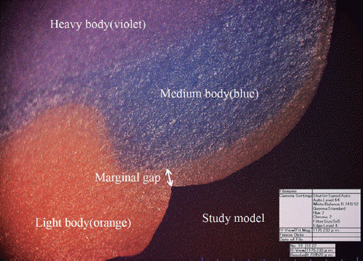
Fig. 2
Measurement of marginal gap by digital microscope at ×160 magnification (orange color: light body silicone; blue color: medium body silicon; violet color: heavy body silicone).
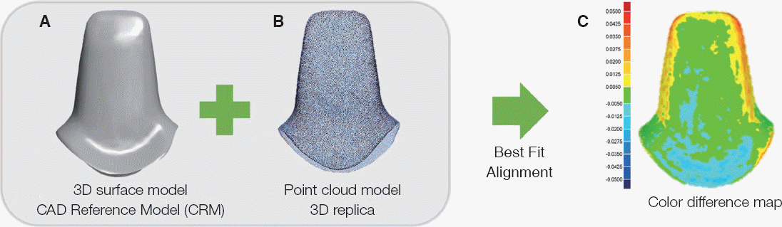
JDRAS_33_063_fig_2.tif
Fig. 3
Three-dimensional replica technique. (A) The 3D surface model from the digitization of the study model used as the control model (CAD reference model; CRM), (B) The point cloud model is the digitization of the light body silicone, (C) The point cloud model is projected onto the surface of the CRM. The distribution of the internal gaps was measured and depicted on the color difference map.
JDRAS_33_063_fig_3.tif
이 두 가지 측정방법은 모두 주 모형이나 보철물의 손상이 없으므로 여러 번 복제가 가능하다. 하지만 전자의 경우 절단 후 인상재 시편을 재사용할 수가 없고, 절단한 부위에 따라 두께가 달라질 수 있으며, 복제된 인상재에서 보철물의 마진과 변연(finishing line)을 확인하는 것이 어렵다. 또한 처음 주모형에서 인상재를 제거할 때 인상재의 찢김이나 변형이 있을 수 있어 정확한 측정이 어려울 수 있다는 점이 한계로 보고되고 있다.27,38,39

3. 3D 스캔 데이터 중첩법(Superimposition of 3D scan data)

컴퓨터 소프트웨어 개발과 CAD 프로그램의 발달로 3차원 디지털 모형을 자유자재로 조작할 수 있게 되었으며, 치아 및 보철물의 불규칙하고 기하학적인 형태를 비교 및 분석하는 다양한 소프트웨어들이 소개되고 있다.40 스캐닝 과정을 통해서 얻어진 3차원 디지털 모형은 기존 석고 모형이나 인상체와 달리 파절 및 분실의 우려가 없고, 데이터베이스 구축이 가능하여 이메일 등을 통해서 전송 및 복사가 가능하기 때문에 최근 많은 보철물이 이와 같은 과정으로 만들어지고 있다.41 3D 스캔 데이터 중첩법은 비파괴적인 방법으로 보철물을 만드는 지대치 석고모델 및 보철물 내면을 치과용 스캐너를 이용해 3D 스캔 데이터를 얻은 뒤 3차원 분석 프로그램을 이용해 중첩시켜 변연간격 및 내면간격을 측정한다(Fig. 4).42
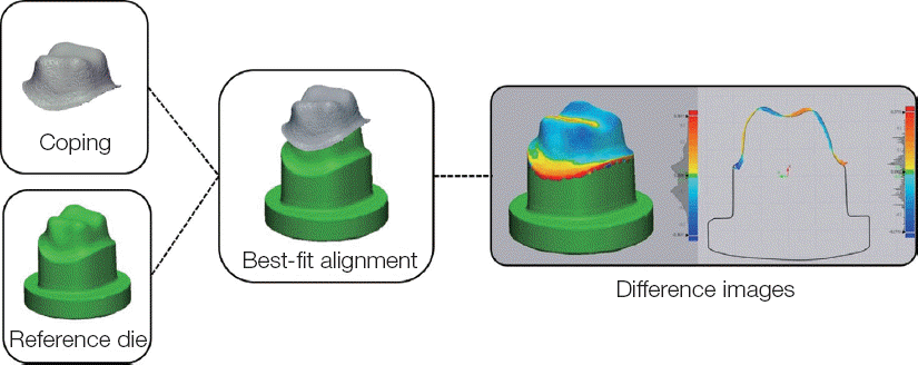
Fig. 4
Three-dimensional analysis procedure. For three- dimensional qualitative evaluation of the discrepancies in the difference between CAD-reference die and the digitized coping data, best-fit alignment was conducted in the program (Control X, Geomagic GmbH, Stuttgart, Germany) and a color-difference map was made with the superimposition process.
JDRAS_33_063_fig_4.tif
중첩 소프트웨어는 보통 두 형상 전체의 오차가 가장적은 값(best-fit)이 되도록 겹치게 하는 것이다.36,43 이런 중첩 방식으로 인해서 한쪽이 정확히 중첩이 되면 그에 따른 보상으로 인해서 다른 쪽에 오차가 발생할 수 있다. 때문에 3D 스캔 데이터 중첩법은 전체적, 상대적인 분석은 가능해도 절대적인 부분에서의 정확도는 판별하기 힘들다는 단점이 있다.44

4. 무게측정법(Weight technique)

무게측정법은 비파괴적으로 적합도를 평가할 수 있는 방법 중 하나로서 삼차원스캔을 통해 지대치의 표면적을 구하고, 실리콘 인상재의 밀도와 무게를 측정함으로써 아래의 공식을 통해서 전체 평균 내면간격을 얻을 수 있는 방법이다(Fig. 5).
Fig. 5
Weight technique procedure. (A) Abutment, (B) Fitting of the crown to the abutment after application of light body silicon, (C) Crown removed from B, (D) Measurement of the weight corresponding to the internal gap.
JDRAS_33_063_fig_5.tif
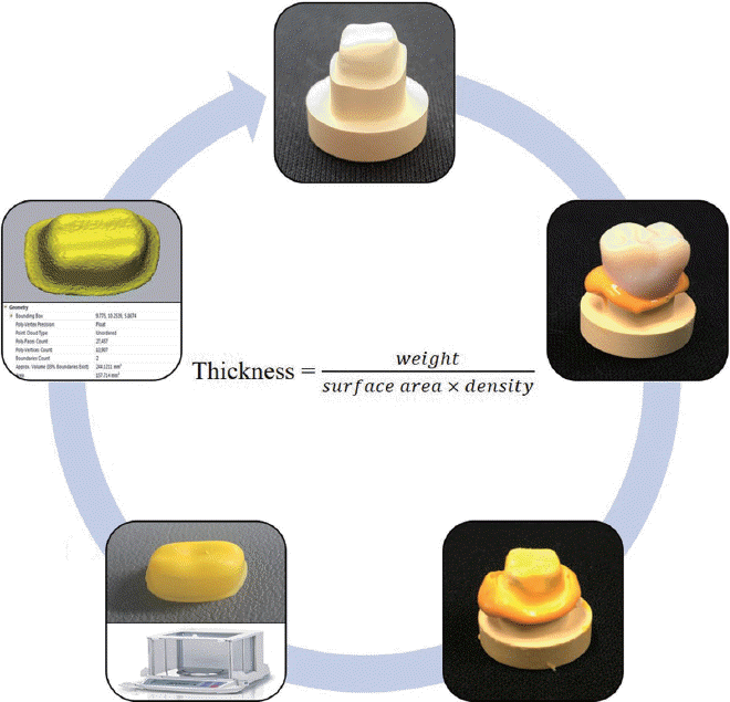
Thickness weight / (surface area × density)
이 방법의 장점은 다른 방법들에 비해 간단하여 시간도 절약되며, 누구나 쉽게 습득할 수 있고 비교적 저렴한 비용으로 측정이 가능하다는 것이다. 또한, 특정부위에 제한된 적합도만 볼 수 있는 다른 방법에 비해 전체 내면의 평균 간격을 간단히 알 수 있다는 것도 큰 장점으로 여겨진다. 그러나 특정부위의 간격을 측정할 수 없어 최대, 최소간격부위 등을 평가할 수 없고, 변연간격을 평가할 수 없다는 것이 단점이다.45-47

5. 마이크로 CT 측정법(Micro-CT)

무게측정법과 함께 또 하나의 비파괴적 측정법으로서 방사선촬영을 통하여 원하는 부위의 영상을 얻는 방법으로 비교적 최근에 많이 활용되고 있다.
2D영상을 통해 원하는 특정부위의 간격을 측정할 수 있을 뿐만 아니라 전체 내면간격의 삼차원적 영상획득도 가능하며 소프트웨어를 통해 자동적으로 측정 및 평균치가 얻어질 수 있어 매우 효과적인 방법이다. 그러나 마이크로 CT의 분해능이 아직 전통적으로 사용되는 현미경의 분해능에 비해 정밀도가 떨어진다는 것과 금속에 대한 허상(artifact)으로 인해 평가가 어려운 경우가 있다는 것이 단점이다. 또한, 고가의 마이크로 CT가 필요하며, 고해상도의 데이터를 얻기 위해서 뿐만 아니라 분석을 위해 많은 시간이 필요하다는 것이 효용성을 떨어뜨리는 이유이다.48-52

결론

보철물의 예후를 결정하는 많은 요인들이 있지만 그중에서 변연 및 내면의 적합도가 중요한 요인이다. 본 논문에서는 보철물의 변연 및 내면의 적합도를 평가하는 방법으로 절단면 측정법(Cross-sectional method), 실리콘 복제 방법(Silicon replica technique), 3D 스캔 데이터 중첩법(Superimposition of 3D scan data), 무게측정법(Weight technique), 마이크로 CT 측정법(Micro-CT)의 실험실 평가법을 소개하였다.
2013년 Nawafleh 등은 보철물 적합도를 평가한 183개 논문 중 70%이상이 직접적으로 변연부를 측정하는 방법과 절단면 측정법이고, 20%는 실리콘 복제 측정법이라고 하였다.53 쉽고 오래된 방법이 높은 비중을 차지하고 있다고 생각할 수 있고, 앞으로는 치과용 소프트웨어와 스캐너의 발달로 인해서 3D 스캔 데이터 중첩법이나 마이크로 CT 측정법 또한 활발히 연구될 것으로 기대된다.
소개한 각각의 방법들은 장단점이 있고, 평가방법이 표준화되어 있지 않아서 연구결과들 간의 직접적인 비교가 어렵고, 잘못된 해석의 가능성도 존재한다. 따라서 각각의 방법들에 대한 정확한 이해 및 분석이 있어야만 좋은 결과를 얻을 수 있으리라 생각되고, 연구들이 활발히 이뤄져 보철물 적합도가 향상 된다면 보철물 실패가 발생하지 않을 수 있으리라 기대한다.

Acknowledgements

이 논문은 2016년도 정부(미래창조과학부)의 재원으로 정보통신기술진흥센터의 지원을 받아 수행 된 연구이며(과제번호: B0101-16-1081, ICT 기반의 의료용 3D 프린팅 응용 S/W 플랫폼 및 서비스 기술개발), 또한 2017년도 산업통상자원부 및 산업기술평가관리원(KEIT) 연구비 지원에 의한 연구임(과제번호: 10062635).

References

1. Kim MY, Lee KW, Moon HS, Chung MK. A study on the gratification of the patient in the Dental Hospital. J Korean Acad Prosthodont. 2008; 46:6582. PMID: 8349002.
2. Rekow ED. High-technology innovations-and limitationsfor restorative dentistry. Dent Clin North Am. 1993; 37:513–24.
3. Hung SH, Hung KS, Eick JD, Chappell RP. Marginal fit of porcelain-fused-to-metal and two types of ceramic crown. J Prosthet Dent. 1990; 63:26–31. DOI: 10.1016/0022-3913(90)90260-J.
4. Schwartz NL, Whitsett LD, Berry TG, Stewart JL. Unserviceable crowns and fixed partial dentures:life-span and causes for loss of serviceability. J Am Dent Assoc. 1970; 81:1395–401. DOI: 10.14219/jada.archive.1970.0398. PMID: 5273607.
5. Grasso JE, Nalbandian J, Sanford C, Bailit H. Effect of restoration quality on periodontal health. J Prosthet Dent. 1985; 53:14–9. DOI: 10.1016/0022-3913(85)90056-3.
6. Holmes JR, Bayne SC, Holland GA, Sulik WD. Considerations in measurement of marginal fit. J Prosthet Dent. 1989; 62:405–8. DOI: 10.1016/0022-3913(89)90170-4.
7. Suárez MJ, González de Villaumbrosia P, Pradíes G, Lozano JF. Comparison of the marginal fit of Procera AllCeram crowns with two finish lines. Int J Prosthodont. 2003; 16:229–32.
8. Pelekanos S, Koumanou M, Koutayas SO, Zinelis S, Eliades G. Micro-CT evaluation of the marginal fit of different In-Ceram alumina copings. Eur J Esthet Dent. 2009; 4:278–92.
9. Christensen GJ. Marginal fit of gold inlay casting. J Prosthet Dent. 1966; 16:297–305. DOI: 10.1016/0022-3913(66)90082-5.
10. Sorensen SE, Larsen IB, Jörgensen KD. Gingival and alveolar bone reaction to marginal fit of subgingival crown margins. Scand J Dent Res. 1986; 94:109–14. DOI: 10.1111/j.1600-0722.1986.tb01373.x.
11. Assif D, Rimer Y, Aviv I. The flow of zinc phosphate cement under a full-coverage restoration and its effect on marginal adaptation according to the location of cement application. Quitessence Int. 1987; 18:765–74.
12. McLean JW, von Fraunhofer JA. The estimation of cement film thickness by an in vivo technique. Br Dent J. 1971; 131:107–11. DOI: 10.1038/sj.bdj.4802708.
13. McLean JW. Polycarboxylate cements. Five years’experience in general practice. Br Dent J. 1972; 132:9–15. DOI: 10.1038/sj.bdj.4802795.
14. Grey NJ, Piddock V, Wilson MA. In vitro comparison of conventional crowns and a new all-ceramic system. J Dent. 1993; 21:47–51. DOI: 10.1016/0300-5712(93)90051-Q. DOI: 10.1016/0300-5712(93)90031-K.
15. Tuntiprawon M, Wilson PR. The effect of cement thickness on the fracture strength of all-ceramic crowns. Aust Dent J. 1995; 40:17–21. DOI: 10.1111/j.1834-7819.1995.tb05607.x.
16. Jorgensen KD, Esbensen AL. The relationship between the film thickness of zinc phosphate cement and the retention of veneer crowns. Acta Odontol Scand. 1968; 26:169–75. DOI: 10.3109/00016356809026130.
17. Passon C, Lambert RH, Lambert RL, Newman S. The effect of multiple layers of die-spacer on crown retention. Oper Dent. 1992; 17:42–9.
18. Sorensen JA, Munksgaard EC. Interfacial gaps of resin cemented ceramic inlays. Eur J Oral Sci. 1995; 103:116–20. DOI: 10.1111/j.1600-0722.1995.tb00128.x.
19. Molin MK, Karlsson SL, Kristiansen MS. Influence of film thickness on joint bend strength of a ceramic/resin composite joint. Dent Mater. 1996; 12:245–9. DOI: 10.1016/S0109-5641(96)80030-3.
20. Kunii J, Hotta Y, Tamaki Y, Ozawa A, Kobayashi Y, Fugishima A, Miyazaki T, Fujiwara T. Effect of sintering on the marginal and internal fit of CAD/CAM-fabricated zirconia frameworks. Dent Mater J. 2007; 26:820–6. DOI: 10.4012/dmj.26.820. PMID: 18203487.
21. Mitchell CA, Pintado MR, Douglas WH. Nondestructive, in vitro quantification of crown margins. J Prosthet Dent. 2001; 85:575–84. DOI: 10.1067/mpr.2001.114268. PMID: 11404758.
22. Shearer B, Gough MB, Setchell DJ. Influence of marginal configuration and porcelain addition on the fit of In-Ceram crowns. Biomaterials. 1996; 17:1891–5. DOI: 10.1016/0142-9612(95)00302-9.
23. Rahme HY, Tehini GE, Adib SM, Ardo AS, Rifai KT. In vitro evaluation of “replica technique” in the measurement of the fit of Procera crowns. J Contemp Dent Pract. 2008; 9:25–32.
24. Tsitrou EA, Northeast SE, van Noort R. Evaluation of the marginal fit of three margin designs of resin composite crowns using CAD/CAM. J Dent. 2007; 35:68–73. DOI: 10.1016/j.jdent.2006.04.008. PMID: 16781043.
25. Tjan AH, Li T, Logan GI, Baum L. Marginal accuracy of complete crowns made from alternative casting alloys. J Prosthet Dent. 1991; 66:157–64. DOI: 10.1016/S0022-3913(05)80041-1.
26. Stappert CF, Dai M, Chitmongkolsuk S, Gerds T, Strub JR. Marginal adaptation of three-unit fixed partial dentures constructed from pressed ceramic systems. Br Dent J. 2004; 196:766–70. DOI: 10.1038/sj.bdj.4811390. PMID: 15220983.
27. Wolfart S, Wegner SM, Al-Halabi A, Kern M. Clinical evaluation of marginal fit of a new experimental all-ceramic system before and after cementation. Int J Prosthodont. 2003; 16:587–92.
28. Okutan M, Heydecke G, Butz F, Strub JR. Fracture load and marginal fit of shrinkage-free ZrSiO4 allceramic crowns after chewing simulation. J Oral Rehabil. 2006; 33:827–32. DOI: 10.1111/j.1365-2842.2006.01637.x. PMID: 17002742.
29. Att W, Komine F, Gerds T, Strub JR. Marginal adaptation of three different zirconium dioxide three-unit fixed dental prostheses. J Prosthet Dent. 2009; 101:239–47. DOI: 10.1016/S0022-3913(09)60047-0.
30. Tinschert J, Natt G, Mautsch W, Spiekermann H, Anusavice KJ. Marginal fit of alumina- and zirconia-based fixed partial dentures produced by a CAD/CAM system. Oper Dent. 2001; 26:367–74.
31. Beschnidt SM, Strub JR. Evaluation of the marginal accuracy of different all-ceramic crown systems after simulation in the artificial mouth. J Oral Rehabil. 1999; 26:582–93. DOI: 10.1046/j.1365-2842.1999.00449.x.
32. Kern M, Schaller HG, Strub JR. Marginal fit of restorations before and after cementation in vivo. Int J Prosthodont. 1993; 6:585–91.
33. Assif D, Antopolski B, Helft M, Kaffe I. Comparison of methods of clinical evaluation of the marginal fit of complete cast gold crowns. J Prosthet Dent. 1985; 54:20–4. DOI: 10.1016/S0022-3913(85)80062-7.
34. Groten M, Axmann D, Pröbster L, Weber H. Determination of the minimum number of marginal gap measurements required for practical in-vitro testing. J Prosthet Dent. 2000; 83:40–9. DOI: 10.1016/S0022-3913(00)70087-4.
35. Luthardt RG, Bornemann G, Lemelson S, Walter MH, Hüls A. An innovative method for evaluation of the 3-D internal fit of CAD/CAM crowns fabricated after direct optical versus indirect laser scan digitizing. Int J Prosthodont. 2004; 17:680–5.
36. Moldovan O, Luthardt RG, Corcodel N, Rudolph H. Three-dimensional fit of CAD/CAM-made zirconia copings. Dent Mater. 2011; 27:1273–8. DOI: 10.1016/j.dental.2011.09.006. PMID: 21983002.
37. Kim KB, Kim JH, Kim WC, Kim HY, Kim JH. Evaluation of the marginal and internal gap of metal-ceramic crown fabricated with a selective laser sintering technology:two- and threedimensional replica techniques. J Adv Prosthodont. 2013; 5:179–86. DOI: 10.4047/jap.2013.5.2.179. PMID: 23755345. PMCID: PMC3675292.
38. Coli P, Karlsson S. Fit of a new pressure-sintered zirconium dioxide coping. Int J Prosthodont. 2004; 17:59–64.
39. Laurent M, Scheer P, Dejou J, Laborde G. Clinical evaluation of the marginal fit of cast crowns-validation of the silicon replica method. J Oral Rehabil. 2008; 35:116–22. DOI: 10.1111/j.1365-2842.2003.01203.x. PMID: 18197844.
40. Kim JH, Kim KB. Evaluation of dimensional stability of digital dental model fabricated by impression scanning method. J Dent Hyg Sci. 2014; 14:1521.
41. DeLong R, Heinzen M, Hodges JS, Ko CC, Douglas WH. Accuracy of a system for creating 3D computer models of dental arches. J Dent Res. 2003; 82:438–42. DOI: 10.1177/154405910308200607. PMID: 12766195.
42. Bae SY, Park JY, Jeong ID, Kim JH, Kim WC. Three-dimensional analysis of marginal and internal fit of copings fabricated with polyetherketoneketone (PEKK) and zirconia. J Prosthodont Res. 2017; 61:106–12. DOI: 10.1016/j.jpor.2016.07.005. PMID: 27484816.
43. Schaefer O, Watts DC, Sigusch BW, Kuepper H, Guentsch A. Marginal and internal fit of pressed lithium disilicate partial crowns in vitro:a three-dimensional analysis of accuracy and reproducibility. Dent Mater. 2012; 28:320–6. DOI: 10.1016/j.dental.2011.12.008. PMID: 22265824.
44. Kim KB, Jung JK, Kim JH. Accuracy of digital impression made from different elastomeric impression materials:three-dimensional superimpositional analysis. J Dent Hyg Sci. 2014; 14:94–100.
45. Nakamura T, Dei N, Kojima T, Wakabayashi K. Marginal and internal fit of Cerec 3 CAD/CAM all-ceramic crowns. Int J Prosthodont. 2003; 16:2448.
46. Lee KB, Park CW, Kim KH, Kwon TY. Marginal and internal fit of all-ceramic crowns fabricated with two different CAD/CAM systems. Dent Mater J. 2008; 27:422–6. DOI: 10.4012/dmj.27.422. PMID: 18717171.
47. Colpani JT, Borba M, Della Bona A. Evaluation of marginal and internal fit of ceramic crown copings. Dent Mater. 2013; 29:174–80. DOI: 10.1016/j.dental.2012.10.012. PMID: 23218747.
48. Borba M, Cesar PF, Griggs JA, Della Bona Á. Adaptation of all-ceramic fixed partial dentures. Dent Mater. 2011; 27:1119–26. DOI: 10.1016/j.dental.2011.08.004. PMID: 21920595. PMCID: PMC3189283.
49. Seo D, Yi Y, Roh B. The effect of preparation designs on the marginal and internal gaps in Cerec3 partial ceramic crowns. J Dent. 2009; 37:374–82. DOI: 10.1016/j.jdent.2009.01.008. PMID: 19282083.
50. Alfaro DP, Ruse ND, Carvalho RM, Wyatt CC. Assessment of the internal fit of lithium disilicate crowns using micro-CT. J Prosthodont. 2015; 24:381–6. DOI: 10.1111/jopr.12274. PMID: 25753858.
51. Rungruanganunt P, Kelly JR, Adams DJ. Two imaging techniques for 3D quantification of precementation space for CAD/CAM crowns. J Dent. 2010; 38:995–1000. DOI: 10.1016/j.jdent.2010.08.015. PMID: 20801185.
52. Kim JH, Jeong JH, Lee JH, Cho HW. Fit of lithium disilicate crowns fabricated from conventional and digital impressions assessed with micro-CT. J Prosthet Dent. 2016; 116:551–7. DOI: 10.1016/j.prosdent.2016.03.028. PMID: 27422237.
53. Nawafleh NA, Mack F, Evans J, Mackay J, Hatamleh MM. Accuracy and reliability of methods to measure marginal adaptation of crowns and FDPs:a literature review. J Prosthodont. 2013; 22:419–28. DOI: 10.1111/jopr.12006. PMID: 23289599 .
TOOLS
Similar articles