

 PDF
PDF Citation
 Print
Print


Introduction
The differential displacement of the teeth and ridge causes a vertical and horizontal movement in the distal extended removable partial denture (DERPD). Because of the cantilever effect, destructive force is concentrated on the abutment teeth and alveolar ridge.1 In this case, implant supported removable partial denture (ISRPD) using implant placed in a biomechanically favorable strategic position can be alternative treatment. As additional support and retention with the implant can be obtained, it is effective for the Kennedy class I, II removable partial denture (RPD).2-4 Implant can be applied for support using healing abutment in ISRPD. And attachment can be connected to implant for retention and support in implant retained partial overdenture (IRPOD).5,6 Although several retrospective ISRPD studies have shown a high survival rate of implant, some mechanical complications can occur. de Freitas et al.7 reported that ISRPD had a high success rate ranging from 95 - 100% and good patient’s satisfaction but showed several complications such as pitting of the healing abutment, replacement of resilient component of the attachment, damage in framework, screw loosening and damage in acrylic denture base.
This clinical report describes the mechanical complication of ISRPD. The patient with mandibular Kennedy class I edentulism was treated with ISRPD placed in the mandibular bilateral first molar area. After 36 months for function, mechanical complications such as wear of healing abutment and screw fracture occurred. Subsequently, healing abutment was replaced with Locator® abutment.
Case Report
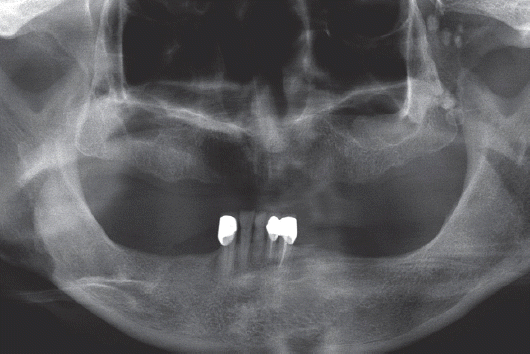
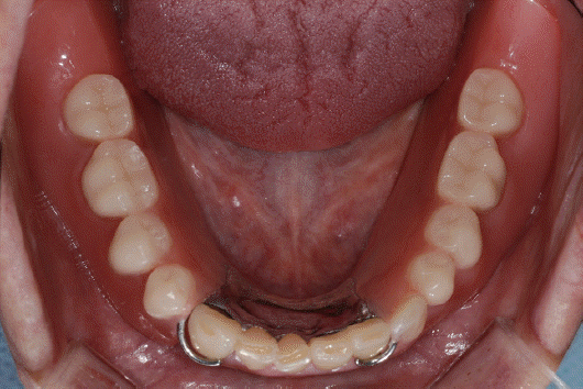
A 65-year-old female with pain of mandibular posterior edentulous ridge and discomfort due to mobility of DERPD, presented to the Department of Prosthodontics, Chonbuk National University Hospital. She had worn maxillary complete denture and mandibular DERPD for 6 years. These dentures showed poor retention, stability and excessive occlusal wear. Mandibular edentulous ridge showed moderate absorption equivalent to Class III edentulous area (Fig. 1).8 She had secondary caries on the right mandibular canine. Specific pathologic findings of jaws and temporomandibular joint were absent on radiographic examination.
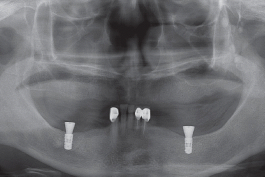
An implant supported fixed partial denture was initially considered as a treatment option. However, due to refusal of extensive surgery and patients’ economic status, maxillary complete denture and mandibular ISRPD with anterior surveyed prostheses were selected. At the initial visit, periodontal treatment was administered for anterior tooth improvement. After determining the implant position,9 computed tomography was taken with old RPD as radiographic stent. Two submerged-type implants (Bone level, RN crossfit®; 4.1 × 8 mm / 4.8 × 8 mm, Straumann AG, Waldenburg, Switzerland) were placed on each left and right mandibular first molar area10 and healing abutment (6.5 mm in diameter, 6.0 mm in height) was connected, showing good initial stability (Fig. 2).
Fig. 2
Postoperative panoramic radiograph. Two short implants were placed in mandibular posterior area.
For two months after surgery, old dentures were relined with resilient material (COE-Soft™, GC Corp., Tokyo, Japan). Mandibular old restorations were removed and dental caries of right canine was treated. Surveyed metal ceramic restorations were fabricated and cemented to the mandibular left central incisor, lateral incisor and right canine (Fig. 3). Subsequently, mandibular ISRPD with linguoplate and wrought wire clasp was fabricated according to conventional procedure (Fig. 4). As the short implants were placed on the mandibular posterior area, the healing abutment was connected for support. Bilateral balanced occlusion was developed for stability of removable prostheses, the metal framework of the RPD was adjusted to minimum contact with the healing abutment of the implant and delivered (Fig. 5). The patient was instructed in hygiene procedures associated with the dentures and examined for fit and occlusion of prostheses after 24 hours. After 1 week, 4 weeks, 6 months and 12 months on the recall check, the patient was satisfied with esthetic and function of prostheses.
Fig. 3
Anterior surveyed prostheses were cemented. No inflammatory signs were observed around healing abutment.
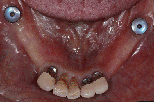
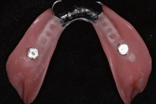
Fig. 4
Definitive removable partial denture was fabricated. Metal framework was adjusted to minimum contact with the healing abutment.
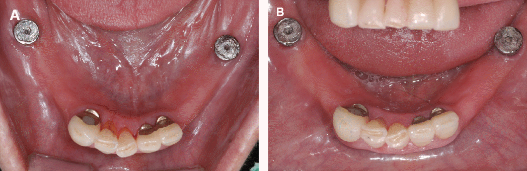
After 18 months of delivery, surface wear of the head of the healing abutment was observed at both sides (Fig. 6A). Metal framework of RPD were adjusted to form the 1-point contact with healing abutment using pressure indicate paste (Fit Checker™, GC Corp.). At recall after 24 months, patient stated that mandibular dentures were loosened. As poor retention and stability were observed, a direct relining was performed with self-curing denture base resin (Rebase II, Tokuyama dental Corp., Tokyo, Japan). At recall after 30 months, no pathological findings were observed except wear of healing abutment (Fig. 6B).
Fig. 6
(A) Occlusal view at 18 months after delivery. Mechanical wear was observed on flat surface of healing abutment, (B) At 30 months after delivery.
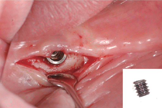
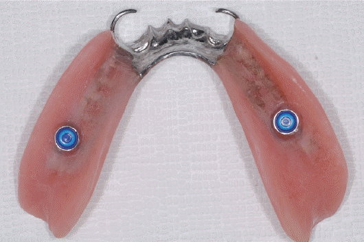
After 36 months, patient experienced side to side rolling of the healing abutment due to fracture of the screw. No mobility or fracture of implant fixture was observed. So, only fragment of screw was removed (Fig. 7). Then, Locator® abutment (ZEST anchors LLC., Escondido, USA) with 4 mm gingival height was tightened to 30 Ncm on both the implants. Metal housing was attached to existing ISRPD and blue nylon patrix was inserted (Fig. 8). Patient was satisfied with the retention and stability of IRPOD.
Discussion
A treatment plan for implant assisted removable partial denture (IARPD) requires consideration of diameter, length, position of the implant and the type of attachment. Cunha et al.9 reported that the amount of displacement of the denture was the smallest when implant was placed on the first molar region; Verri et al.10 strongly recommended the use of long and wide diameter implants because of favorable stress distribution. In this case, the healing abutments were connected to the implants placed in the area of the right and left mandibular first molars to provide additional support of the ISRPD.
Considering complications such as pitting corrosion of healing abutment, screw loosening and fractures, periodic recall appointments should be arranged.7,11 At 18 months recall appointment after delivery, the wear of healing abutment was observed. On the basis of studies on displacement and stress distribution of DERPD associated implant attachment,12 it is conceivable that the metal framework of denture was repeatedly in contact with healing abutment due to vertical and horizontal movement of the ISRPD during function. In addition, because the healing abutment with a flat head was used, the denture framework was in close contact with the healing abutment, and rotational movement of denture was prevented. It is likely that destructive lateral stress was transferred to the healing abutment. To prevent this problem, a dome shaped abutment that is designed to allow the denture and abutment 1-point contact or allow the denture rotation, is recommended.11 In order to obtain support and additional retention from the implant, attachment can be connected.13
The attachment might cause slightly more stress on the implant fixture than would be the case with the healing abutment. However, it can prevent friction between the metal structure and surface of healing abutment. And using resilient attachment component such as nylon insert can release the lateral stress by the movement of the RPD.12 Attachments can improve the satisfaction of patient for providing adequate retention and reduce the mobility of RPD.5 Elsyad et al.14 reported that ball and socket attachment decreases the stress around abutment teeth but increases the stress around implant due to lack of vertical resiliency. On the other hand, as Locator® attachment can appropriately redistribute the stress between the implant and the abutment teeth for allowing the vertical and horizontal movement of RPD by resilient nylon insert.5 In addition, due to its low vertical profile, it effectively reduces the destructive lateral stress around the implant for lowering rotational center.5,15 This is supported by Macros et al., who reported less marginal bone loss from implants with locator attachment resulting in better effects on implants.16
For preventing the mechanical problem of ISRPD, it is important to select the type of attachment for reducing the lateral stress and excessive contact with the RPD. In addition, it is important to fabricate a precise prosthesis for adaptation. Continuous recall for the fit and occlusion of the denture is necessary. Also, the early signs of mechanical and biological complications, such as mechanical wear of the abutment, require careful observation to ensure good long-term prognosis.
Conclusion
For preventing the mechanical problem of ISRPD, it is important to select the type of attachment for reducing the lateral stress and excessive contact with the RPD. In addition, continuous recall for the fit and occlusion of the denture is necessary. Also, the early signs of mechanical and biological complications, such as mechanical wear of the abutment, require careful observation to ensure good long-term prognosis.
ORCID
Jung-Yun Choihttp://orcid.org/0000-0002-4849-9635
Jung-Jin Lee http://orcid.org/0000-0002-7381-5230
Kwang-Yeob Song http://orcid.org/0000-0003-4283-1278
Ju-Mi Park http://orcid.org/0000-0003-1910-1525
Kyoung-A Kim http://orcid.org/0000-0002-2923-5351
Jae-Min Seo http://orcid.org/0000-0001-5095-4046
 XML Download