

 PDF
PDF Citation
 Print
Print


Introduction
Light-cured resin composites have good mechanical properties, but polymerization shrinkage and shrinkage stress, which lead to internal microcrack, gap formation, marginal microleakage,1 are major limitations of composite resin materials. Managing the polymerization shrinkage stress of the composite resin material is important to ensure adequate marginal integrity and, therefore, the long-term stability of the restoration.
Recently, bulk fill composites have been more widely used because materials with improved curing,2 controlled polymerization contraction stresses,3,4 and reduced cuspal deflection5 have been developed. Clinical recommendations suggest that placing these in 4-mm bulk increments, resulting in adequate polymerization. Bulk filling a cavity reduces some of the technical disadvantages associated with layering conventional composite resins, such as the incorporation of contamination or voids between the layers, and improves the chairside efficiency.6
The potential for increased contraction stress and inadequate cure depth are the major remaining concerns when curing bulk increments.7 Although several previous reports have suggested that this is an acceptable technique,2,4,8-10 others have suggested that bulk filling may produce undercured resin composites.11-13
Light-cured resin composites require adequate light exposure in order to achieve a proper degree of conversion and good physical properties. This is because the amount of free radical production is directly proportional to the amount of absorbed light irradiance. At the bottom of the cavity, the light intensity is insufficient for initiation of polymerization.14 Thus, at an increased exposure time, more light will consequently reach the deeper layer at the beginning of irradiation.
The curing time plays a crucial role in determining the stresses produced during the contraction of dental composites. The mechanical properties of the material formed are superior when the number of monomers converted in the polymers is larger.15,16 However, as the degree of conversion is increased, the volumetric shrinkage is also increased.17 High intensity light curing produces high hardness values as well as high shrinkage. Therefore, it is important to balance both of these effects by choosing the proper curing time.18
The advisability of rapid photocuring and bulk filling as an option for shortening the chairside time needs to be examined. However, this topic requires further exploration because few studies have investigated the interaction between marginal leakage and the irradiation time in bulk fill flowable composite resins.
Lately micro-CT analysis has been used as a reliable alternative to the conventional sectioning method for evaluating marginal leakage. To our knowledge, only a few previous investigations comparatively evaluated microleakage by using micro-CT.
Therefore, the purpose of this study was to evaluate the marginal leakage of bulk fill flowable composite resin filling with different time by using microcomputed tomography technology. The hypothesis of this study was that the increasing the light curing time would affect the microleakage of the bulk fill flowable composite resin.
Go to : 
Materials and Methods
Tooth selection
A total of 30 previously extracted human molars were selected. The calculus, soft tissue, and other tooth debris were cleaned and the teeth were stored in thymol solution (0.1%) and saline until use. The root base was embedded with self-curing acrylic resin.
Sample preparation
The design of the experimental groups was based on the following factors: 1) restorative system (SonicFill and SureFil SDR flow) and 2) different curing time (20 seconds, 40 seconds, and 60 seconds). The groups are described in Table 1.
Class II cavities (vertical slot cavities) in human molars were prepared by using methods modified from Dewaele and others.19 The cavities were prepared under water cooling in the approximal surfaces of the molars, with the following dimensions (±0.5 mm) : width, 4 mm; height, 4 mm; depth, 2 mm. The gingival floor was measured, 4.0 mm gingivally, located slightly occlusal and/or apical in proximity to the cementoenamel junction (CEJ), depending upon the proximal surface anatomy of each tooth. The dimensions of the cavities were verified with a periodontal probe.
A new bur was used for each cavity preparation. One operator performed all cavity preparations. Subsequently, the teeth were then randomly divided into 6 groups based upon the restorative system and curing time. The materials and light curing units utilized in this study are described in Table 2 and Table 3.
Table 2
Materials used in this study
![]()
An individual metallic matrix was used to build up the proximal wall. The cavity was conditioned by using 37.5% phosphoric acid gel etchant (Kerr Corporation, Orange, USA) for 15 seconds. Optibond Solo Plus adhesive agent (Kerr Corporation) was applied to the cavity, and followed by light polymerization for 20 seconds.
SonicFill composite system (Kerr Corporation) and SureFil SDR flow (Dentsply International, Milford, USA) was inserted into the cavity by using 1 bulk increment (4-mm thickness), followed by light polymerization for the specified curing time (20 seconds, 40 seconds, or 60 seconds). Light irradiation was performed by placing the light tip directly on the top of the resin with a 0-mm distance.
The SonicFill was inserted into the cavity by using the SonicFill handpiece (Kavo, Biberach, Germany). The SonicFill handpiece was used to dispense automatically the rheologically matched filling materials that are contained in the SonicFill unidose tips into the cavity by the action of sound and pressure under a frequency of 5 - 6 kHz.
The curing unit was a Bluephase 20i (Ivoclar Vivadent, Schaan, Liechtenstein) with a light intensity of 1200 mW/cm2.
Aging procedure
After the restorative procedures, all samples were artificially aged using thermal cycling for 24 hours. The specimens were thermocycled for 5,000 cycles in water baths between 5 ± 2°C and 55 ± 2°C at a dwell time of 30 seconds in each bath and a transfer time of 5 seconds between baths.
Silver nitrate immersion
The entire tooth surface was covered with 2 layers of nail varnish within 1 mm of the bonded interface. In order to allow the varnish to dry, the specimens were left undisturbed for 1 day.
Subsequently, the teeth were immersed in a 50% weight/weight silver nitrate aqueous solution for 24 hours at room temperature. The silver-impregnated teeth were thoroughly rinsed with distilled water, placed into a photo-developing solution for 8 hours, and rinsed again with water.
Micro-CT
After being placed and fixed in the specimen holder, each tooth was scanned individually by using micro-CT at a resolution of 12 μm.
Micro-CT scanning was performed by using the SkyScan 1272 (Bruker, Kontich, Belgium). The settings of the micro-focus X-ray source were as follows: 80 kV, 125 μA, 1-mm thick aluminum plate filter, and 0.4° rotation step with 180° rotation.
The cross-sections were collected by sample and the raw data were converted to 16-bit-mapped image film with a resolution of 1,632 × 1,092 pixels. NRecon version 1.6.1 software (Bruker) was used for reconstruction of all images.
Microleakage assessment
Microleakage of the silver nitrate solution at the resin-dentin interface was assessed by one evaluator. The 3D image of each leakage around the restoration was reconstructed by using CT-Analyser V.1.14.4 (Bruker) and was visualized by using CTvol V2.2.3 (Bruker). The region of interest was set up along the cervical wall and coronal wall of each restoration, including their respective extensions at the axial wall (Fig. 1). The volume of leakage was measured by using CT-Analyser V.1.14.4 (Bruker).
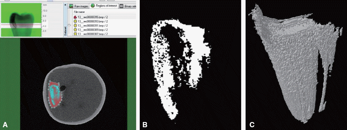 | Fig. 1Microleakage assessment. (A) The region of interest was set up along the cervical wall and coronal wall of each restoration, including their respective extensions at the axial wall. (B) 2D image of leakage around the restoration (C) 3D image of leakage around the restoration (reconstructed by using CT-Analyser).
|
Statistics
The means and standard deviations of the leakage volumes were calculated. The data showed a nonnormal distribution; therefore, the mean differences in the microleakage were statistically analyzed by using the Mann-Whitney test for multiple pairwise comparisons of groups.
The statistical analysis was performed by using SPSS 20.0 for Windows (IBM Corp., New York, USA). The level of significance was set to P <0.05.
Go to :
Results
The leakage volume around each restoration as measured by using micro-CT is shown in Table 4. Significant differences were observed between the light curing times, but no significant differences were found between the bulk fill flowable composite resins. Increasing the photoactivation time increased microleakage in all of the experimental groups. Groups 3 and 6 showed the highest mean microleakage values. Means and standard deviations (SD) in mm3 (n = 5).
Table 4
Marginal leakage in the experimental groups
| Group | Bulk fill composites | |
|---|---|---|
| Curing time | SonicFill | SureFil SDR flow |
| 20 seconds | 0.06 (0.03)Aa | 0.03 (0.05)Aa |
| 40 seconds | 0.68 (0.27)Ab | 0.54 (0.52)Ab |
| 60 seconds | 2.12 (0.83)Ac | 1.95 (0.90)Ac |
![]()
The lowercase letters indicate significant differences at the 5% significance level in the groups in the vertical columns.
The capital letter indicates no significant difference at the 5% significance level in the groups in the horizontal columns.
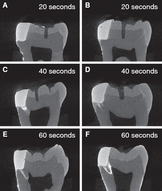
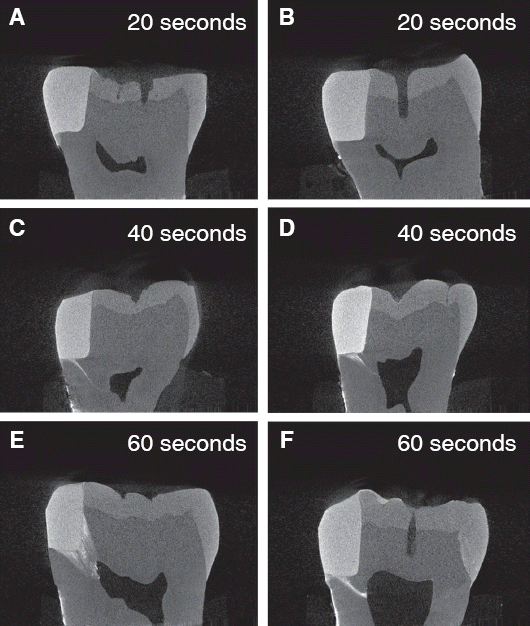
Representative images of each of the experimental groups are shown in Fig. 2 and 3. Silver nitrate infiltrated from the gap and was identifiable by white spots or high-intensity clusters. In groups 2, 3, 5 and 6, the silver nitrate infiltrated into the dentinal tubules and penetrated toward the pulp chamber. As expected, most restorations had more leakage in the cervical region than in the coronal region in terms of both depth and area. In addition, some leakage extended to the axial wall from the cervical wall of the cavty.
Representative images for several sections are shown in Fig. 4. Although no microleakage of silver nitrate solution was observed in the mid-longitudinal sections of some specimens, leakage was detected in the other sections of the specimens.
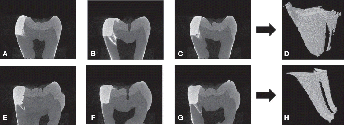 | Fig. 4Representative images of the mid-longitudinal sections and other sections of the restorations (Group 3). (A), (B), and (C): section images for 1 specimen, (E), (F), and (G): section images for another specimen. (B) and (F): images forthe mid-longitudinal sections, (A), (C), (E), and (G): images for the other sections. (D) and (H): reconstructed 3D images of microleakage. (B) shows microleakage toward the pulp chamber, whereas (F) shows no microleakage.
|
Go to :
Discussion
In this study, we found statistically significant differences between the curing time and microleakage values. Moreover, the mean microleakage values increased as photoactivation time increased.
The results of this study may be explained by the following 2 factors: 1) polymerization shrinkage stress and 2) polymer elastic modulus.
Higher radiant exposure induces greater stress because of contraction of the dental composites.20 In the case of lower radiant exposure, the reaction occurs more slowly and fewer polymer centers will form on the top surface. This slow reaction results in a polymeric structure that is more linear in the composite resins.21 Thus, lower irradiance will delay the increase in the viscosity of the resin composite during polymerization and decrease the polymerization stress.
Further, the quality of the polymer formed after photocuring may influence the microleakage values. Linear polymers with low cross-linking density have a low elastic modulus, and this is associated with relieving stress from contraction. Therefore, a lower elastic modulus may be related to lower microleakage values.22
A prominent increase in marginal leakage was observed as the photoactivation time increased, and the relationship between the exposure time and marginal leakage was strong. Visvanathan et al. reported that the mechanical properties of different curing regimens were similar, but the shrinkage stress differed significantly.18 Bouschlicher et al. also showed that the exposure time was important for determining the polymerization shrinkage force.23 Similarly, the use of a single curing method (light exposure for 20 seconds with a 0-mm distance to the resin) with both the SonicFill and SureFil SDR flow induced a cure degree of 4 mm.24 Therefore, light irradiation for a 20-second exposure period appears to produce appropriate polymerization as well as minimal microleakage.
In our study, 2 bulk fill flowable composite materials were used for evaluating marginal leakage. For these composite resins, the increased depth of cure might be due to the increased translucency, as well as the photoinitiator systems and modified monomers.11 SonicFill and SureFil SDR flow increase translucency because of the large filler size. In addition, the manufacturer states that the polymerization modulator of the SureFil SDR flow synergistically interacts with the camphoroquinone photoinitiator, thereby accelerating the polymerization process.25 Bulk fill composites with greater translucency can cure outside of the indirect light path because of internal scattering, so it may be less important how the light guide is positioned and oriented with these composites.26
For the study design, we used 3 different curing times (20, 40, and 60 seconds). The manufacturer recommends that the SonicFill and SureFil SDR flow can be placed in a 4-mm bulk increment with a light exposure of 20 seconds. Several studies reported that the SureFil SDR flow properly cured in 4-mm increments for the recommended curing time (20 seconds), but the SonicFill failed to meet the curing requirements under these conditions.27-29 Ilie and Stark demonstrated that 3 curing regimes (0-mm exposure distance, standard power mode, and 20- or 40-second exposures; or 7-mm exposure distance, standard power mode, and 40 seconds) induced depth of cure values of 4 mm or larger with the SonicFill.24
In our study, we used micro-CT to assess marginal leakage. Micro-CT is non-destructive method that enables the visualization of continuous images and detection of the deepest marginal leakage point.30 Furthermore, the length, area, and volume can be measured quantitatively by using analysis software.31 A previous study demonstrated that micro-CT analysis was consistent with stereomicroscopy in 92.5% of observations when assessing with silver nitrate staining.32
As shown in Fig. 4, no microleakage of silver nitrate solution occurred in the mid-longitudinal sections of some specimens, whereas microleakage did occur in the other sections of specimens. The leakage measured for a selected area did not represent the entire area. Detecting all of the leakage around the restoration for 3D reconstruction was possible with micro-CT. The limitations of the conventional sectioning method are that the interfacial staining is viewed on a limited number of tooth slices, and the cutting procedure inevitably involves damage to the specimens. In addition, the sectioning method is time consuming and prevents further use of the specimen.33
For the micro-CT method, the radiographic contrast agents used to display leakage should be highly radiopaque in relation to the restorative material and tooth structure. The atomic number of silver is much higher than that of the elements that exist in tooth hard tissues and composite resin fillings. Thus, silver is capable of presenting a good radiopaque contrast when it is dense.31 Further, the silver nitrate method of measuring microleakage is an acceptable technique because the silver ion is extremely small (0.059 nm) compared to the size of a typical bacterium (0.5 - 1.0 μm), thereby making it more penetrative.34 The silver ion could be trapped in the dentinal tubules during the course of infiltration.
However, the drawback of micro-CT is lower sensitivity when identifying leakage in the enamel. Dentin is less radiopaque than enamel because of its lower hydroxyapatite content, so it provides a good contrast to silver leakage. Therefore, some peripheral areas of silver leakage near the enamel structure cannot be detected because of their lower radiopaque value, which results in a 3D image of leakage that is smaller than the actual leakage.32
The current study had several limitations. First, our study conditions could not perfectly imitate actual condition. Numerous factors such as the interference of matrix bands, oral anatomy, limited opening by the patient, curing light design, and variable light curing technique by the dentist or assistant will affect the quantity of the delivered light to the composite resin. Such clinical limitations may require consideration of a greater curing time to achieve ideal photo-polymerization. Second, the sample size for each test was small. Greater statistical discrimination among the factors could have been identified if the sample size had been larger.
Go to :
Conclusion
Within the limitations of this study, we conclude that increasing the photoactivation time is the factors that may increase the marginal microleakage of the bulk fill flowable composite resins, and no significant differences in marginal leakage were found between SonicFill and SureFil SDR flow. Further, micro-CT can nondestructively detect the leakage around the resin composite restoration in three dimensions.
Go to :
ORCID
Eun-Ji Kim http://orcid.org/0000-0003-0514-2406
Kyu-Bok Lee http://orcid.org/0000-0002-1838-7229
Myoung Uk Jin http://orcid.org/0000-0001-9263-047X
Go to :
 XML Download