

 PDF
PDF Citation
 Print
Print


Introduction
Malignant lymphomas are malignant neoplasms of the lympho-reticular system; they have an overall mortality rate of more than 55%.1-3 In the oral cavity, malignant lymphomas constitute about 5% of all lymphomas. The most frequently affected sites are the palate and Waldeyer’s ring. Gingivae, floor of the mouth, paranasal sinuses, salivary glands, cheeks, and buccal sulcus are rarely affected.3-5
Malignant lymphomas are divided into two groups: Hodgkin’s disease and non-Hodgkin’s lymphomas (NHL). Diffuse large B-cell lymphoma (DLBCL), a subtype of NHL, rarely affects the head and neck area as primary lesion sites (> 1%).1-3 DLBCL occurs predominantly in males from the 7th decade of life and is characterized by a fast growing mass with symptoms.5-7 The etiology of DLBCL is not clearly defined but it is thought to be caused by immunosuppression or a virus.5 Radiotherapy, chemotherapy, and concurrent chemoradiotherapy are possible treatment modalities for DLBCL.8
Here we report a case of primary DLBCL that mimicked a dental abscess and maxillary sinusitis.
Case Report
A 76-year-old female patient was referred to the oral and maxillofacial surgery office for evaluation of facial swelling in the right infraorbital region and gingival swelling in the right maxillary posterior area after functional endoscopic sinus surgery from the otorhinolaryngology office. She had endoscopic surgery twice during 2 months for previously diagnosed right maxillary sinusitis.
She had no history of smoking or alcohol abuse. Medically, she had hypertension, diabetes mellitus, and osteoarthritis of the knee. She had dull pain during palpation and eating. There was no history of bleeding or discharge in the right maxillary region.
Extraoral examination detected swelling, redness, and heating sense in the right infraorbital area (Fig. 1). Intraoral examination detected a slightly elevated vestibular gingiva covered with intact mucosa without ulceration in the right maxillary posterior region. No exposure of necrotic bone was detected. In the dental panoramic view, no bony irregularity was found (Fig. 2). Computed tomography (CT) revealed a loss of bony continuity of the lateral wall and the upper wall (infraorbital region) of the right maxillary sinus (Fig. 3). Magnetic resonance imaging (MRI) revealed a mildly enhanced large mass in the right maxillary sinus and pterygopalatine fossa with an extension into the subperiosteal space of the right orbit. Bony destruction of the right zygoma, maxillary anterior and posterior walls, and the inferior orbital wall was detected. The size of the mass was approximately 5.4 cm × 4.0 cm × 4.0 cm (length × width × height) (Fig. 4). Reactive enlarged lymph nodes were detected in the neck area. Positron emission tomography/computed tomography (PET/CT) revealed a large hypermetabolic lesion in the right maxillary area and in the colon or small bowel, which was suspected to be a metastatic lesion (Fig. 5). In laboratory analysis, white blood cell count and high-sensitivity C-reactive protein were within the normal range. A slightly elevated level of blood sugar (127 mg/dL) was detected, which was due to diabetes mellitus.
Fig. 1
Swelling and redness on the right infraorbital area was detected in the clinical photography.
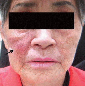
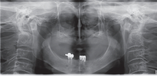
Fig. 3
Computed tomography (CT) revealed loss of bony continuity of lateral wall and upper wall of the right maxillary sinus.

Fig. 4
In the magnetic resonance imaging (MRI) revealed mild enhancing large mass in the right maxillary sinus (T2 weighted axial image).
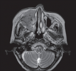
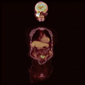
Fig. 5
A positron emission tomography/computed tomography (PET/CT) revealed large hypermetabolic lesion in the right maxillary area and in the colon or small bowel.
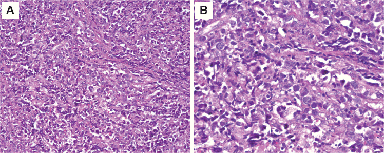
Incisional biopsy was performed under local anesthesia. An incision was made in the vestibular sulcus in the right maxillary posterior region and mass was incised with adjacent normal tissue. Through the destroyed lateral wall of the right maxillary sinus, a small amount of the mass in the maxillary sinus was also incised. Histological analysis revealed DLBCL (Fig. 6, 7). According to our routine treatment protocol, the patient was treated with concurrent chemoradiotherapy.
Discussion
NHL that occurs in the paranasal sinus constitutes 9 - 13% of all NHL cases and is predominantly B-cell lymphoma. It is rare and has poor prognosis because of delayed correct diagnosis, which is complicated by its non-specific symptoms.9
DLBCL is aggressive extranodal disease (> 40%) with fast tumor growth.10 Rhinosinusitis, pain, facial and mucosal swelling, nasal obstruction, fever, epistaxis, and weight loss are the main symptoms of paranasal DLBCL.11,12
In Western populations, lymphoma of the maxillary sinus is more prevalent than lymphoma of the nasal cavity. However, in Asian populations, the latter is more prevalent.3 In most cases of lymphoma of the paranasal sinus, CT examination reveals a large mass occupying the paranasal sinus. The best tool for radiologic diagnosis of a soft tumor extension is MRI. In MRI, a lymphoma has slightly elevated signal intensity and appears as a heterogeneous solid mass.12 PET/CT is also a helpful diagnostic imaging tool for lymphoma.2 In our case, the mass showed high signal intensity and had a heterogeneous solid form in a T2 weighted image, and a high uptake signal was observed in the right maxillary sinus on a PET/CT image.
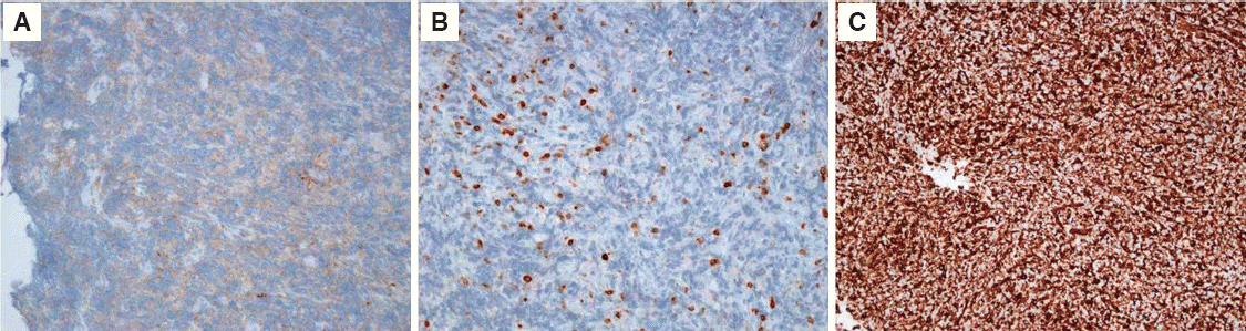
Histologically, DLBCL is characterized by diffuse proliferation of large neoplastic B-cells. The size of the nuclei is more than twice that of normal lymphocytes and is equal to or exceeds the size of normal macrophage nuclei.1,13 CD3, CD45RO, CD56, and TIA-1 are the most important immunohistological diagnostic markers of lymphoma.14 In our case, histological analysis revealed diffuse proliferation of large neoplastic B cells with large nuclei; these cells were slightly positive for CD45 and CD3 and strongly positive for vimentin in immunohistological staining. Therefore, the tumor was diagnosed as DLBCL.
Although DLBCL of the maxillary sinus is rare, its differential diagnosis with some aggressive inflammatory or infectious diseases (such as invasive fungal sinusitis and dental abscesses) is necessary.12,13
The current treatment strategy for DLBCL is chemotherapy with multiple agents such as cyclophosphamide, prednisone, oncovin, and hydroxydoxorubicin, whereas surgical resection plays an extremely limited role.13
Conclusion
In conclusion, because of non-specific symptoms, diagnosis of the early stages of DLBCL is often delayed. Misdiagnosis of an early stage of DLBCL in the maxillary sinus as maxillary sinusitis or a dental abscess is frequent. Therefore, surgical biopsy may be necessary for the final diagnosis together with using radiologic tools, such as CT, MRI, and PET/CT.
 XML Download