

 PDF
PDF Citation
 Print
Print


Introduction
The principle purpose of root canal treatment is to reduce discomfort and pain, eliminate bacteria from the root canal and prevent reinfection. A definite understanding of root morphology and canal anatomy is essential to achieving success, and perfect obturated root canal system.
The variation of root canal system morphology, especially multi-rooted teeth represents a continuous challenge to endodontic diagnosis and therapeutics. Anatomic characteristics of permanent maxillary molars are described as a group of teeth with three roots, one palatal and two buccal, each root with one root canal. Most of the clinical reports deal with fourth mesiobuccal canal of maxillary molar, but this is also common to other roots.
Case reports of maxillary molars with four or more canals also describe maxillary molars with three buccal roots maxillary molars with five canals, and maxillary molars with two canal in the palatal root.1
An anomalous root morphology which occurs infrequently, such as two palatal roots on maxillary molar, is rarely mentioned in textbooks. In 1952, Diamond2 shows two extracted teeth that are identified as first molars and have two widely divergent palatal roots.
Slowey3 also reported the treatment of first maxillary molar with two palatal roots and showed a second maxillary molar with four independent roots. Libfeld and Rostein4 examined 1,200 molars and found a 0.4% incidence of maxillary molar with four roots. Stone and Stroner1 examined more than 500 extracted molars and found less than a 2% incidence of more than one palatal canal when both anomalies were included.
Radiographs are an important and necessary aid in root canal treatment. Therefore an accurate radiographic techniques and proper interpretation are essential for accurate diagnosis and treatment. The use of preoperative radiograph is the best way to detect and evaluate tooth canal morphology and anatomy. Further radiographs should be taken at different angles to confirm any variation, in the case of, computed tomography (CT) is useful.
The purpose of this clinical report is to describe the unusual anatomy of maxillary molar with 2 separate palatal roots and the application of CT for successful endodontic treatment.
Case Report
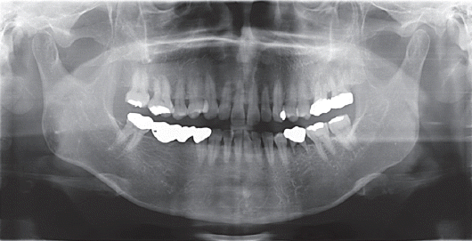
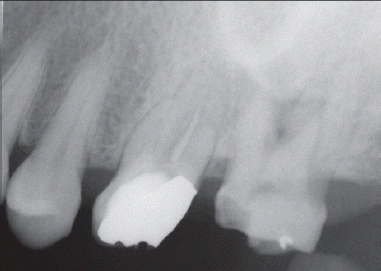
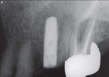
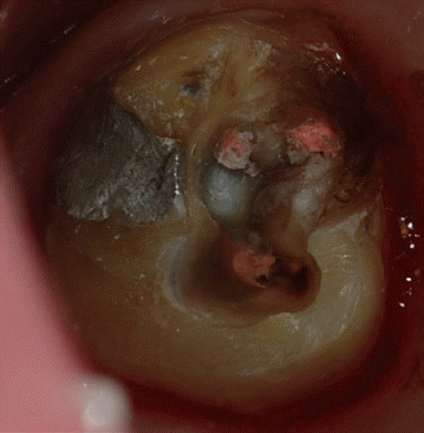
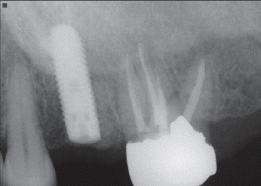
A 54-year-old woman visited Seoul Veterans Hospital for a dental examination. The medical history was noncontributory. The chief complaint of the patient is discomfort of left maxillary molar area. During clinical examination, left maxillary first molar and second molar were found to be restored with gold crown. In radiographic examination, left maxillary first molar seemed to be in incomplete endodontic treatment state and diagnostic radiograph revealed radiolucency in the periapical area (Fig. 1). The patient was diagnosed with chronic periapical periodontitis. After removing previously existed bridge and clinical examination, severe secondary decay on the left maxillary first molr was detected (Fig. 2) and the left maxillary first molar was extracted.
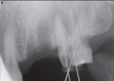
At the second appointment, left maxillary second molar was anesthetized and isolated with cotton roll. The access to the pulp chamber was achieved using a round diamond bur (ISO 801001016, Komet, Lemgo, Sweitzerland). Three orifices were located, and working lengths were determined by electronic apex locator (Root ZX, Morita, Tokyo, Japan) then confirmed by a radiograph (Fig. 3). The root canals were initially instrumented with stainless steel hand K-file and Gate Glidden drill under irrigation with 2.5% NaOCl. All canal enlarged to a size #40 using K3 Ni-Ti file (K3 file, Sybronendo, Orange, USA). The root canals were dried with sterile paper points, and calcium hydroxide was applied as an intracanal medicament.
The patient returned in 10 days with no symptoms. The root canals were filled using the continuous wave technique with Elements Obturation Unit (Sybronendo) (Fig. 4). The tooth was restored with a resin core and gold crown. After two weeks, Cone Beam Computed Tomography (Kavo 3D Exam, Icat, Hatfield, USA) was taken for implant surgery on left maxillary first molar.
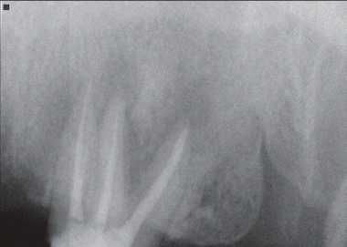
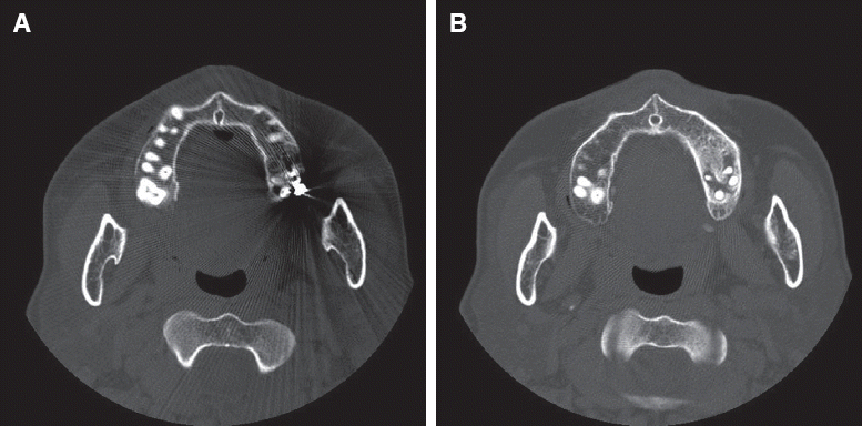
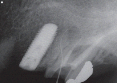
CT examination revealed a 2 palatal roots two separated foramina at the apex by chance (Fig. 5). After implantation of left maxillary first molar in OroMaxillaFacialSurgery (OMFS), further radiograph was taken at different angles to confirm two separated palatal canals (Fig. 6). The gold crown and resin core were removed and access was gained using a round diamond bur. Pulp extirpation was carried out and the 4th root canal orifice were clearly identified (Fig. 7). The working length was determined by electronic apex locator (Root ZX, Morita, Tokyo, Japan) then confirmed by a radiograph (Fig. 8). The 4th canal was cleaned and prepared by hand with nickel titanium files. The 4th canal was filled using the continuous wave technique with Element Obturation Unit (Fig. 9).
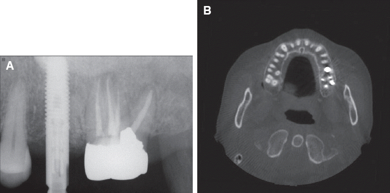
Fig. 5
Cone beam computed tomography showing secondary palatal root. (A, B) Revealed a 2 palatal roots two separated foramina at the apex of maxillary second molar.
After completion of root canal treatment, the left maxillary second molar was restored with resin core (Luxacoresmartmix dual, CE, Hamburg, Germany) and gold crown. During a 5-month follow-up, the patient remained asymptomatic (Fig. 10).
Fig. 10
(A) Periapical standard radiograph: 5-month follow-up after 4th canal filling, (B) Cone beam computed tomography: 5-month follow-up after 4th canal filling.
The patient used the treated teeth for 5 years without any symptoms and follow-up check after treatment (Fig. 11).
Discussion
Root canal anatomy can vary in number. Christie et al.5 reported 16 cases of maxillary molars with two palatal roots found during 40 years of daily clinical practice. Christie classified these teeth according to the shape and root separation as type I, II, III. Type I maxillary molars have two widely divergent palatal roots, which often are long and tortuous. The buccal roots often are “cow-horn” shaped and less divergent. Four separate root apices are seen on the radiograph. Type II maxillary molars have four separate roots, but the roots often are shorter, run parallel, have buccal and lingual root morphology, and have blunt root apices. Type III maxillary molars also are constricted in root morphology with the mesiobuccal, mesiopalatal, and distopalatal canal engaged in a web of root dentin. The distobuccal root in these cases seems to stand and may even diverge to the distobuccal.
Recently, Shin et al.6 and Barbizam et al.7 reported case of unusual root canal anatomy in permanent maxillary molars using microscope. Tomazinho et al.8 reported maxillary first molar showing unusual anatomy (four roots and six root canals).
This clinical report covered type I maxillary molar. Conventional radiography do not show four roots. After three canal endodontic treatment, for pre-surgical assessment of maxillary left first molar for implant, CT revealed fourth root by accident. In pre-surgical assessment, CT showed conventional type I maxillary molar and resin core was removed. The fourth palatal canal was treated with conventional endodontic treatment. After finishing the root canal treatment, at 3-month routine check-up, the patient remained asymptomatic.
The management of endodontic problem is reliant on radiographs to assess the anatomy of the tooth and its surrounding anatomy.9 Recently, most of this core information would be obtained from conventional radiographs. However, such images have inherent limitation. The lack of three-dimensional information and masking of areas of interest by overlying anatomy are of particular relevance in endodontics. Cone beam computed tomography reconstructed scans are invaluable for assessing teeth with unusual anatomy, such as teeth with an unusual number of root, dilacerated teeth and dens in dente. The exact location and anatomy of root canal system can be assessed, allowing successful management of the case.10
Conclusion
The variation of root canal system morphology, especially multi-rooted teeth represents a continuous challenge to endodontic diagnosis and therapeutics. An accurate radiographic techniques and proper interpretation are essential for accurate diagnosis and treatment. The use of cone beam computed tomography (CBCT) can overcome the limitation of the periapical standard radiography.
 XML Download