

 PDF
PDF Citation
 Print
Print


Introduction
Knowledge of root canal anatomy and variations are essential for clinicians to carry out effective root canal treatment. Predicting and negotiating a Cshaped root canal configuration is challenging. The failure of the Hertwig’s epithelial root sheath to fused the lingual or buccal root surface was main cause of C-shaped root. The C-shaped canal is an anatomical varition of a root fusion. Failure on the buccal side will in a lingual groove.1-3 C-shaped canal is most frequency found in the mandibular 2nd molar although it can also occur in maxillary molars and mandibular premolars.2
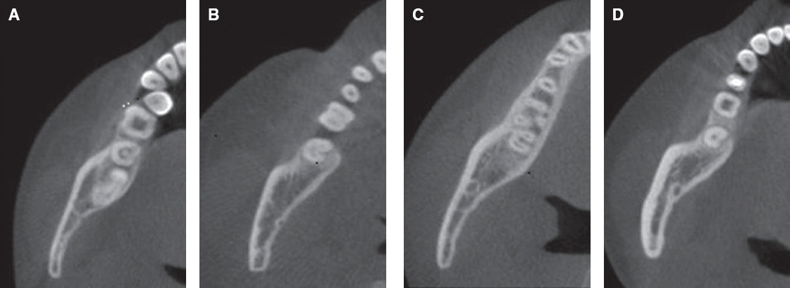
The C-shaped canal system can have many variations in its configuration. Cooke and Cox were first to describe the clinical significance of C-shaped canals, which have difficulties with respect to their debridement and obturation.4 Melton proposed a classification of C-shaped canals based on their cross-sectional shape.5 The canal configuration of C-shaped roots were determined and categorized based on Melton’s classification with the modified proposed by Fan et al.6,7 The canals were classified as follow; 1) C1 (continuous C-shaped canal: characterized by a C-shaped outline with no separation 2) C2 (semicolon-shaped canal: characterized by dentin separating one distinct canal from another C-shaped buccal or lingual canal) 3) C3 (separating canals: two or more discrete and separate canals) 4) C4 (a single round or oval canal) 5) C5 (no canal lumen could be observed) (Fig. 1). However, it has been pointed out that this shape can vary along the root so that the clinical crown morphology or the appearance of the canal orifice may not be exact feature of the actual canal anatomy.
As a non-invasive three-dimensional (3D) imaging technique cone beam computed tomography (CBCT) was reported to be sufficiently precise for morphological analysis. Compared with conventional medical computed tomography, studies using CBCT can be accomplished with a substantially lower effective dose and shorter working time.8
This study aimed to evaluate the prevalence and types of C-shaped canals in permanent mandibular 2nd molars in a Koreans people by using CBCT. The cross sectional characteristics of roots with C-shaped canals were evaluated by gender and tooth position. The unilateral and bilateral occurrence of C-shaped canals were also evaluated.
Go to : 
Materials and Methods
Cone beam computed tomograph images of 824 individuals were identified in the veterans health service medical center in 2013 (2015-09-026). The image were taken as part of routine examination, diagnosis and treatment planning for dental implantation. In this study, 711 mandibular 2nd molars were examined. Mandibular molars included in this study had fully formed apices and excluded root canal fillings, posts and crown restorated teeth. Of the study population, 43 individuals had bilateral molars.
Axial, coronal and sagittal section image were displayed and inspected by two dentist. The frequency of C-shaped canal and correlation with gender and tooth position were determined and assessed by the chi-squared test. The bilateral and unilateral appearance of C-shaped canals were evaluated.
The cross-sectional image at the level of canal orifice, coronal third portion. Coronal middle portion and apical third portion were assessed and classified for each according to the classification by Fan et al.(2004)6
The Cone beam computed tomography equipment is a KaVo 3D exam (Kavo Dental Excellence, Hatfield, USA). The Analysis program used the On Demand 3D™ (CyberMed International, Seoul, Korea).
Go to :
Results
The 824 individual with mandibular 2nd molar were examined. Among these teeth, 648 were male and 63 were female with the age ranging from 21 to 87. The average of age (Median) was 67 years (IQR 63 - 70). In the 824 people, 122 people had a C-shaped canal system (14.8%). In the 711 teeth, 154 teeth had a C-shaped canal system (21.5%) (Table 1).
Table 2, 3 show the distribution of C-shaped canal systems in the mandibular 2nd molars by gender and tooth position. No statistical difference were observed in the tooth position. But, there was the significant difference in the gender.
Table 2
Frequency of C-shaped canals in mandibular 2nd molars by gender
| Male n = 626 | Female n = 85 | |
|---|---|---|
| Number of teeth | 126 | 28 |
| No. of teeth Frequency | 20.1% | 32.9%* |
![]()
The cross sectional shaped of C-shaped canals at the different root levels was shown in Table 4. The most frequently observed root canal pattern was C1 at the orifice level and C3 at the apical level.
Table 4
Cross sectional canal shapes of C-shaped canals at different levels
| Root level | C1 | C2 | C3 | C4 | Total |
|---|---|---|---|---|---|
| Orifice | 137 (89%)* | 0 (0.0)% | 7 (4.5%) | 10 (6.5%) | 154 (100.0%) |
| Coronal | 46 (29.9%) | 9 (5.8%) | 84 (54.5%)* | 15 (9.7%) | 154 (100.0%) |
| Middle | 7 (4.5%) | 13 (8.4%) | 119 (77.3%)* | 15 (9.7%) | 154 (100.0%) |
| Apical | 4 (2.6%) | 3 (1.9%) | 129 (83.8%)* | 18 (11.7%) | 154 (100.0%) |
![]()
The distribution of unilateral and bilateral occurrence of C-shaped canals in patients with both mandibular 2nd molars is shown in Table 5. The bilateral occurrence in patients with C-shaped canals in mandibular 2nd molars was 74.4%.
Table 6 show the changes in root canal crosssectional classification at the different levels. The classification type of 89.6% of the C-shaped canals changed between two adjacent levels.
Go to :
Discussion
The C-shaped root canal system has an anatomical variant. This canal system has the continuous isthmus connects individual root canals.9 This morphology is most frequency seen in the mandibular 2nd molar and Asians.10,11 Because of the complex of Cshape canal system, the comprehension of root canal anatomy is important for clinical dentists.
There is an ethnic difference in the incidence of C-shaped canals in molars. It has been shown that C-shaped canals are most common in Asian populations. There is reported the highest incidence 44.5%12 In several study, the C-shaped canal was most common in a Korean population, with 31 - 45% prevalence.10 However, in this study, the prevalence C-shaped canal was 21.5%. Probably, because of in Veteran Health Service Medical Center most patient’s age were more than 60 years and missing molar ratio gets higher with age. The age profile was not analyzed because the age of patients was heavily weighted in the range of 60s to 70s. Also, due to the nature of Veterans Health Service Medical Center, most of patients were male. It seems statistically significant that expression frequency of C-shape canal of female was higher than that of male due to small number of female patients. The bilateral 2nd molars were rare according to CBCT analysis. Most of 2nd molar were unilateral and treated with root canal therapy and crown restoration. And those were excluded from analysis. Because CBCT was taken for diagnosis and treatment of implant, furthermore patients had a large number of missing tooth, patients with intact 2nd molars were excluded unfortunately. As CBCT analysis were performed according to vertical cross section of jaw, not vertical cross section of tooth axis, C2 and C3 of Meltons’s classification were not clearly distinguished. It made analysis more difficult that C2 could transform to C3 according to cross-sectional surface.
Another study reported that when present on one side, a C-shaped canal may be found in the contralateral tooth in over 70% of individuals.13 Similarly, we found that C-shaped canals in this study were 74.4% bilaterally. So if the C-shaped canal system in 2nd mandibular molar observed, we should be assumed that a C-shaped canal is existed in the opposite 2nd molar.
The C-shaped canal system was more complex. In this study, the cross sectional canal shape was analyzed at four root level including canal orifice, coronal, middle and apical third of the root. The result showed that only 16 teeth (10.4%) did not change canal sectional type from orifice to apical area. Our study corresponded the previous study. Seo and Park reported that there was no consistent change between two adjacent levels.
CBCT provide an excellent, nondestructive imaging option with the potential to detect most anatomical variations. Also CBCT can provide an accurate representation of the external and internal dental anatomy. The quality of CBCT is sufficiently high to visualize root canal morphology for clinical endodontic treatment at low radiation and dosimetry. However, even with all the benefits and information that provides, we should always consider the as low as reasonably amount of radiation that is possible to gain the most useful information for proper diagnosis.
Go to :
Conclusion
There was a high prevalence of C-shaped canals in the mandibular 2nd molars in Korean people. The C-shaped canal was very complex. Thus clinicians need to know the presence of C-shaped root canal systems and their configuration characteristics in order to increase the success rate of endodontic treatment. The CBCT analysis could be a noninvasive and clinically effective method for determining root and canal morphology. However, CBCT must not be used routinely in all cases of endodontic treatment, considering the as low as reasonably achievable concept.
Go to :
ORCID
Miyeon Kim http://orcid.org/0000-0001-5795-978X
Jeonghee Kim http://orcid.org/0000-0002-9075-1347
Go to :
 XML Download