

 PDF
PDF Citation
 Print
Print


INTRODUCTION
Spinal contusion injury is a complex condition that impacts all aspects of an individual's life. A traumatic spinal contusion injury is defined as “the occurrence of an acute, traumatic lesion of neural elements in the spinal canal resulting in temporary or permanent motor or sensory deficit or bladder dysfunction”. In India, about 20 persons out of every 1 million are suffering due to spinal cord injury, and with this rate, about 18,000 new patients will be added each year [1].
The spinal contusion injury may be complete or incomplete. A complete spinal contusion injury is associated with complete loss of motor and sensory function below the level of the injury, whereas an incomplete contusion injury may produce partial loss of function below the injury level [2]. Spinal contusion injury in the cervical region is very severe as it affects the arms (mainly), legs (less commonly), the middle part of the body. In some cases, it may also even affect the respiration of patients [3]. The damage to the spinal cord at the thoracic level mainly affects the legs. In some cases, it may also affect the blood pressure. The damage at the lumbar level mainly suggests the movement of one or both legs. However, in severe cases, such patients may also develop bladder or bowel dysfunction.
In order to mimic the spinal cord contusion of humans in animals, scientists have developed various animal models of spinal cord contusion injury. Two types of animal models have been developed i.e. bilateral contusion injury models [345] and unilateral contusion injury models [678]. Since in humans, spinal cord injury may occur at any level of the spinal cord i.e. the cervical, thoracic, lumbar, coccygeal, etc., therefore, different models have been developed by inducing contusion of the spinal cord at different levels [489]. The present review discusses these different animal models of spinal contusion injury in different animals.
Go to : 
MAIN BODY
1. Bilateral contusion models
1) Cervical contusion injury models
(1) C5 contusion injury model
In this model, Sprague–Dawley rats were anesthetized and a dorsal laminectomy was performed on the fifth cervical vertebra (C5). The vertebral column was stabilized by clamping the rostral and caudal segment of the exposed spinal cord with stabilizing forceps. Contusive injury of variable degree, such as mild and moderate injury, was produced by varying the force of the impactor. Mild injury was produced by delivering 200 kdyn of force, and moderate injury was produced by delivering 250 kdyn of force through an impactor having a 3.5 mm diameter. Animals with mild contusion showed locomotor impairment for 1–2 days whereas animals with moderate contusion showed locomotor impairment for 3–7 days. The moderate contusions produced greater locomotor impairment than mild contusions immediately post injury, and hind limb sensory function was more severely impaired than forelimb sensory function [3].
In another model, Sprague-Dawley rats were anesthetized and partial laminectomies over C5 and C6 were performed on the dorsal surface of the spinal cord to create a 3-mm-diameter opening. The clamps were attached to vertebrae C4–C7 of the injured rat and the animal was placed into an MR rig scanner. Contusion spinal cord injury was induced by operating the pneumatic actuator of an MR rig scanner at a velocity of 1100 mm/s, and the injury was sustained for 35 m [10].
(2) C7 contusion injury model
In this model, Sprague–Dawley rats were anesthetized. A C6–8 vertebral laminectomies were performed and the C7 spinal cord segment was exposed without the opening of the dura mater. To induce the contusion injury, 160 kdyn of force was applied to the C7 spinal cord using a 1.5 mm diameter impactor. The behavioral alterations were noted by performing different tests such as the cylinder test and grid walk test [11].
2) Thoracic contusion injury models
(1) T8 contusion injury model
In this model, Lewis rats were anesthetized and a midline incision was made along the thoracic vertebrae to open the skin. The paravertebral muscles at the region of T6–T12 were removed and a laminectomy was performed at the T8 thoracic vertebra. The stabilization clamps were attached at the T6 and T12 vertebrae to stabilize the spinal column. The contusive injury at the T8 level was produced by dropping a 10 g rod from the height of 12.5 mm. The locomotor test was performed the day of the injury, as well as the 4th, 7th, 10th, 13th, 16th, 19th, 22nd, 25th and 28th day post-injury in order to detect behavioral alterations. The maximum locomotor impairment was observed at the 28th day after injury [12].
In another model, Sprague–Dawley rats were anesthetized and a laminectomy was performed at the T8 thoracic vertebra. The contusive injury at T8 was produced by d ropping an impactor having a 10 g weight from a height of 2.5 cm. The locomotor function parameters such as joint movement, trunk posture, stepping and weight support ability, as well as paw position and tail position were noted at the 14th, 28th, 42nd, 56th, 70th and 84th day post-injury. The animals with a contusion injury demonstrated hind limb paralysis until 7 days after injury, which was followed by a progressive recovery in locomotor function over the next 35 days [13].
In another model, Sprague Dawley rats were anesthetized and a skin incision was made at the T5–T12 vertebrae, and a laminectomy was performed at the T7–T10 vertebral level. The contusion injury was induced at the T8 level by dropping a 5 g weight from a height of 8 cm onto the exposed spinal cord. The behavioral alterations were noted with the assessment of hind limb motor function for the first 3 days and followed for 1–4 weeks after injury. The levels of neurofilament protein and brain-derived neurotropic factor were increased at the 1st, 48th, and 72nd h after injury, which is an indicator of the progression of spinal cord injury [14].
In another model, Sprague-Dawley rats were anaesthetized. A laminectomy was performed at the T12– L1 spinal cord level and contusive injury was induced at T8 by dropping a 10 g metal rod with a 2 mm diameter from a height of 25 mm. Then, the wound was closed by suturing the muscles and skin. The behavioral alterations were noted with the assessment of locomotor activity at the 3rd, 7th, 14th, 21th and 28th day post-injury [1516].
(2) T9 contusion injury model
In this model, Sprague-Dawley rats were anesthetized. A longitudinal midline incision was made along the T8–T10 vertebrae and a T8 selective laminectomy was performed. The contusive injury at T9 of variable degree was produced by varying the force of impactor. In a 100 (mild injury) animal group, 104.9 ± 2.08 kdyn of force was applied; whereas animals from the 200 (moderate injury) group received a force of 209.14 ± 3.61 kdyn from the impactor. The locomotor test was performed at the 14th, 28th, and 42nd day post injury to detect hind paw and forepaw movements. The maximum dysfunction in forepaw movement in the 100 kdyn animal group was observed at the 42nd day, and in 200 kdyn animal group, was observed at 28th day post injury. The maximum hind paw displacement in the 100 kdyn animal group was observed at the 28th day, and in 200 kdyn animal group was observed at 14th day post-injury [17].
In another model, Sprague-Dawley rats were anesthetized and a partial T9 laminectomy was performed to remove the periosteum, but not the dura mater. Contusion was produced of variable degree by varying the force of impactor, time of dwelling, and diameter of the impactor.
In one of the models, a contusion of the spinal cord at the midline of T9 was induced, using the Infinite Horizon device, by delivering 150 or 200 kdyn of force, with a 0 s dwell time with a 1.5 mm impactor. In a second model, a contusion at the midline of T9 was induced by delivering 150 kdyn of force, with a 1 s dwell time, and a 1.5 mm impactor. The behavioral alterations were noted with the assessment of locomotor activity at the 1st, 4th, 7th, 10th, and 14th day post-injury, followed by weekly assessments. Female rats, subjected to 150 kdyn of force, with a 0 s dwell time showed less hind limb impairment at the 1st, 7th, 35th, and 42nd days after injury as compared to females that received 200 kdyn of force. Both the 150 and 200 kdyn injury groups also displayed heat hypersensitivity at the 14th, 28th, and 35th day and the 28th, 35th and 42nd day after injury, respectively. In the second model (150 kdyn, with 1 s dwell), female rats did not show mechanical allodynia, but male rats showed mechanical allodynia at the 14th day, which persisted for 42 days after injury. However, both male and female rats developed thermal hyperalgesia at the 14th, 35th, 42nd day after injury [4].
Rhesus monkeys (Macaca mulatta) have also been used to produce spinal contusion injuries. A 9–10 cm longitudinal midline incision was made to expose the T8–11 vertebrae and the paraspinal muscles were separated. Stainless steel arms were fixed to the T9 facets bilaterally to stabilize the spine. A laminectomy of the T9 was performed and the T10–11 segments of the spinal cord were exposed. The monkeys were then placed in the Louisville Injury System Apparatus-Large (LISA-L) impact device to produce contusive injury at the T9 vertebral level.
Contusion injury of variable degree was produced, such as mild (1.0 mm displacement) and moderate (1.5 mm displacement) by using an impactor having a 3.2 mm diameter, with 1.32 ± 0.05 m/sec peak velocity and 30-psi compressed air. The contact duration of the impactor against the spinal cord was set at 0.25 ± 0.05 s. The behavioral alterations such as hind limb motor function was noted a day before injury, 2 days after injury, once a week for 4 weeks, and once a month for 4 months after injury. The 1.0 mm injury group showed better hind limb motor function than the 1.5 mm injury group during the 4-month-observation period. The major advantage of this method is that the forelimb hanging and swinging of the monkeys was restricted within the corridor. One limitation of this method is that the injury did not produce bilateral tissue damage, possibly due to the use of a relatively small diameter tip in relationship to the large cord size [1819].
In another model, Sprague-Dawley rats were anesthetized. The contusion injury at the T9 spinal segment was produced by dropping a rod with 10 g of weight and a 2.0 mm diameter from a height of 25 mm onto the exposed spinal cord. After injury, the overlying muscles and skin were closed in layers. The behavioral alterations, such as mechanical paw withdrawal thresholds and thermal paw withdrawal latencies, were noted at the 30th, 31st, 32nd, 33rd, 34th and 35th day post-injury. The maximum reduction in mechanical paw withdrawal thresholds were observed at the 33rd day, and the maximum reduction in thermal paw withdrawal latencies was observed at the 31st day post-injury [20].
(3) T10 contusion injury model
In a very recent study, Sprague-Dawley rats were anesthetized. A laminectomy at the T9 vertebra was performed to expose the spinal cord and a circular incision of 2.5 mm in diameter was made. Contusion at the T10 was produced of a variable degree by varying the height of the impactor. An impactor having a 2.5 mm diameter and 10 g of weight was used to produce injury. Mild injury was produced by dropping the impactor from a height of 6.25–12.5 mm, moderate injury was produced by dropping the impactor from a height of 25 mm, and severe injury was produced by dropping an impactor from a height of 50 mm [2122].
In another model, Sprague-Dawley rats were anesthetized. A cut was made into the skin and the surrounding muscles were removed to expose the T9–T10 in the spinal cord level. Contusion at T10 was produced, of variable degree, by varying the height of the impactor. An impactor having a 2.5 mm diameter and 10 g weight was used to produce injury. Mild injury was produced by dropping an impactor from a height of 6.25–12.5 mm, moderate injury was produced by dropping the impactor from height of 25 mm, and severe injury was produced by dropping the impactor from a height of 50 mm. The behavioral alterations were noted with the assessment of locomotor activity on the day of injury, and on the 1st, 3rd, 7th, 14th and 28th day after injury [2123].
In another model, Sprague-Dawley rats were anesthetized. A midline dorsal skin incision was made between the spinous processes of C2 and T2 and the underlying paravertebral muscles of C4–C6 were removed. A bilateral laminectomy was performed at the fifth cervical (C5) vertebra to expose the dorsal aspect of the spinal cord. The spinal column was stabilized by clamping the C2 vertebra and the spinous process of T2 using forceps. A cervical hemi-contusion injury at T10, of variable degree was produced by varying the force of the impactor. In the 100 (mild injury) animal group, 103.4 ± 1.0 kdyn force was applied. In the 200 (moderate injury) animal group, 213.6 ± 2.8 kdyn of force was used, and in the 300 ( severe i njury) animal group, 312.6 ± 3.0 kdyn of force was applied through the impactor.
The behavioral alterations were noted on the day of injury, and on the 7th, 14th, 21st, 28th and 35th day post-injury by performing different tests such as the paw preference test (an unskilled forelimb function assessment), cat walk gait analysis (unskilled locomotor function), vermicelli handling test (skilled forelimb function), and horizontal ladder test (skilled locomotor function) as indicators of locomotor activity.
In paw preference test, the animals in the severe (300) injury group showed significant increase in contralateral paw use as compared to animals in the mild (100) injury group 14–35 days after injury and the moderate (200) injury group 14–28 days after injury. In the vermicelli handling test, rats in the 300 injury group made fewer ipsilateral forepaw adjustments than in the 100 injury group at 21–35 days. In the cat walk gait analysis test, the 300 injury group had smaller paw print areas than the animals in the 100 injury group on the 14th, 21st, and 35th day following injury.
In the horizontal ladder test, all injury groups used a higher percentage of rungs (a horizontal support on a ladder for the use of the feet) than the uninjured control group. The severe (300) and moderate (200) injury group showed an increase in ipsilateral forepaw error scores on all days after injury, and a graded decrease was observed with injury severity [524].
In another model, Fisher 344 rats were anesthetized and a laminectomy was performed at T10 to expose the dorsal portion of the spinal cord. The animals were stabilized by attaching forceps to the rostral T9 and caudal T11 vertebrae. A 2.5 mm impactor was placed approximately 3–4 mm above the surface of the exposed spinal cord. The contusion injury at T10 was induced by applying a force of 200 kdyn through the impactor to the exposed spinal cord. The impactor was operated at a velocity of 130 mm/s. Overlying muscle layers and skin were sutured. Then, the MR imaging of the injured spinal cord was done at the 14th, 42nd and 56th day after injury [2425].
In another model, Sprague-Dawley rats were anesthetized. A dorsal midline incision was made over the thoracic spinal cord. A laminectomy was performed at the T10 vertebra. The contusion injury was produced by dropping a 10 g weight from a height of 50 mm on to the exposed spinal cord. The behavioral alterations were noted with the assessment of locomotor activity from 1–7 days after injury. The injured rats exhibited dramatic and bilateral hind limb paralysis with no movement or only slight movements of the joint after injury [26].
In another model, Sprague-Dawley rats were anesthetized. A longitudinal dorsal incision was made to expose the T6–T10 spinous processes and a laminectomy was performed at the T8–T9 vertebrae. The injury was induced by dropping a metal rod with a weight of 25 g and a diameter of 2 mm from a height of 3 cm at the T 10 vertebra of rat's exposed spinal cord. To detect behavioral alterations, the hind-limb function of the rats was assessed on the day of injury, and on the 7th, 14th, 21st, 28th, 35th, 42nd, 49th and 56th day after injury [2728].
In another model, Sprague-Dawley rats were anaesthetized and a longitudinal incision was made through the lower thoracic skin to expose the T9–T11 vertebrae and a laminectomy was performed at the T10 vertebra. The clamps were attached at the T9 and T11 vertebral spines to stabilize the vertebral column. The tip of the impactor was placed at approximately 2 mm deep in the T10 vertebra. The contusion injury at the T10 vertebra was produced by operating the impactor at a velocity of 5 m/s for a 100 ms dwell time.
The behavioral alterations were noted by performing locomotor assessment tests such as the ledged beam test (to assess the total number of ledge uses by the hind limbs) and the random rung horizontal ladder test (to assess the total number of hind limb foot slips). These behavioral alterations were noted at the 14th, 28th, 42nd, 56th and 70th day post injury. In the ledged beam test, the spinal cord injured rats made significantly more use of the ledge with their hind limbs; whereas in the random rung ladder test, they made many hind limb slips through the missing rungs. In the ledged beam test, the maximum number of ledge uses were observed at the 42nd day and in the random rung ladder test, the maximum number of hind limb foot slips were observed at the 56th day [2930].
In another model, Sprague-Dawley rats were anesthetized. An incision was made over the T8–T11 spinous processes, exposing the underlying paravertebral muscles. A blunt dissection was made to expose the transverse processes at T9–T11, and a complete laminectomy was performed at T10. Then the T9 and T11 spinal segments were clamped and stabilized. The contusion injury was produced by delivering 250 kdyn of force through the impactor [31].
(4) T11 contusion injury model
In this model, Sprague-Dawley rats were anesthetized and a laminectomy was performed at the T10 vertebra. A contusive injury was induced at T11 by dropping a 10 g weight onto the spinal cord from a 12.5 mm height. To detect motor function, a behavioral test (locomotor activity) was performed before and after injury at the 1st, 4th, 7th, 10th, 14th, 21st, 28th and 35th day [9].
(5) T12 contusion injury model
In this model, Wistar rats were anesthetized and a laminectomy was performed and the T12 spinal segment was exposed. Rats were mounted on a spinal cord impactor frame and animals were stabilized by holding the dorsal processes on the rostral (T9) and caudal (T11) vertebral segments. An injury was induced with the impactor by delivering 200 kdyn of force with a 30 s dwell time.
In this model, behavioral alterations such as the total number of food pellets retrieved and eaten, total number of forelimb placements, and number of steps on a ladder were observed, which are indicators of locomotor activity. These behavioral parameters were noted on the day of injury, and maximum pain developed on the 14th day, which persisted for 42–56 days [8].
2. Unilateral contusion models
1) Cervical contusion injury models
(1) C4 and C5 contusion injury model
Apart from rats, mice have also been employed for studying contusion injury. In this model, C57Bl/6 mice were anesthetized and an incision was made between the spinous processes of C2 and T1 to expose the cervical region of the spinal cord. Then the paravertebral muscles overlying C4–C6 were removed.
A unilateral laminectomy on the right side at the C4 and C5 levels was performed, and mice were subjected to a double spinal contusion injury using the Infinite Horizon impactor at both the C4 and C5 levels. At the C4 spinal cord level, injury was induced with the impactor by delivering 55.3 ± 1.6 kdyn of force with a velocity of 121.1 ± 0.6 mm/s. A C5 level injury was induced by delivering 53.6 ± 1.6 kdyn of force with a velocity of 123.0 ± 0.6 mm/s. The neurophysiological recordings, such as Phrenic nerve compound muscle action potential (CMAP) and electromyography (EMG) recordings, were noted at the 14th and 42nd day after injury. CMAP amplitudes were reduced at the 14th and 42nd day post-injury. Analysis of EMG signals showed that there was a significant decrease in inspiratory burst frequency at the 42nd day post-injury [6].
In another model, Sprague-Dawley rats were anesthetized and placed in a stereotaxic frame in a prone position. A dissection was made through the dorsal musculature, and the muscles overlaying the laminae of C4–C7 were scraped off. A 4–5 cm dorsal midline incision was made to insert a clamp (tilted at a 22.5° angle off the horizontal plane) at C4–C6 and the animal is placed in an Infinite Horizon impactor. A force of 150 kdyn is delivered through the impactor to induce injury at the C5 level.
In this model, behavioral alterations such as the total number of food pellets retrieved and eaten, total number of forelimb placements, and number of steps on a ladder were observed, which are indicator of locomotor activity. These alterations were noted to develop at 0 and 14th day and persisted for 42–56 days. The number of food pellets retrieved with the ipsilateral forelimb was decreased after the spinal contusion injury. The maximum reduction in food pellet retrieving capability, total number of forelimb placements, and number of steps on a ladder were observed at the 42nd, 28th and 14th day after injury, respectively [832].
In another model, Sprague-Dawley rats were anesthetized and a partial laminectomy was performed at the C4 cervical. The vertebral column was stabilized by clamping the C3 and C5 vertebral bodies. An impactor tip having a 1.6 mm diameter was positioned between the dorsal vein and the right lateral edge of the spinal cord. A unilateral C4 contusion was produced by delivering 200 kdyn of force through the impactor to induce injury. The impactor tip was positioned 3–4 mm above the spinal cord. The behavioral alterations were noted by performing different tests such as single-pellet test training, staircase test training, and forelimb locomotor score [733].
2) Thoracic contusion injury models
(1) T13 contusion injury model
In this model, Sprague-Dawley rats were anesthetized and a partial laminectomy at T13 was performed. A contusion injury at T13 was induced by delivering 100 kdyn of force, with a 1 s dwell time, and the impactor having a 1.0 mm diameter on left side. The animals showed mechanical allodynia in both ipsilateral and contralateral hind paws at the 28th day, and at the 21st and 28th day after injury, respectively [4].
3. Summarized discussion
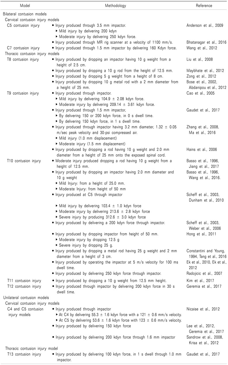
To mimic spinal cord contusion in humans, various animal models of spinal contusion injury have been developed. These animal models have been developed in rats [3811], mice [6], and monkeys [19] (Table 1). Two types of animal models have been developed, i.e. bilateral contusion injury models [312] and unilateral contusion injury models [46]. The spinal cord contusion injury led to development of behavioral alterations such as heat hypersensitivity, mechanical allodynia, thermal hyperalgesia, reduction in food pellet retrieving capability, total number of forelimb placements, and number of steps on a ladder, which are indicators of locomotor activity [48]. These behavioral changes are generally evident from the day of surgery to the 14th day afterwards [89], and are at their peak after 28–56 days of surgery [48].
Spinal cord contusion injury can be induced either by a weight drop method [1112] or impactor method [34]. In the weight drop method, a specific weight or a rod having a specific weight and diameter is dropped from a specific height on to the exposed spinal cord, and in the impactor method, injury is produced through an impactor by delivering a specific force to the exposed spinal cord area. The weight drop method is a crude one, and there are more chances of variation. The impactor method is more specific, accurate, and precise than the weight drop method. It is easier to induce injury through the impactor than the weight drop method [8]. In these methods, injury can be produced in animals of different intensities: low intensity, moderate intensity, and high intensity.
In the weight drop method, low intensity injury can be produced by dropping a 5 g weight from a height of 8 cm [14], moderate intensity injury can be produced by dropping a 10 g weight from a height of 12.5–25 mm [916], and high intensity injury can be produced by dropping a 25 g weight from a height of 50 mm [26]. In the impactor method, mild injury can be produced by delivering 100 ± 5 kdyn of force, moderate injury can be produced by delivering 200 ± 10 kdyn of force, and severe injury can be produced by delivering 300 ± 10 kdyn of force through the impactor [5].
Each method described above has its own advantages and disadvantages. Weight-based injury methods are less precise, while impactor-based methods are more precise. The choice of opting for the best animal model of spinal cord injury amongst the described methods actually depends on the purpose of experiments.
The following points may be kept in mind while selecting a model: ① These methods are different from one another on the basis of site of injury i.e. cervical, thoracic or lumbar. Therefore, the experimenter may choose the model depending on his/her aim for the experiment i.e. whether to work with cervical injury, lumbar, or thoracic injury. Furthermore, the site of injury within the region may also be varied, i.e. the lower lumbar region or upper lumbar region. Thus, the choice of the site of injury in the spinal cord typically depends on the objective of the experiment. ② Moreover, use of either a unilateral model or bilateral model depends on the aim of the experiment. In bilateral methods, the symptoms related to locomotor impairment are produced on both sides of the body. However, in the unilateral method, the symptoms are restricted to a single side of the body. ③ Furthermore according to the aim of the experiment, the intensity of injury may be selected, i.e. mild injury, moderate injury, or severe injury. In mild injury models, less weight or less impactor force is used; while for high intensity, high weight, or impactor of high force has to be used.
Go to :
CONCLUSIONS
Since in humans, spinal cord injury may occur at any level of the spinal cord i.e. cervical, thoracic, lumbar, coccygeal etc., therefore, different animal models have been developed by inducing contusion of the spinal cord at different levels, using either a weight drop method or impactor method. Moreover, the impactor method is more specific, accurate, and precise than the weight drop method.
Go to :
 XML Download