Abstract
5-aminosalicylic acid (5-ASA) is used widely to treat ulcerative colitis. The common side effects of 5-ASA include nausea, vomiting, abdominal pain, headache, and skin rash. 5-ASA-induced myocarditis is a rare side effect, and few cases have been reported. 5-ASA-induced myocarditis usually occurs within 2-4 weeks of drug use and causes chest pain and dyspnea. This paper reports 5-ASA-induced myocarditis in a 31-year-old male patient who took 5-ASA for 20 days prior. The patient was hospitalized with dyspnea that worsened when lying down, with chest pain radiating to the left neck, fever, and vomiting. Myocarditis was suspected. The work-up included electrocardiogram, transthoracic echocardiogram, cardiac MRI, and laboratory investigations. The patient’s signs and symptoms improved within a few days after withdrawing 5-ASA. This case shows that an evaluation including the possibility of myocarditis should be performed when patients with ulcerative colitis receiving 5-ASA present with cardiac problems, such as dyspnea and chest pain.
5-Aminosalicylic acid (5-ASA) is a commonly used first-line drug for inducing and maintaining remission of mild to moderate ulcerative colitis.1 Although the mechanism of action of 5-ASA has not been elucidated, it is believed to inhibit the cyclooxygenase and lipoxygenase enzymes, reducing the formation of pro-inflammatory prostaglandins and leukotriens.2 5-ASA can activate the peroxisome proliferator-activated receptor-γ and has antioxidant and free-radical scavenging properties.3 The most common side effects of 5-ASA include nausea, vomiting, and abdominal pain, but rare side effects include pancreatitis, nephrotoxicity, and cardiotoxicity. Myocarditis is a very rare side effect from the use of 5-ASA.4 This paper describes the first case in Korea of 5-ASA-induced myocarditis in a young male patient with atypical ulcerative colitis. Wonkwang University Hospital (IRB No. WKUH 2021-09-008) approved this case report and waived the requirement for informed consent due to the case report nature of the study.
A 31-year-old male patient was admitted to the emergency department with chest pain and dyspnea. At the time of admission, his vital signs were a blood pressure of 87/46 mmHg, pulse rate of 118 beats per minute, respiration rate of 20 beats per minute, body temperature of 40.2℃, and oxygen saturation of 90%. Twenty days before presentation, the patient started taking 5-ASA 2.4 g/day because of a sigmoidoscopy performed 2 months earlier at a local clinic for a history of abdominal pain and bloody stools indicating ulcerative colitis. After taking 5-ASA, the patient reported improved abdominal pain and bloody stools. On the other hand, the patient reported dyspnea that worsened when lying down and chest pain radiating to the left neck that began 3 days before presentation.
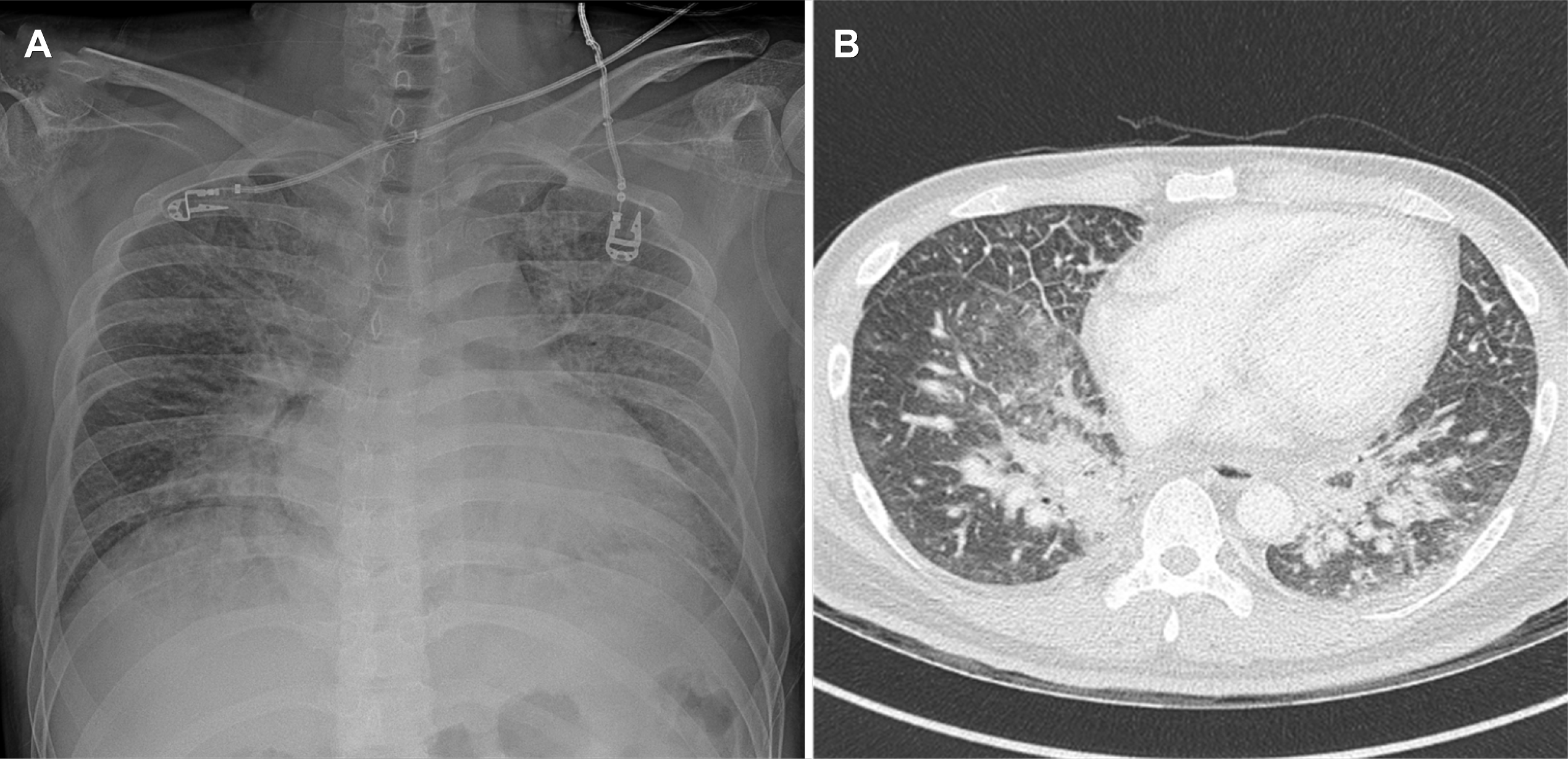
The patient showed an acute ill-looking appearance, and a physical examination revealed crackles in both lung fields on auscultation. The laboratory investigations showed an increased white blood count (21,280/μL) and neutrophils (18,690/μL). The inflammatory markers, erythrocyte sedimentation rate (ESR), and CRP were increased (ESR 46 mm/hour, CRP 214.29 mg/L), and the cardiac biomarkers troponin T and brain natriuretic peptide (BNP) were increased (troponin T 2,150 pg/mL, BNP 1,514.3 pg/mL). Chest radiography and chest CT revealed pulmonary edema and a small amount of pleural effusion in both lungs (Fig. 1). Abdominal CT showed wall thickening of the descending colon and sigmoid colon, pericolic vascular engorgement, and multiple lymph node enlargement. No specific findings other than sinus tachycardia were observed on the electrocardiogram (EKG). A transthoracic echocardiogram (TTE), performed to evaluate heart failure, revealed a moderate global left ventricular systolic dysfunction, an ejection fraction of 40.1%, and edematous myocardium.
The 5-ASA was discontinued because of potential 5-ASA-induced myocarditis. In addition, antibiotics (levofloxacin and metronidazole), methylprednisolone 60 mg/day, and human IgG 0.5 g/kg were used without excluding the extra-intestinal manifestations of inflammatory bowel disease and infectious myocarditis. Furosemide was also administered to treat pulmonary edema.
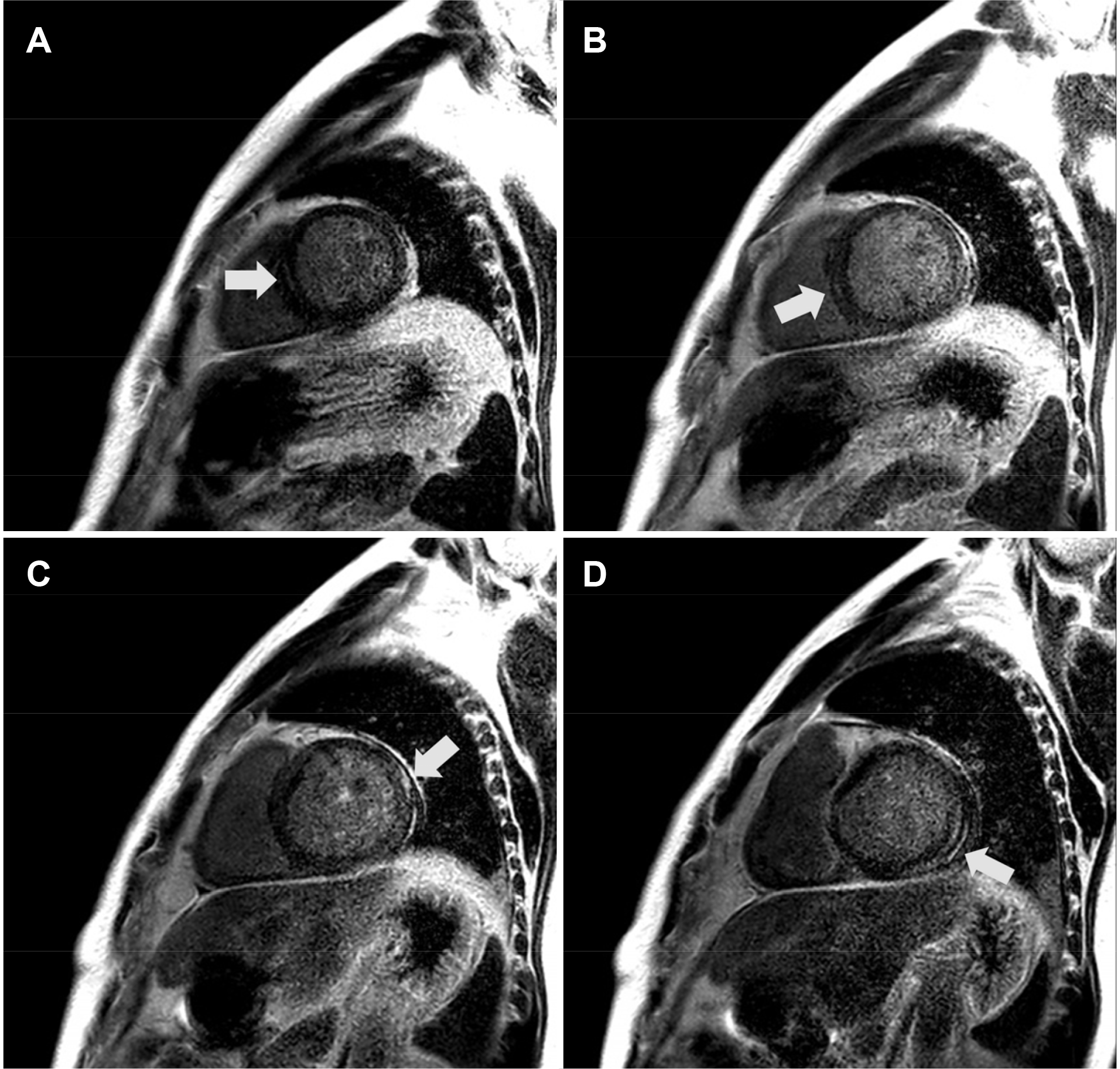
On hospital day 2, the patient’s clinical symptoms and signs were improved, and the previously observed pulmonary edema in both lung fields was almost resolved on chest radiography. After the patient was hemodynamically stable on hospital day 3, cardiac MRI was performed to diagnose myocarditis. Midwall and subepicardial delayed gadolinium enhancement was observed in the septal, inferolateral, and lateral segments at the basal level and in the septal segment at the mid-ventricular level. High signal intensity was observed in the T2-weighted images in the same range (Fig. 2).
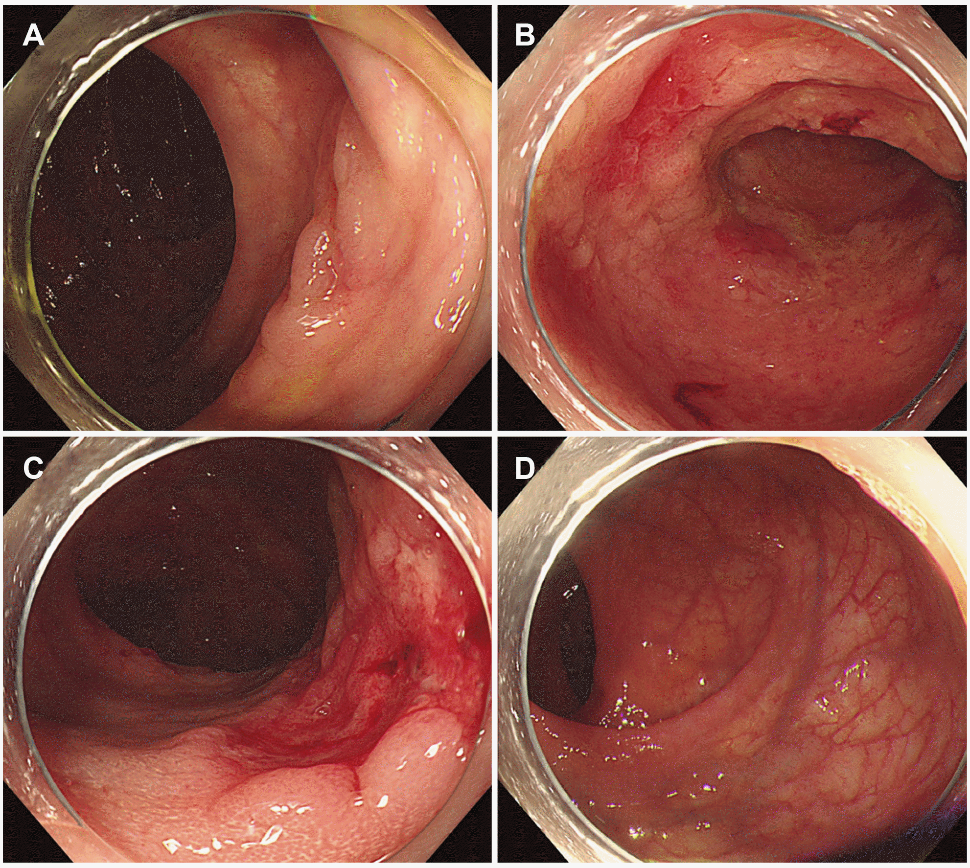
On hospital day 6, pulmonary edema was absent on chest radiography, and left ventricle systolic function recovered completely on the follow-up TTE, with an ejection fraction of 60%. In laboratory investigations on hospital day 7, the leukocytes and neutrophils returned to normal, and the CRP (35.74 mg/L), troponin T (201 pg/mL), and BNP (519 pg/mL) decreased. A colonoscopy performed to evaluate ulcerative colitis revealed obliteration of the vascular pattern, mucosal friability, and multiple deep ulcerations throughout the descending colon and sigmoid colon, but the rectum was spared (Fig. 3). Multiple biopsies were performed on the ulcerative lesions. Fecal calprotectin was highly elevated at 5,515 mg/kg. The viral infection and tuberculosis tests were negative, and colon biopsy results were reasonable for ulcerative colitis.
On hospital day 8, the patient was discharged with 40 mg/day prednisolone. He is being followed up as an out patient, and biologics are planned after tapering the steroid.
5-ASA is a commonly used first-line drug for inducing and maintaining remission in mild to moderate ulcerative colitis.1 The common side effects of 5-ASA include nausea, vomiting, abdominal pain, headache, and skin rash. Interstitial nephritis, renal failure, male infertility, atrioventricular block, myocarditis, and pericarditis can occur in rare cases.4 Myocarditis caused by 5-ASA is a very rare but life-threatening side effect. Although the mechanism of 5-ASA-induced cardiotoxicity is unclear, direct toxic effects, allergic reactions mediated by IgE, cell-mediated hypersensitivity reactions, and humoral antibody responses have been hypothesized.5
The symptoms of 5-ASA-induced myocarditis are observed within 2-4 weeks after use, and patients often complain of chest pain and dyspnea.6 In laboratory investigations, leukocytosis, elevations of inflammatory markers (ESR and CRP), and elevated cardiac biomarkers (creatinine kinase-MB and troponin T) are often observed, as is an elevation of BNP. In EKG, ST-segment elevation or ST-segment depression can be observed, as shown in myocarditis.7,8 A systolic dysfunction and pericardial effusion can be observed in TTE.6 Cardiac MRI can be a useful test for diagnosing 5-ASA-induced myocarditis.9 These test results are non-specific. Hence, a diagnosis of 5-ASA-induced myocarditis requires a temporal relationship between the 5-ASA intake and symptom onset and the exclusion of other causative diseases (extra-intestinal manifestations of inflammatory bowel disease, viral myocarditis, and vasculitis).10 In the present case, after taking 5-ASA, the patient reported chest pain and dyspnea. In addition, leukocytosis and elevated ESR, CRP, troponin T, and BNP levels were observed. His EKG showed no specific findings other than sinus tachycardia, and TTE revealed systolic dysfunction and pericardial effusion. Cardiac MRI showed results suitable for myocarditis, and there was no evidence of viral myocarditis or vasculitis.
The treatment of 5-ASA-induced myocarditis is the discontinuation of the drug. Most symptoms improve quickly, and patients recover within a few days. Many clinicians have attempted to use corticosteroids to control inflammation and the symptoms of myocarditis, but it is unclear how corticosteroids affect the course of treatment.6
This patient was diagnosed with 5-ASA-induced myocarditis from a rectal sparing type of ulcerative colitis, which is the first reported case in Korea and was not reported worldwide. The onset time and symptoms of 5-ASA-induced myocarditis in patients with atypical ulcerative colitis were similar to those of typical ulcerative colitis, and there was no difference in the treatment response.
In conclusion, although the use of 5-ASA in patients with inflammatory bowel disease is generally safe, potentially life-threatening cardiotoxicity with heart failure can occur. Therefore, careful observation and differentiation from other causes are required for patients who develop dyspnea and chest pain within 2-4 weeks after initiating treatment with 5-ASA. 5-ASA should be discontinued in cases of 5-ASA-induced myocarditis, after which complete recovery is achieved in most cases within a few days.
REFERENCES
1. Bergman R, Parkes M. 2006; Systematic review: the use of mesalazine in inflammatory bowel disease. Aliment Pharmacol Ther. 23:841–855. DOI: 10.1111/j.1365-2036.2006.02846.x. PMID: 16573787.

2. Ye B, van Langenberg DR. 2015; Mesalazine preparations for the treatment of ulcerative colitis: are all created equal? World J Gastrointest Pharmacol Ther. 6:137–144. DOI: 10.4292/wjgpt.v6.i4.137. PMID: 26558148. PMCID: PMC4635154.

3. Sood A, Ahuja V, Midha V, et al. 2020; Colitis and Crohn's Foundation (India) consensus statements on use of 5-aminosalicylic acid in inflammatory bowel disease. Intest Res. 18:355–378. DOI: 10.5217/ir.2019.09176. PMID: 32646198. PMCID: PMC7609395.

4. Sehgal P, Colombel JF, Aboubakr A, Narula N. 2018; Systematic review: safety of mesalazine in ulcerative colitis. Aliment Pharmacol Ther. 47:1597–1609. DOI: 10.1111/apt.14688. PMID: 29722441.

5. Kaiser GC, Milov DE, Erhart NA, Bailey DJ. 1997; Massive pericardial effusion in a child following the administration of mesalamine. J Pediatr Gastroenterol Nutr. 25:435–438. DOI: 10.1097/00005176-199710000-00015. PMID: 9327378.

6. Brown G. 2016; 5-Aminosalicylic acid-associated myocarditis and pericarditis: a narrative review. Can J Hosp Pharm. 69:466–472. DOI: 10.4212/cjhp.v69i6.1610. PMID: 28123193. PMCID: PMC5242279.

7. Perrot S, Aslangul E, Szwebel T, Gadhoum H, Romnicianu S, Le Jeunne C. 2007; Sulfasalazine-induced pericarditis in a patient with ulcerative colitis without recurrence when switching to mesalazine. Int J Colorectal Dis. 22:1119–1121. DOI: 10.1007/s00384-007-0310-2. PMID: 17440739.

8. Park EH, Kim BJ, Huh JK, et al. 2012; Recurrent mesalazine-induced myopericarditis in a patient with ulcerative colitis. J Cardiovasc Ultrasound. 20:154–156. DOI: 10.4250/jcu.2012.20.3.154. PMID: 23185660. PMCID: PMC3498314.

9. Tschöpe C, Cooper LT, Torre-Amione G, Van Linthout S. 2019; Management of myocarditis-related cardiomyopathy in adults. Circ Res. 124:1568–1583. DOI: 10.1161/CIRCRESAHA.118.313578. PMID: 31120823.

10. Paschalis T, Paschali M, Mandal AKJ, Missouris CG. 2019; Plasma N-terminal pro-B-type natriuretic peptide (BNP) in mesalazine-induced myopericarditis. BMJ Case Rep. 12:e229142. DOI: 10.1136/bcr-2018-229142. PMID: 30975785. PMCID: PMC6506044.

Fig. 1
Chest radiography and chest computed tomography (CT) images. (A) Chest radiograph demonstrated homogenously increased pulmonary parenchymal opacification in both lung fields. (B) Chest CT demonstrated peripheral ground-glass opacities and thickening of bronchovascular bundles.


 PDF
PDF Citation
 Print
Print


 XML Download