

 PDF
PDF Citation
 Print
Print


INTRODUCTION
Common conceptions of clinical pain tend to emphasize such benign disorders as headache or back pain or to focus on pain associated with cancer. It surprises many individuals to learn that TMDs are the most common chronic orofacial condition and that they affect a very sizeable proportion of the population [1]. In the craniofacial region, TMDs affect 4-12% of the population, with masticatory muscle pain often labeled as myofascial pain dysfunction syndrome (MPDS) or myofascial pain (MFP) being the most frequent (66%) patient complaint [2]. TMDs are often not restricted to the TMJ, but frequently include pain and tenderness of the masticatory muscles designated as group I in the Research Diagnostic Criteria (RDC) for TMDs [3].
Myofascial pain syndrome is a trigger point induced regional musculoskeletal pain disorder affecting one or more muscles or group of muscles [4]. A myofascial trigger point is defined as a hyperirritable spot, usually within a taut band of skeletal muscle or in the muscle fascia, which is painful on compression and can give rise to characteristic referred pain, motor dysfunction and autonomic phenomenon [4].
For more than 40 years, the contributing factors and actual underlying etiology for temporomandibular joint (TMJ) pain and myofascial pain (MFP) have been the subject of debate. Chronic pain is the most serious problem in this patient clientele, making the prevention and management of persistent pain the most important clinical challenge [5]. The etiology of MPDS is multidimensional. Biomechanical, neuromuscular, biopsychosocial, and neurobiological factors may contribute to the disorder [6]. This suggests that interventions such as cognitive behavioral therapy (CBT) targeting cognitive, emotional, and behavioral factors may be effective in modifying the expression of pain and dysfunction. For the development of such treatments, a comprehensive insight into the complex interactions between physical symptoms and psychosocial factors is essential [5].
Psychometric tests, as well as endocrine measurements, have been used to evaluate some of these factors and have sometimes linked psychological features to certain subgroups. According to biopsychosocial models, future research should emphasize both physiological and psychological factors and the relationship between them [7]. Physiological stress is known to induce various adaptational responses of physiologic systems, including increased activity in the hypothalamic-pituitary-adrenocortical (HPA) system, which promotes cortisol secretion from the adrenal cortex [8], with many bodily effects. Cortisol, also known as a stress hormone, has been used as an indicator in stress evaluation studies [9]. The assessment of cortisol in saliva has gained interest in studies for evaluating anxiety, because it provides measurement of unbound cortisol compared to serum [10]. Collecting saliva is a relatively stress free and non - invasive procedure and does not require trained personnel, and changes in environmental conditions like temperature, motion and growth of organisms do not alter the concentration of cortisol in saliva samples [10].
The present cross-sectional study aimed to conduct an investigation by evaluating the relationship between anxiety and salivary cortisol in patients with myofascial pain dysfunction syndrome (MPDS) using both a psychological testing instrument (Hamilton's anxiety scale) and a physiological testing instrument (salivary cortisol).
Go to : 
MATERIALS AND METHODS
The study was conducted in the department of Oral Medicine and Radiology, Kamineni Institute of Dental Sciences, India, after approval by the institutional ethics committee. The study and control population of approximately 40 subjects were considered for this study after informed consent was given. Twenty subjects fulfilling the research diagnostic criteria for temporomandibular disorders (RDC/TMD) Axis I, group I criteria [11] without any other joint symptoms and radiographic findings (11 females 55%, 9 males 45%) were recruited as the study group. Twenty age and sex matched healthy subjects were recruited as the control group. None of the above patients had any systemic disease (endocrine and metabolic) nor were they using any type of medication including corticosteroids and oral contraceptives. All patients were non-smokers. None of the patients had any type of parafunctional habits and malocclusion.
MPDS patients were diagnosed only based on clinical findings (RDC/TMD Axis I group I) and also subjected to transcranial projections to rule out any internal joint problems. Such patients were given an appointment at 9:00 AM. Saliva samples were collected for cortisol and anxiety questionnaire forms were filled.
Saliva samples from both the study and control groups were collected between 9 to 9:15 am, before their meal without stimulation by spitting method directly into a sterile glass tube. Patients were allowed to spit saliva until 5 ml were collected. All participants were asked to wash their mouth properly before sample collection. The collected salivary samples were centrifuged for 15 minutes at 3,000 rpm and frozen at -20℃ until shortly before assay. During the assay, the samples were thawed at 37℃.
Salivary cortisol was measured by competitive enzyme linked immunosorbent assay (ELISA) method, with cortisol EIA (Diametra kit, Korea). The normal cortisol concentrations that were given as a guideline according to the kit are in the range of 3-10 ng/ml in the morning time and 0.6-2.5 ng/ml in the evening collected samples.
After saliva collection, the patients were subjected to psychological evaluation. Anxiety levels were measured with the "Hamilton's anxiety scale [12]" that provides the measures of overall anxiety, psychic anxiety (mental agitation and psychological distress) and somatic anxiety (physical complaints related to anxiety).
T-test was used to compare the anxiety and salivary cortisol levels between patients with MPDS and the control group. Pearson's correlation analysis was used to study the correlation among anxiety and salivary cortisol levels in patients with MPDS. Logistic regression analysis was used to assess the variables related with MPDS. Logistic regression analysis was used to calculate each variable's independent contribution to the dependent variable. The dependent variable must always be dichotomous, as group membership. MPDS was a dependent variable, and salivary cortisol and anxiety levels were independent variables in this model.
Go to :
RESULTS
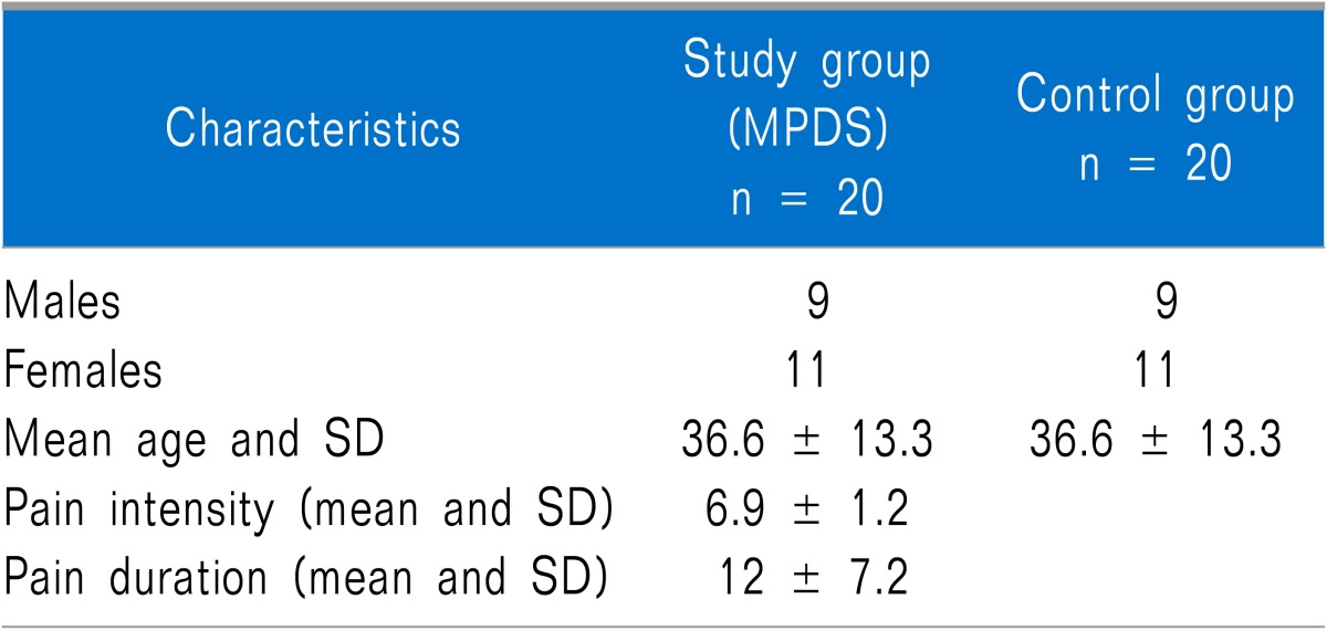
Forty patients were enrolled in this trial. The MPDS group was comprised of 9 males and 11 females and the mean age was 36.6 years (range 18-51 years). The control group was comprised of the same number of age and sex matched individuals (Table 1).
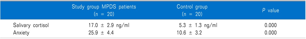
The mean salivary cortisol levels were 17.0 ± 2.9 ng/ml in the MPDS patients and 5.3 ± 1.3 ng/ml in the controls with a P value of 0.000. The mean anxiety levels in the MPDS group were 25.9 ± 4.4 and 10.6 ± 3.2 in the control group (p < 0.001) presented in Table 2.
We found that salivary cortisol and anxiety levels were significantly higher in the MPDS group compared to the control group. There was a highly significant positive correlation (p < 0.001) between the salivary cortisol levels and anxiety.
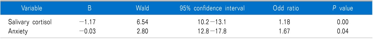
A logistic regression model in which the MPDS was taken as the dependent variable and salivary cortisol and anxiety levels were taken as the independent variables were performed. Salivary cortisol levels and anxiety scores were found significantly related with MPDS. Group membership of each individual can be predicted correctly with 89 % probability in this model (Table 3).
Go to :
DISCUSSION
The results of this study suggest that both salivary cortisol and anxiety levels were increased in the MPDS patients compared to the control group. Literature data on TMDs clearly support the existence of an association with a number of psychosocial disorders [13]. In particular, some early works suggested that populations of patients with the muscular form of TMDs were the most compromised from a psychosocial viewpoint [14,15].
It is considered that persons exposed to stress are under increased risk of the occurrence and progression of MPDS, as stress and unpleasant life experiences are more frequent in patients with dysfunctions [16]. One of the most frequently suggested mechanisms causing myofascial pain associated with TMDs is hyperactivity of the masticatory muscles [8]. When exposed to stress, these patients respond with increased masticatory muscle activity, rather than with a general increase in body muscle tonus. Such activity whether centrally generated or peripherally manifested as parafunctional habits, or both, can result in muscular fatigue and spasm, leading to MPDS [17].
The main system in the human body associated with stress psychobiology is the HPA axis, and the major marker found in saliva associated with the HPA axis is cortisol. Cortisol activates various energy sources within the body to initiate an automatic fight or flight response [18]. This process takes place through the breakdown of proteins, conversion of amino acids and lactic acid to glucose and through the breakdown of triglycerides [19]. This activation of energy sources eventually results in fatigue, wear and tear on bodily muscles and steroid diabetes [20].
Jones et al. [7] showed a significantly higher cortisol response to experimental stress in TMDs group than the control group. Korszun et al. [21] evaluated plasma cortisol levels in patients with muscular TMDS and compared with controls concluded that increased cortisol levels were found in the muscular group of TMDs patients than in the controls. These studies are supported by another study conducted by Yoshihara et al. [8] who concluded that plasma cortisol, adrenaline, nor adrenaline were significantly higher in patients suffering from MFP than in healthy controls. Our study results also showed that anxiety and salivary cortisol levels were statistically significantly higher in MFP patients compared to the control population, matching with the results of previous studies. In contrast to these studies, Nilsson et al. [22] concluded that patients with TMDs, irrespective of diagnosis, appeared to be more psychologically distressed than controls, but did not find any corresponding differences in salivary cortisol levels.
Some studies evaluated only psychosocial profiles in TMD patients by using different questionnaires and showed the existence of a close association between pain and psychosocial disorders in TMDs patients and also showed that myofascial pain patients differ from those with other TMDs in relation to some psychopathological symptoms and MFP patients were the most compromised from a psychosocial view point compared to any other group of TMDS [5,13,15,23-25]. Our study also showed statistically significant higher anxiety levels compared to the controls matching with the results of these studies.
In conclusion, this study showed a relationship between psychological and endocrine variables in MPDS patients; therefore, it is important to take an integrated approach that covers the whole biopsychosocial spectrum. These results suggest that teamwork between clinicians, psychiatrists and dentists is needed for a successful outcome.
Go to :
 XML Download