

 PDF
PDF Citation
 Print
Print


Herpes zoster is a disease where grouped vesicobulous skin lesions appear along the ipsilateral one or two dermatome and is accompanied by pain. This disease is caused by the reactivation of the varicella-zoster virus (VZV), which may be dormant in the sensory ganglia of the cranial nerve or in the dorsal root ganglia after a previous varicella infection. A disseminated herpes zoster infection can be diagnosed when 20 or more blisters develop systemically within a week of showing skin symptoms found in typical herpes zoster infections. This occurs very rarely compared to the general herpes zoster infection, where most cases develop in immune-compromised elderly people, patients with hematological malignancies, or in human immunodeficiency virus infections, along with patients who are undergoing chemotherapy. This study reports a case where a disseminated herpes zoster infection, with accompanying Ramsay Hunt syndrome, was treated in an elderly patient with no clear evidence of immunosuppression.
CASE REPORT
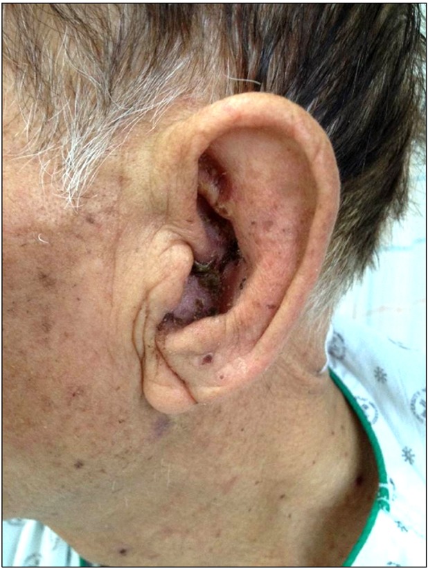
A 75 year old male patient visited the pain clinic at our hospital complaining of stabbing pain and grouped erythematous vesicles in the external auricle and the C2 dermatome of the ipsilateral temporal region, which started approximately 10 days before (Fig. 1). At the initial occurrence of the symptoms, the patient had been diagnosed with an acute herpes zoster infection at the local ENT and dermatology hospital, so was taking oral antiviral agents for more than a week with no improvements in the symptoms. According to his premedical history, the patient had been undergoing treatment for diabetes mellitus and angina, while taking aspirin, 100 mg/day, and cilostazol, 200 mg/day, (pletaal@). At the time of the hospital visit, the visual analogue scale was 7/10, so the patient was hospitalized. Aspirin and cilostazol were temporarily discontinued and oral drug treatment was started with pregabalin, 150 mg/day, nortriptyline, 10 mg/day, and acetaminophen, 750 mg/day. For the relief of sympathetic mediated pain, a stellate ganglion block was performed daily using 0.375% ropivacaine 6 cc with a paratracheal approach technique on the left C6 level. According to the routine blood tests taken at the time of hospitalization, there were no abnormal observations other than an erythrocyte sedimentation rate (ESR) of 39 mm/hr and a high sensitive C-reactive protein (hs-CRP) of 5.81 mg/dl.
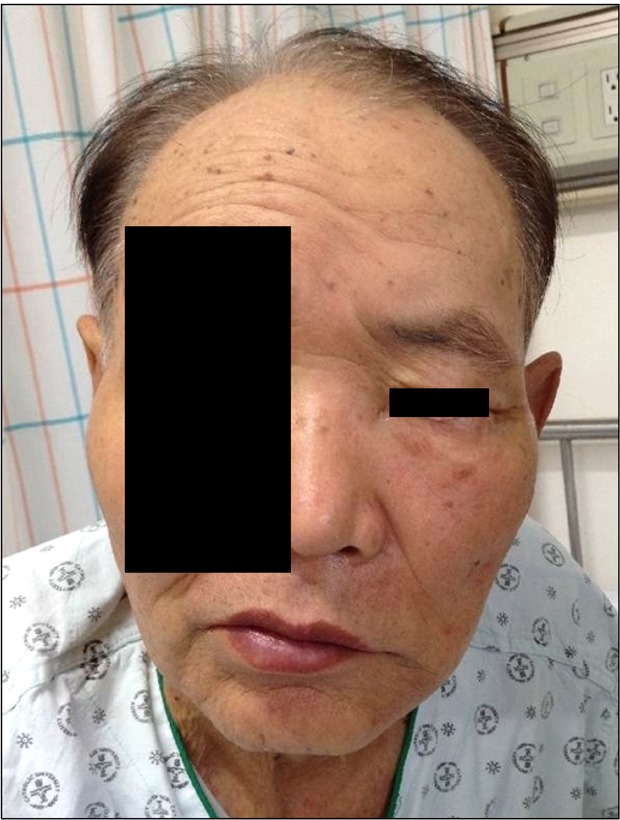
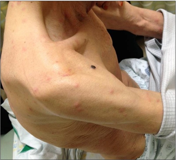
On the morning of the second day of hospitalization, skin eruptions began to appear in numerous areas along the bilateral scalp, posterior neck, shoulder, upper arm, and post upper back (Fig. 2, 3). The patient complained of an itching sensation, but no pain in the newly blistered areas. At the same time, there were symptoms of left facial palsy, hearing impairment, otalgia, and tinnitus, and the patient was unable to walk independently due to vertigo with accompanying mild cognitive disability (Fig. 4). His vital signs showed that his blood pressure, heart rate, and respiration rate were normal, but the temperature had risen to a maximum of 38.0℃, while there were no headaches, nausea, or Kernig's signs. Hence, a disseminated herpes zoster infection with accompanying Ramsay Hunt syndrome in the left facial area was suspected, so a blood culture, urine culture, tumor marker (CEA, CA 19-9, PSA), serum test for anti-HIV antibody (ELISA) and anti-VZV IgM antibody (ELISA), and chest x-ray were performed first to differentiate the cause of infection and immune suppression. Immediately after the above symptoms were observed, nephrologists, neurologists, dermatologists, and otolaryngologists were consulted, with the resulting diagnosis being a disseminated herpes zoster infection. In concern for renal injury from old age, the BUN and creatinine figures were observed every 2 days, and acyclovir, 1,500 mg/day, was IV administered for a week. Pregabalin was stopped, as it may have caused the cognitive disability and vertigo, and was replaced with neurontin, 900 mg/day. The stellate ganglion block was continued. Considering the possibility of cerebrovascular disease or myelopathy, a brain stroke MRI was taken on the 3rd day of hospitalization, followed by a T2 sagittal whole body spine MRI on the 5th day of hospitalization, but no abnormalities were observed. The possibility of meningitis from the virus was also suspected, so a CSF culture (body fluid examination, gram stain, fungus, acid fast bacilli smear) was performed, but again no abnormalities were observed. The administration of steroids was considered for the Ramsay Hunt syndrome, but it was not attempted due to concerns of immune suppression. No abnormalities were observed in the additionally performed cultures (blood, urine, CSF), tumor markers, anti-HIV antibodies, and anti-VZV IgM antibody tests. The hs-CRP was high at 2.44 mg/dl, but it was lower than at the time of hospitalization, so there was a gradually decreasing trend that recovered to normal figures before discharge from the hospital. While maintaining drug therapy and observing the progress, all vital signs returned to normal on the 4th day and the skin lesions did not develop further after the 6th day of hospitalization. Gradually, the vertigo and tinnitus symptoms also improved, so by the 10th day the patient recovered enough to walk independently. The patient began to recover from the facial palsy and hearing impairment from the 9th day, so the patient was discharged according to his wishes on the 12th day while maintaining outpatient treatment at the clinic. The patient visited the clinic every week for two months after his discharge, and there were nearly no complications.
Go to : 
DISCUSSION
The reactivation of VZV is seen to be connected to the decrease in VZV-specific T-cell immunity, and with an increase in age the cell mediated immunity is weakened, thus there is a higher incidence rate of herpes zoster and the possibility of it developing into postherpetic neuralgia [1]. The incidence rate of disseminated herpes zoster infections is very high, at 10-40% in patients with decreased immunity, especially in patients with HIV infections, hematological malignancies, and those who are undergoing chemotherapy, since they have a decreased number and decreased activity of T lymphocytes. The recurrence rate is also high, so 10 mg/kg (or 500 mg/m2) of acyclovir is IV administered every 8 hours and replaced with oral agents (valacyclovir, famciclovir) when symptoms are controlled, or patients with less severe immune suppression can be administered with oral agents from the beginning [2-6].
VZV lies dormant extensively along the sensory ganglia, so herpes zoster infections can occur on any part of the body. When it occurs along several dermatomes, as in disseminated herpes zoster infections, the virus can be spread through interconnections between several ganglia or hematogenous spreading. In our case, the dorsal root ganglia of the geniculate, petrous, accessory, jugular, and C2-C3 were all connected anatomically, so if inflammation occurred in any one of the ganglia, it could spread to other adjacent ganglia [7-9].
Ramsay Hunt syndrome is also known as otic herpes zoster, which is when VZV is reactivated in the geniculate ganglia and causes skin lesions in the face and ears, accompanied with peripheral facial palsy. VZV also invades the 8th cranial nerve causing tinnitus, vertigo, and hearing impairment. Generally, the infection first appears as neurosensory abnormalities 2-15 days after the first occurrence of skin eruptions, but in some cases the symptoms appear before the skin lesions or when there are no skin lesions, making it difficult to diagnose. Facial palsy is most severe 1 week after the occurrence and then gradually recovers, with poor prognosis being observed in cases with advanced age, elevated arterial blood pressure, vertigo, or diabetes. Treatment is generally the administration of high dose corticosteroids and antiviral agents, but for this case, steroids were not administered due to concern for immune suppression. Fortunately, there was early recovery and no severe complications. There are numerous studies which report that there were good nervous functions and improved recovery rates in groups where antiviral agents were administered together than in groups where corticosteroids were administered exclusively. When there is paralysis, a high dose of corticosteroids plays the role of accelerating the recovery from facial palsy by decreasing the degeneration of the facial nerve through the reduction of neural edema and by the alleviation of vascular spasms [10-15].
Neurological complications, which can occur from VZV infections other than Ramsay Hunt syndrome, are myelitis, aseptic meningitis, acute or chronic encephalitis, Bells' palsy, granulomatous cerebral angiitis, and Gullain-Barre syndrome, and although occurrences are rare, it can result in severe consequences. In immune-suppressed patients, disseminated herpes zoster can occur due to viremia of VZV, and when seeding occurs in the internal organs, such as the lungs, liver, intestines, or brain, the fatality rate is reported to be 5%-15%, despite antiviral therapy [5].
In this case, a disseminated herpes zoster infection was observed along the left geniculate ganglia and the superior dorsal root ganglia in an elderly patient. Hence, the possibility of severe complications was anticipated to be high compared to general cases of Ramsay Hunt syndrome, where a single cranial ganglion is affected by a herpes zoster infection. Therefore, various tests were performed in order to find evidence of immunity disorders while performing antiviral therapy and a stellate ganglion block. However, contrary to expectations, no abnormalities were observed and the patient was successfully treated without any severe complications in a time frame similar to the pathogenesis and regression of general acute herpes zoster infections. Currently, there are no standard criteria or diagnostic indices to predict the possibility or prognosis of disseminated herpes zoster infections in aged patients without clear evidence of immune suppression. However, in the case of occurrence, there is a high possibility for systemic complications and the development of postherpetic neuralgia, so intensive treatments, including antiviral therapy and sympathetic nerve block, should be performed as early as possible.
Go to :
 XML Download