Abstract
Femoroacetabular Impingement (FAI) arises from morphological abnormalities between the proximal femur and acetabulum. Impingement caused by these morphologic abnormalities induces early degenerative changes in the hip joint. Furthermore, FAI patients complain of severe pain and limited range of motion in the hip, but a guideline for treatment of FAI has not yet been established. Medication, supportive physical treatment and surgical procedures have been used in the treatment of the FAI patients; however, the efficacies of these treatments have been limited. Here, we report the diagnosis and treatment for 3 cases of FAI patients. Intra-articular (IA) steroid injection of the hip joint was performed in all three patients. After IA injection, pain was reduced and function had improved for up to three months.
Femoroacetabular Impingement (FAI) is a syndrome from the abnormal collision of hip joint which changes the morphology. FAI progresses gradually to secondary hip osteoarthritis (OA) in young patients because structural abnormalities induce impingement. FAI is classified into two typical types, and there are combinations of the two types. The first is the cam type in which the radius of the femur head is increased due to excess bone at the head-neck junction or to an unusual shaped pistol grip deformity or asphericity of the femur head. The second is the pincer type which is caused by an abnormality of the acetabulum and by the orientation of the acetabulum within the pelvis [1] (Fig. 1). Repetitive trauma causes an early hip OA in the beginning of the disease; thereafter, acetabular labrum tearing and excruciating pain develop [1]. There is no established guideline for treatment although medication, supportive physical treatment and surgical procedures have used in FAI patients. On the other hand, the effect of intra-articular (IA) steroid injection in hip OA has been proven in previous studies [2,3]. The main effects of IA steroid injection in hip OA are pain control and reduced synovial hypertrophy [3]. It was reported that these effects persist over 8 weeks [4]. Therefore, we thought that IA steroid injection would be beneficial in FAI because it has an anti-inflammatory effect. We report here the experience of diagnosing and treating 3 cases of FAI.
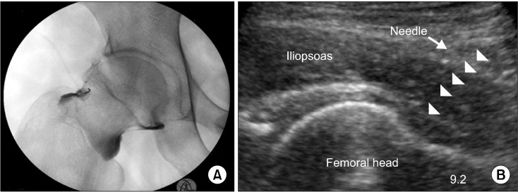
A 42-year-old male patient (height 170 cm, weight 63.7 kg) visited our pain clinic with complaint of severe right groin pain. Two years earlier, the patient had been diagnosed with bilateral cam type FAI and underwent arthroscopic surgery in the right hip after hip magnetic resonance imaging which showed fibrocystic change of the right femur and superior labral tear and minimal joint effusion on both hips. The recurrent pain began about 1 year prior to his visit and walking was impossible. The degree of pain was 10/10 on the visual analogue score system (VAS, ranging from 0 = no pain to 10 = absolutely intolerable pain) and the Oxford hip score (OHS, function of hip joint, excellent = below 19, good function = 19-26, fair = 27-33, poor = 33 or more) was 47/60. During the physical examination, the straight leg raising test (SLR) was right 45° and left 80°. The FABER test and anterior impingement test (flexion and internal rotation of knee) were all positive at the right hip. The frog lateral view of the X-ray showed left superior labral calcification and an osteophyte at the right femur neck. In addition, the head-neck offset of the left femur was decreased (Fig. 2A). The alpha angle in the translateral view was 78.2° (Fig. 3). The ultrasonographic finding showed mild effusion and capsular thickness. The patient had no previous past medical history and took tramadol 200 mg, NSAIDS 200 mg and gabapentin 600 mg per day. We decided to perform IA steroid injection under C-arm guidance. Written informed consent was received after sufficient explanation about the procedure and related complications. With the patient in the supine position, C-arm fluoroscopy was focused on the hip joint in the anterolateral view. After insertion of a 22 gauge spinal needle, 0.75% ropivacaine 5 ml and triamcinolone 40 mg injection was achieved in the right hip joint (Fig. 4A). The patient visited our clinic again checking his VAS, OHS and satisfaction scores (5-point Likert scale; 5 = very satisfied, 4 = somewhat satisfied, 3 = neither satisfied nor dissatisfied, 2 = somewhat dissatisfied, 1 = very dissatisfied) at 2, 4, 8 and 12 weeks after injection (Table 1). The patient took NSAIDS 200 mg intermittently during the 12 weeks. Although moderate right hip pain remained, the patient could walk and return to work.
A 59-year-old female patient (height 161 cm, weight 59 kg) visited our pain clinic with left hip joint pain. The patient had been taking medications for rheumatoid arthritis. The patient had been diagnosed with pincer type FAI about two years prior to her visit. The hip AP (anteroposterior) view of the X-ray showed suspicious FAI, with labral calcification and excessive coverage by the superior margin of both acetabula (Fig. 2B). The pain site was in the lateral and subgluteal area of only the left hip joint. The degree of pain was 7/10 on the VAS and the OHS was 26/60. During the physical examination, the SLR was right 90° and left 90°. FABER test was positive on the left side. However, the anterior impingement test was negative for both hips. No abnormality was found in the ultrasonographic image. We decided to perform ultrasound guided IA steroid injection. Written informed consent was received after sufficient explanation of the procedure and related complications. With the patient in the supine position, the hip was internally rotated about 15-20°. An ultrasound convex probe (2-5 MHz, MicroMAXXTM, Sonosite, USA) was aligned with the long axis of the femoral neck, including the acetabulum and the femoral head. A 22 gauge spinal needle was then advanced under direct ultrasonographic guidance into the anterior synovial recess at the junction of the femoral head and neck (Fig. 4B). Injection of 0.75% ropivacaine 5 ml and triamcinolone 40 mg in the left hip joint was performed. The patient visited our clinic again checking her VAS, OHS and satisfaction scores at 2, 4, 8 and 12 weeks after injection (Table 1). The patient took her previous medications for rheumatoid arthritis during the 12 weeks and additional analgesics were not prescribed. Three months after the injection, the pain in the trochanteric area was partially reduced.
A 50-year-old male patient (height 175 cm, weight 73 kg) visited our pain clinic with complaint of pain in both hip joints. The pain had been ongoing for one year prior to his visit. The patient had been prescribed NSAIDS and muscle relaxants at a local pain clinic whenever he felt pain. The pain in the right anterior groin area was more severe than that of the left side. Because of the hip pain, the patient could not sit crossed-legged on the floor. The degree of pain was 3/10 on the VAS scale and the OHS was 21/60. During the physical examination, the SLR was right 90° and left 90°. The FABER test and anterior impingement test were positive on both sides. The frog lateral view of the X-ray showed the possibility of mixed type FAI of the right hip (Fig. 2C). The head-neck offset of the right femur was decreased and there was excessive coverage by the superior margin of the right acetabulum. The alpha angle in the translateral view was 75.3°. No abnormality was found in the ultrasound image. We performed C-arm fluoroscopy guided injection of 0.75% ropivacaine 5 ml and triamcinolone 40 mg in the right hip joint just like case 1. His symptom was much improved after the injection. The patient visited our clinic again checking his VAS, OHS and satisfaction scores at 2, 4, 8 and 12 weeks after injection (Table 1). The patient took NSAIDS 200 mg during the first month after injection and thereafter, was discontinued. The patient could sit crossed-legged on the floor 2 weeks after the injection although mild right hip pain remained.
In this case series report, the first case was a typical cam type in a young patient and the second case was a pincer type in a rheumatoid arthritis patient. The third case was a mixed type (cam and pincer type). Three months after IA steroid injection, the patient in the first case complained of moderate pain although function of the hip joint was improved. On the other hand, the patient in the second case complained of partially decreased pain (change from VAS 7 to 5) and improved hip joint function. The patient in the second case did not take additional analgesics after IA injection and had high satisfaction. Therefore, we consider the patient in the second case as a good responder. The patient in the third case complained of mild pain.
The patient in the first case had advanced hip OA although the labral tear had been removed through arthroscopic surgery. Therefore, the morphological abnormalities resulted in the recurrence of FAI and impingement leading to an early degenerative change of the hip joint which could cause injuries to the labrum in young patients. Early diagnosis of FAI is important; however, the risk factors of FAI and the current prevalence rate in the general population are not well known.
FAI patients have two problems simultaneously, pain and disability. The pain begins insidiously with or without trauma and impingement occurs by flexion of the hip during exercises. The patient complains of sharp pain with flexion and internal rotation of the hip joint. Although the pain depends on the location and size of the lesion, the pain is mainly located in the groin. Limited terminal hip motion is a typical feature in FAI patients and the pain can worsen while sitting for a long time or from climbing stairs [5]. FAI patients have a difficult time to squat and a snapping or clicking sensation is common [1,5]. On the other hand, the specificity of the physical exams for FAI is low. The physical exams induce impingement between the acetabular rim and femur head-neck junction. The anterior impingement test consists of passive hip flexion and internal rotation, and adduction causes deep anterior pain and decreased motion [5]. The FABER or Patrick's test is performed by flexing, abducting and externally rotating the tested leg. However, the positive results for those tests are not specific to FAI; abnormalities of intra-articular, psoas, or sacroiliac lesions can show positive results, too [5].
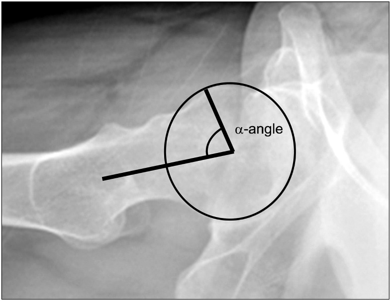
The most important point in the diagnosis of FAI is the bony abnormal morphology. AP pelvic, 45° Dunn, and frog lateral radiographs could be used as diagnostic method for most FAI patients without CT. The cam type in cases 1 and 3 showed a decreasing head-neck offset in the frog lateral view. The pincer type in case 2 showed excessive bony coverage by the acetabular rim in the hip AP view. Additionally, plain radiography of the femoral head alpha angle for the cam type had higher sensitivity than that of CT [6]. The alpha angle is the angle between two lines; one line is up to the point of no sphericity of the femoral head from the center of the femoral head and the other line is up to the center of the femoral head from the center of the femoral neck at the narrowest point [7] (Fig. 3). If an alpha angle greater than 50° was measured, the camtype FAI deformity was considered [8,9]. The alpha angles in cases 1 and 3 in the translateral view were 78.2° and 75.3° (Fig. 3). However, magnetic resonance imaging is required to evaluate the labrum and articular cartilage [10].
First line treatment of femoroacetabular impingement is physiotherapy or anti-inflammatory therapy, but there have been many questions about their effectiveness [11]. Surgical treatment of the morphological changes of the femoral head and acetabulum is also done. Ganz argued that a delay in surgical correction of symptomatic patients may lead to disease progression and that FAI requires surgery [12]. FAI patients with early primary OA who received hip arthroscopic labrectomy had relative improvement in pain score and showed higher satisfaction [13]. However, hip arthroscopic surgery of FAI patients with severe OA showed relatively less improvement and only a temporary pain reduction [13,14]. Surgical dislocation was effective in patients with early degenerative changes but had no benefit in patients with advanced degenerative changes or extensive cartilage damage [15]. On the other hand, intra-articular steroid injection as a treatment for OA is known to be effective. It is effective in reducing pain and synovial hypertrophy and could be a relatively safe treatment [3]. Therefore, we assumed that IA steroid injection for FAI with secondary OA would be effective. IA steroid injection has an important role in the treatment of FAI, which is an anti-inflammatory effect. Its anti-inflammatory effect decreases the inflammation caused by OA. This effect was explained such that patients with hip joint effusion show a better response to IA steroid injection [16]. According to the American College of Rheumatology (ACR) recommendations for IA steroid injection, we chose a steroid (triamcinolone) dose of 40 mg mixed with local anesthetics [17]. We used OHS to evaluate quality of life. OHS is simple, has a high follow-up rate, and can be easily analyzed. It consists of twelve questions, and each question is composed of five scores from 1 (none) to 5 (extreme). The total score is calculated by summing each score and the minimum is 12 and the maximum is 60 [18]. The OHS scores for all 3 patients decreased meaning the patients were gradually getting a better quality of life. As a result, the two main problems of FAI, disability and pain, were improved through IA steroid injection. The limitation of IA steroid injection is a low response to FAI with severe OA. Another author reported corticosteroid injections in fifty-two patients with symptomatic hip osteoarthritis were effective for 3 months. Although it showed good efficacy in moderate and mild OA, it showed only a modest effect in severe OA [2]. Treatment for FAI due to severe OA has low effectiveness for not only IA steroid injection but also for surgery. We think that the good responses to the IA steroid injections in these cases were associated with the severity of OA. The patient with severe OA (case 1) showed good functional improvement, but moderate pain remained after injection. Whereas the patients with mild OA showed both good functional improvement (case 3) and pain relief (cases 2 and 3). Therefore, FAI, a leading cause of secondary hip OA in young adults, should be detected early to prevent disease progression. Moreover, it is important to be aware of the symptoms and signs associated with FAI. However, complications from IA steroid injection such as infectious arthritis, osteonecrosis and necrotizing fasciitis have been reported in previous studies [19,20]. IA steroid injection in weight-bearing joints based on the recommendation of the ACR should not be performed more than once per month or more than 4 times per year.
In conclusion, IA steroid injection reduced the pain and improved function in the 3 cases with FAI, and it could be an effective treatment for mild hip OA more than for severe hip OA. However, it is important to be aware of complications from IA steroid injection. A large number of studies have not been conducted and there are limited long-term studies for the treatment of FAI. Further research for novel treatments and evidence-based algorithm for FAI should be conducted in the future.
References
1. Reid GD, Reid CG, Widmer N, Munk PL. Femoroacetabular impingement syndrome: an underrecognized cause of hip pain and premature osteoarthritis? J Rheumatol. 2010; 37:1395–1404. PMID: 20516017.

2. Lambert RG, Hutchings EJ, Grace MG, Jhangri GS, Conner-Spady B, Maksymowych WP. Steroid injection for osteoarthritis of the hip: a randomized, double-blind, placebo-controlled trial. Arthritis Rheum. 2007; 56:2278–2287. PMID: 17599747.

3. Micu MC, Bogdan GD, Fodor D. Steroid injection for hip osteoarthritis: efficacy under ultrasound guidance. Rheumatology (Oxford). 2010; 49:1490–1494. PMID: 20338889.

4. Atchia I, Kane D, Reed MR, Isaacs JD, Birrell F. Efficacy of a single ultrasound-guided injection for the treatment of hip osteoarthritis. Ann Rheum Dis. 2011; 70:110–116. PMID: 21068096.

5. Philippon MJ, Stubbs AJ, Schenker ML, Maxwell RB, Ganz R, Leunig M. Arthroscopic management of femoroacetabular impingement: osteoplasty technique and literature review. Am J Sports Med. 2007; 35:1571–1580. PMID: 17420508.

6. Nepple JJ, Martel JM, Kim YJ, Zaltz I, Clohisy JC. ANCHOR Study Group. Do plain radiographs correlate with CT for imaging of cam-type femoroacetabular impingement? Clin Orthop Relat Res. 2012; 470:3313–3320. PMID: 22930210.

7. Nötzli HP, Wyss TF, Stoecklin CH, Schmid MR, Treiber K, Hodler J. The contour of the femoral head-neck junction as a predictor for the risk of anterior impingement. J Bone Joint Surg Br. 2002; 84:556–560. PMID: 12043778.

8. Beaulé PE, Zaragoza E, Motamedi K, Copelan N, Dorey FJ. Three-dimensional computed tomography of the hip in the assessment of femoroacetabular impingement. J Orthop Res. 2005; 23:1286–1292. PMID: 15921872.

9. Johnston TL, Schenker ML, Briggs KK, Philippon MJ. Relationship between offset angle alpha and hip chondral injury in femoroacetabular impingement. Arthroscopy. 2008; 24:669–675. PMID: 18514110.

10. Zaragoza E, Lattanzio PJ, Beaule PE. Magnetic resonance imaging with gadolinium arthrography to assess acetabular cartilage delamination. Hip Int. 2009; 19:18–23. PMID: 19455497.

11. Leunig M, Beaulé PE, Ganz R. The concept of femoroacetabular impingement: current status and future perspectives. Clin Orthop Relat Res. 2009; 467:616–622. PMID: 19082681.

12. Leunig M, Ganz R. FAI - concept and etiology. Orthopade. 2009; 38:394–401. PMID: 19407990.
13. Hwang DS, Rhee KJ, Kwon ST, Kim KC, Lee CH, Yang JH. Radiologic and clinical findings of anterior femoroacetabular impingement in early osteoarthritis of the hip. J Korean Orthop Assoc. 2005; 40:630–634.

14. Guanche CA, Bare AA. Arthroscopic treatment of femoroacetabular impingement. Arthroscopy. 2006; 22:95–106. PMID: 16399468.

15. Beck M, Leunig M, Parvizi J, Boutier V, Wyss D, Ganz R. Anterior femoroacetabular impingement: part II. Midterm results of surgical treatment. Clin Orthop Relat Res. 2004; (418):67–73. PMID: 15043095.
16. Micu MC, Bogdan GD, Fodor D. Steroid injection for hip osteoarthritis: efficacy under ultrasound guidance. Rheumatology (Oxford). 2010; 49:1490–1494. PMID: 20338889.

17. Hochberg MC, Altman RD, Brandt KD, Clark BM, Dieppe PA, Griffin MR, et al. American College of Rheumatology. Guidelines for the medical management of osteoarthritis. Part II. Osteoarthritis of the knee. Arthritis Rheum. 1995; 38:1541–1546. PMID: 7488273.
18. Kalairajah Y, Azurza K, Hulme C, Molloy S, Drabu KJ. Health outcome measures in the evaluation of total hip arthroplasties--a comparison between the Harris hip score and the Oxford hip score. J Arthroplasty. 2005; 20:1037–1041. PMID: 16376260.

19. Kruse DW. Intraarticular cortisone injection for osteoarthritis of the hip. Is it effective? Is it safe? Curr Rev Musculoskelet Med. 2008; 1:227–233. PMID: 19468910.

20. Cheng J, Abdi S. Complications of joint, tendon, and muscle injections. Tech Reg Anesth Pain Manag. 2007; 11:141–147. PMID: 18591992.

Fig. 1
Type of femoroacetabular impingement. (A) Normal hip joint. (B) Cam type (arrow: decreased head-neck offset of femur). (C) Pincer type (arrow: excessive bony coverage by the acetabular rim). (D) Mixed type.

Fig. 2
Simple radiographic classification of femoroacetabular impingement. (A) Cam type (frog lateral view of hip). (B) Pincer type (anteroposterior view of hip). (C) Mixed type (frog lateral view of hip).

Fig. 3
Measurement of the alpha angle of the hip. The alpha angle is the measured angle between the line connecting the point of no sphericity of the femoral head from the center of the femoral head and the other line extending up to the center of the femoral head from the center of the femoral neck at the narrowest point. Translateral view of hip is used.

Fig. 4
(A) Fluoroscopy guided intra-articular (IA) injection of the hip joint (case 1). (B) Ultrasound guided IA injection of the hip joint (case 2).

Table 1
Changes of Treatment Effect in the Three Femoroacetabular Impingement Patients

VAS: visual analog scale, ranging from 0 = no pain to 10 = absolutely intolerable pain, Oxford Hip Score (OHS): excellent (below 19), good (19-26), fair (27-33), and poor (33 or more), satisfaction, 5-point Likert scale: 5 (very satisfied), 4 (somewhat satisfied), 3 (neither satisfied nor dissatisfied), 2 (somewhat dissatisfied), 1 (very dissatisfied).


 PDF
PDF Citation
 Print
Print


 XML Download